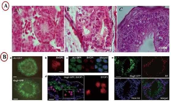
Figure 2. Experimental methods schematic presentation of in vitro spermatogenesis using culture of seminiferous tubules. A) Histological section of the testis tissue fragment cultured for (A) 8 weeks, (B) 10 weeks, and (C) 12 weeks. Stars and the arrow demonstrate spermatogonial cell and spermatocyte cell during culture, respectively (Alrahel et al., 2018). B) a, 10% KSR induced the expression of both Acr-GFP and Gsg2-GFP in 2.5 and 0.5 dpp mouse testes, respectively. b, Immunostaining with anti-SYCP3 antibody. c, Acr-GFP-expressing cells (green) at the pachytene stage were also stained with SYCP1 (red). d, In a cryosection of a Gsg2-GFP testis tissue, SYCP1 was demonstrated in cells (red) outside the Gsg2-GFPpositive cells (green). Hoechst (blue). e, Gsg2-GFP-expressing testis tissue, originating from 2.5 dpp mice and cultured for 21 days, was cryosectioned and stained with antibodies against GFP, AR, and counterstained with Hoechst dye (Sato et al., 2011).