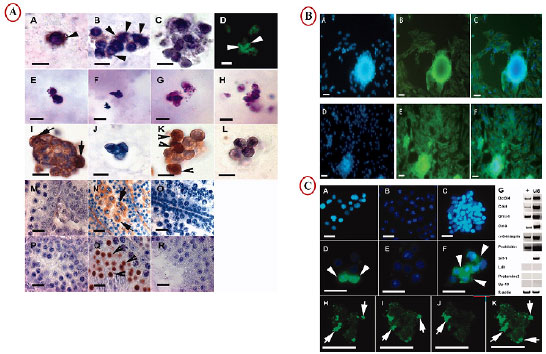
Figure 4. Advanced procedures of 3D culture to induction of spermatogenesis. A) Expression Gfrα-1 (A, B, D) in SACS-cultured cells. An image of a control labeling omitting a primary antibody is shown in C. Expression of the Boule protein (E-I) and Crem (K) in SACS-cultured cells and in the murine testis at different developmental stages (M-R) (Stukenborg et al., 2008). B) Representative immunofluorescence images (A) staining nucleus with DAPI (blue) and (B)showing SCP3 positive cells (green) (C) merge in control group (D, E, F) in co-culture group (Khajavi et al., 2014). C) A heterogeneous cell suspension containing single cells is observed in the unsorted cell fraction after digestion of cells before the separation procedure (A) and in the depleted fraction after MAC sorting with Gfrα-1 (B). A homogeneous cell suspension is observed in the enriched fraction (C). The mentioned fractions are shown after immunofluorescent labeling with anti-Gfrα-1 (unsorted fraction [D], depleted fraction [E], and enriched fraction [F]; arrowheads: Gfrα-1-positive cells [FITC]). Expression analysis of different spermatogenic marker genes (murine spermatogonial stages: Oct3/4, C-kit, Gfrα-1, Cd-9, and α-6-integrin; murine meiotic stages: Prohibitin and Srf-1; murine postmeiotic stages: Ldh, Protamine-2, and Sp-10; positive control: β-actin before (G; unsorted [us] lane) and after sorting with anti-GFRα-1 (G; enriched [+] lane). Confocal microscopy images of fluorescent-labeled cells showed expression of Gfrα-1 (arrows) on the cell surface of spermatogonial cells (H-J; overlay K) (Stukenborg et al., 2008).