JBRA Assist. Reprod. 2025;29(2):282-288
ORIGINAL ARTICLE
doi: 10.5935/1518-0557.20240106
Apolipoprotein A1: A potential biomarker in the secretome of euploid and aneuploid human embryos
1Department of Anatomical Sciences, School of Medicine, Zanjan University of Medical Sciences, Zanjan, Iran
2Department of Embryology, Reproductive Biomedicine Research Centre, Royan Institute for Reproductive Biomedicine, ACECR, Tehran, Iran
3Department of Genetics, Reproductive Biomedicine Research Centre, Royan Institute for Reproductive Biomedicine, ACECR, Tehran, Iran
CONFLICTS OF INTEREST
The authors declare that there is no conflict of interest that could be perceived as prejudicing the impartiality of the research reported.
ABSTRACT
Objective: Morphologic assessment of an embryo is a valuable indicator for determining embryo health; however, it does not provide information on the chromosomal status of an embryo. Therefore, this study aimed to investigate the levels of Apolipoprotein A1 secreted by day-5 embryos in the spent media of euploid and aneuploid human embryos.
Methods: This study utilized 131 spent culture media samples from 22 infertile couples who were referred to the fertility clinic of Royan Institute. Following ovulation induction, retrieved oocytes were fertilized by intracytoplasmic sperm injection. For pre-implantation genetic diagnosis, embryos were frozen and thawed on days 2 to 3 and a single blastomere was isolated from each embryo for the assessment of chromosomal abnormalities by fluorescence in situ hybridization. Five days after fertilization, the levels of Apolipoprotein A1 were determined in the spent media of normal embryos, aneuploid embryos (with chromosome abnormalities), and the control group (medium without any embryos) using enzyme-linked immunosorbent assay.
Results: The Apolipoprotein A1 levels in the secretome of euploid cleavage-arrested embryos were significantly lower than those in the control group (p<0.04). However, Apolipoprotein A1 levels increased significantly in groups of euploid blastocysts, aneuploid cleavage-arrested embryos, aneuploid morulae, and aneuploid blastocysts compared to the control group (p<0.04). Furthermore, the Apolipoprotein A1 levels in the spent media of euploid early blastocysts were significantly higher compared to euploid hatching blastocysts and aneuploid blastocysts in the early, mid, and late stages (p<0.03).
Conclusions: This study highlights the significant potential of Apolipoprotein A1 as a developmental bi-omarker to distinguish between euploid and aneuploid embryos.
Keywords: aneuploidy screening, Apolipoprotein A1, blastocyst, conditioned media, non-invasive
INTRODUCTION
Despite many improvements in assisted reproductive technology (ART), the success rate remains relatively low (Zargar et al., 2021). The selection of a high-quality embryo to transfer to the mother’s uterus is a crucial factor in enhancing the success rate of ART (Zmuidinaite et al., 2021). Furthermore, transferring only one embryo helps reduce the risks associated with multiple pregnancies. Selecting a high-quality embryo is a challenging task and needs the expertise of an experienced embryologist or the application of specialized lab techniques (Mains et al., 2011). Currently, in most fertility clinics, the assessment of embryo quality is carried out according to the morphological criteria (Ebner et al., 2003; Kim et al., 2022); however, morphological assessment alone may not identify chromosome aneuploidies (Bielanska et al., 2005).
Increasing maternal age is one of the important causes of chromosomal abnormalities in human oocytes and embryos (Sawarkar et al., 2021). Approximately 50% of abortions occur during the first trimester of pregnancy due to aneuploidy (Maxwell et al., 2016). Therefore, transferring a euploid embryo (with all 23 pairs of human chromosomes) to the uterus can enhance implantation and live birth rates while reducing the risk of spontaneous pregnancy loss (Sullivan-Pyke & Dokras, 2018).
Pre-implantation genetic diagnosis (PGD) is widely used for aneuploidy screening of an embryo; however, blastomere biopsy for PGD is invasive and may potentially affect the embryo growth and the outcome of in vitro fertilization (IVF) (Chen et al., 2021; Navarro-Sánchez et al., 2022). In contrast to PGD, omics technologies, including genomics (study of genes), transcriptomics (study of mRNA), proteomics (study of proteins) and metabolomics (study of metabolome) offer a noninvasive approach to aneuploidy screening (Anagnostopoulou et al., 2022; Egea et al., 2014). Although the number of omics technology is increasing, transcriptomics, proteomics, and metabolomics are widely used in assisted reproductive technologies. Omics techniques can be used to design non-invasive diagnostic methods that are applicable in fertility clinics. For instance, they can be applied for selecting a high quality embryo, particularly in single embryo transfer protocols. Noninvasive analysis of the embryo secretome (proteins and metabolites secreted by an embryo into the surrounding conditioned media) can detect a set of aneuploidy biomarkers, leading to noninvasive viability screening (Cortezzi et al., 2011; Schoolcraft et al., 2010; Sousa & Monteiro, 2022).
Apolipoprotein A1 (APOA1) is a primary apolipoprotein found in high-density lipoprotein (HDL) and has been identified in human blastocysts (Telford et al., 1990). The levels of APOA1 are positively correlated with the quality of human blastocysts and the rate of implantation. Apparently, cholesterol metabolism has a direct relationship with the quality of blastocysts due to the crucial role of cholesterol in the synthesis of cell membranes of the pre-implantation embryo (Mains et al., 2011). Given the indication that APOA1 levels are linked to human embryo quality, we hypothesized that the quantity of APOA1 in the secretome may be correlated with aneuploidy. Therefore, this study aimed to investigate the APOA1 protein levels in the secretome of human embryos with chromosome abnormalities (aneuploid) and normal embryos (euploid) in a non-invasive manner.
MATERIALS AND METHODS
This study was carried out at the Reproductive Biomedicine Research Center at the Royan Institute and involved analyzing conditioned spent media surrounding embryos from PGD candidates who provided informed consent. The research protocol was approved by the Ethics Committee of Royan Institute, Tehran, Iran (reference number: EC/93/1081).
Patients
The study included women aged between 25 and 40 years who were PGD candidates. The exclusion criteria were as follows: (i) endometriosis, (ii) ovarian polycystic syndrome, and (iii) sperms extracted from the testes or epididymis by surgical procedures. The spent media of embryos that were first vitrified and then underwent PGD were included in this study. The experimental design of this study is summarized in a diagram indicated in Figure 1.
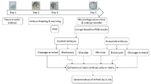
Figure 1. The schematic image indicates the study design in brief.
Embryo culture
Human embryos were produced through Intracytoplasmic Sperm Injection (ICSI) after oocyte retrieval using a standard GnRH agonist long protocol as previously described (Rezazadeh Valojerdi et al., 2009). The newly formed embryos were cultured in G-1 medium (Vitrolife, Kungsback, Sweden), supplemented with 10% recombinant human serum albumin (rHA, Vitrolife) in an environment of 5% CO2 in air at 37°C for 2 - 3 days. On either day 2 or 3, the embryos were cryopreserved and thawed when the endometrium was prepared to receive the embryo (Rezazadeh Valojerdi et al., 2009).
For vitrification, embryos were initially incubated in equilibration solution of 7.5% ethylene glycol (EG) (Sigma-Aldrich, Steinleim, Germany) and 7.5% dimethyl sulphoxide (DMSO) (Sigma-Aldrich) in Ham’s F-10 medium supplemented with 20% Albuminal-5 for 5-15 min. Then equilibrated embryos were exposed to the vitrification solution of 15% EG, 15% DMSO and 0.5 M sucrose in Ham’s F-10 medium supplemented with 20% Albuminal-5 for 50-60 s and then loaded on the tip of the cryotop (Kitazato, Japan) and immediately plunged into the liquid nitrogen. For warming, the embryos were incubated in thawing solution (1M sucrose in Ham’s F-10 medium supplemented with 20% Albumin-5) for 50-60 s and then transferred into dilution solution of 0.5 M sucrose for 3min, followed by another dilution solution of 0.25M sucrose for 3min.
Cryopreserved-thawed embryos (72 hours after fertilization) underwent PGD to detect chromosomal abnormalities. For the embryo biopsy procedure, the embryos were incubated in Ca2+/Mg2±free G-PGDTM biopsy medium (Vitrolife, Sweden) for 1-2 minutes. To detect chromosomal abnormalities, fluorescence in situ hybridization (FISH) was carried out on the isolated blastomere (Bazrgar et al., 2016). Following the biopsy of one blastomere, the embryos were placed in individual 50 µL droplets of G-2 medium (Vitrolife, Kungsback, Sweden) supplemented with 10% recombinant human serum albumin and incubated until day 5. Simultaneously, a 50 µL droplet of G-2 culture media without any embryo was incubated for 48 hours under the same conditions as a blank control.
Grading the embryos on day 5 post-fertilization
On day 5 post-fertilization, all embryos were examined under an inverted microscope (Olympus, Japan) by an expert embryologist, and the embryos were classified into three main groups: embryos arrested at the cleavage stage, embryos at the morula stage, and embryos at the blastocyst stage. Blastocysts were also classified into four subgroups: early blastocyst (grade I) where the blastocoel cavity occupied less than 50% of the embryo volume, mid blastocyst (grade II) where the blastocoel cavity filled 50% or more of the embryo volume, late blastocyst (grade III) where the blastocoel cavity filled the embryos and the zona pellucida (ZP) became thinner, and hatching/hatched blastocysts (grade IV) representing embryos that had hatched from the ZP (Hardarson et al., 2012).
Collection of spent embryo culture media
A total of 131 spent embryo culture media samples from 22 patients with an average maternal age of 31.36±1.07 years were collected. After the receipt of PGD results, the normal embryos (euploid) were transferred to the uterus on day 5 post-fertilization, and their spent culture media were collected. Also, the spent embryo culture media of embryos with two or higher chromosomal abnormalities were collected for the study. In total, all spent embryo culture media, where the embryos were incubated for about 48 hours, from both euploid (41 samples) and aneuploid (90 samples) embryos, along with the blank control culture media (media without embryos, 20 samples) were collected and kept individually in labeled cryotubes at -80°C for further analysis.
Determination of APOA1 levels using the enzyme-linked immunosorbent assay (ELISA) method
The APOA1 levels in spent embryo culture media and blank control media were determined using a quantitative sandwich ELISA kit (ab108803, Abcam Ltd, Cambridge, UK). The minimum detectable dose of APOA1 for this kit was 3 ng/mL. Serial dilutions of the APOA1 protein standard were performed using a diluent to achieve a concentration range from 200 to 3.13 ng in a final volume of 50 µL. The culture media were thawed and diluted to a final volume of 50 µL. Each well in the ELISA plate was coated with polyclonal anti-APOA1 (anti-human APOA1). Also, 50 µL of all standards, diluted samples, and blank control (only diluent) were incubated on the antibody-coated plate at room temperature. All standards, diluted samples, and the blank control were tested in duplicate. After 2 hours, the plate was rinsed, and then the biotinylated APOA1 antibody was added and incubated for 1 hour. Following the plate rinse, the streptavidin conjugate antibody (streptavidin-peroxidase complex) was applied and left to incubate for 30 minutes. Subsequently, the unbound conjugates were washed off. A chromogenic substrate, TMB (3,30,5,50-tetramethylbenzidine), was then used to measure the streptavidin-peroxidase enzymatic reaction. After about 15 minutes, the reaction was halted by adding an acidic stop solution, resulting in a yellow-colored product. The intensity of the yellow color was then measured at 450 nm using a spectrophotometer to determine the APOA1 concentration in all the samples used. Each duplicated media sample was tested three times, resulting in six estimates. These six estimates were analyzed simultaneously to indicate the relative levels of APOA1 compared to the standard curve.
Statistical analysis
After testing the normality of the data through histograms and statistical tests, such as Shapiro-Wilk and Kolmogorov-Smirnov tests, the normally distributed data were subjected to one-way ANOVA and Bonferroni post-hoc test using SPSS 16 software (IBM, Chicago, USA) to assess the significance of differences. Differences were deemed significant if p=0.05, and statistical comparisons between specific groups were carried out using the student’s t-test. The results are expressed as mean±standard error (SE).
RESULTS
APOA1 levels in spent culture media of euploid and aneuploid embryos
APOA1 levels in all aneuploid embryos, including cleavage-arrested embryos (44.74±6.5ng/mL), morulae (50.74±4.2ng/mL), and blastocysts (51.07±3.6ng/mL), as well as in euploid blastocysts (59.58±8.12ng/mL) were significantly higher than those in the control group (29±4.59ng/mL) (p=0.04) (Figure 2). APOA1 levels were significantly lower in euploid cleavage-arrested embryos (14.67±2.63ng/mL) compared to all other groups (p=0.02) and the control group (p=0.04). However, this reduction was not significant compared to the euploid morulae group (35.86±10.03ng/mL) (Figure 2).
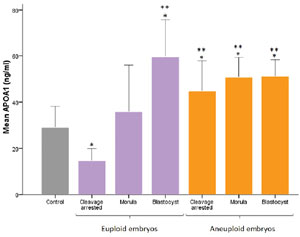
Figure 2. APOA1 level in spent embryo culture media for groups of control (without embryos), euploid embryos and aneuploid embryos. Data are reported as Mean±SE. *: Significant difference between groups (p=0.04) vs. control. **: Significant difference between groups (p=0.02) vs. euploid cleavage arrested.
APOA1 levels in spent culture media of euploid and aneuploid blastocysts at different grades
APOA1 levels were notably higher in euploid early blastocysts (77.67±9.13ng/mL) compared to euploid hatching blastocysts (41.60±3.96ng/mL) and aneuploid blastocysts, including early (44.59±7.2ng/mL), mid (52.75±4.8ng/mL), and late (50±5.43ng/mL) blastocysts (p=0.03) (Figure 3). APOA1 levels changed significantly among euploid blastocysts of different grades, with late-stage blastocysts (71±1ng/mL) showing significantly higher levels compared to mid-stage (61±1ng/mL) and hatching blastocysts (41.6±3.96ng/mL) (p=0.03) (Figure 3). However, the APOA1 levels did not show significant changes in aneuploid blastocysts across different grades (Figure 3).
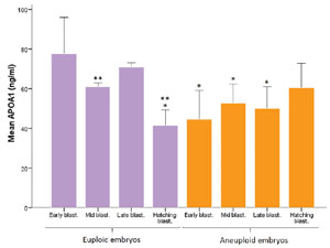
Figure 3. APOA1 level in spent embryo culture media for groups of euploid blastocysts and aneuploid blastocysts at different grades. Data are reported as Mean±SE. *: Significant difference between groups (p=0.03) vs. euploid early blastocysts. **: Significant difference between groups (p=0.03) vs. euploid late blastocysts. Blast.: blastocyst.
DISCUSSION
Finding a noninvasive, low-cost, and time-consuming method for aneuploidy screening of an embryo can help embryologists select a healthy embryo without any chromosomal abnormalities for embryo transfer. Additionally, a specific biomarker in pre-implantation embryos may guide embryologists to transfer only one high-quality embryo to the endometrium, increasing the chance of embryo implantation and avoiding the risk of multiple gestations. Recently, some molecules including ubiquitin (Katz-Jaffe et al., 2006) and lipocalin 1 (McReynolds et al., 2011) have been introduced as biomarkers indicating a healthy state of an embryo. However, to date, no distinct biomarker has been found. Indeed, the complexity of embryo development involves many molecules and factors, making it challenging to identify a single potential biomarker for noninvasive aneuploidy screening, therefore a set of biomarkers is required for the selection of a high-quality embryo.
APOA1 is a main apolipoprotein found in high-density lipoprotein (HDL) (Santos et al., 2008), which plays an important role in cholesterol metabolism during cell division, cell membrane synthesis, and embryo implantation (Brosens et al., 2010). Although there are many studies on APOA1 concerning the female reproductive system, particularly endometrium, there remains insufficient information regarding the synthesis and function of APOA1 in pre-implantation embryos. Given the indication that APOA1 levels are linked to human embryo quality, we hypothesized that the quantity of APOA1 in the secretome may be correlated with aneuploidy.
The reduction in APOA1 in follicular fluid leads to increased embryo fragmentation (Browne et al., 2008) and the occurrence of cleavage-arrested embryos (Trigatti et al., 1999), indicating the importance of cholesterol and APOA1 in pre-implantation embryo development. In women with recurrent implantation failure (RIF), the level of APOA1 in the endometrium was found to be 4.2 times higher than in healthy women. APOA1 levels decrease in the endometrium during the mid-secretory phase under the influence of LH/hCG and also diminish during embryo implantation (Brosens et al., 2010). Therefore, the elevation of APOA1 levels in the secretome of euploid blastocysts, as indicated by the findings of this study, along with the reduction of APOA1 expression in the endometrium during embryo implantation (Brosens et al., 2010), may serve as a signaling mechanism. This signaling could potentially aid in the embryo’s interaction with the mother’s endometrium, leading to a more favorable response of the endometrium to the implanting embryo.
Familial APOA1 deficiency causes a remarkable reduction in HDL levels (Bekaert et al., 1991; Santos et al., 2008), and mice with impaired APOA1 synthesis exhibit very low HDL levels (Williamson et al., 1992). Intracellular cholesterol plays a crucial role in cytokinesis during mitosis (Simons & Ikonen, 2000); thus, cholesterol depletion in urchins results in incomplete cytokinesis (Ng et al., 2005). Also, cholesterol is vital for embryo development as it aids in energy production through oxidative pathways and supports rapid membrane synthesis during cellular proliferation (Sturmey et al., 2006). Therefore, the increase in APOA1 levels in the secretome of euploid blastocysts compared to euploid morulae may be linked to the significance of cholesterol metabolism and phospholipids in pre-implantation embryo development. As the embryo develops from the morula stage (16-32 blastomeres) to the blastocyst stage (100-120 blastomeres), rapid mitosis divisions and a rise in cell numbers occur within only 1-2 days, facilitated by cholesterol metabolism and the synthesis of cell membranes for the newly formed blastomeres. However, minimal and insignificant changes were observed in APOA1 secretion among aneuploid embryos in the current study. Embryos with chromosomal abnormalities likely encounter disruptions in the expression and synthesis of proteins, including APOA1.
Mains et al. (2011) reported that the APOA1 levels in the spent embryo culture media of high-quality blastocysts are higher than in low-quality blastocysts, demonstrating that cholesterol metabolism can be an important factor for embryo developmental competence. This observation could also be linked to the antioxidant properties of HDL in the presence of APOA1 (Goulinet & Chapman, 1997; Kontush & Chapman, 2010; Moss et al., 2012). Therefore, the increase in APOA1 levels in the secretome of euploid blastocysts compared to aneuploid blastocysts may help in scavenging the free radicals generated during ART procedures (Agarwal & Majzoub, 2017).
The evaluation of human embryo secretome presents challenges because of differences in protocols, reagents, and culture media used in fertility clinics. Many fertility clinics utilize serum protein substitutes to supply the culture media, which contain many undesired proteins, including albumin, potentially leading to discrepancies in secretome assessments (Mains et al., 2011). In other words, the accuracy of measurement tools, various embryo culture media, and the actual and precise levels of secreted proteins (varying orders of magnitude) (Ménézo et al., 2006; Sargent et al., 2007) can result in conflicting data. Virant-Klun et al. (2016) reported findings contradicting those of Mains et al. (2011). They found that the APOA1 levels in the spent embryo culture media of high-quality blastocysts are lower than low-quality blastocysts, morulae, and degenerated embryos, supporting the findings of Nyalwidhe et al. (2013). However, this comparison may not be entirely accurate as they measured APOA1 levels in the secretome of blastocysts, whereas Nyalwidhe et al. (2013) measured them in the secretome of day-2 and day-3 embryos.
Nyalwidhe et al. (2013) reported a negative correlation between APOA1 levels in the secretome of 2- and 3-day embryos and the quality of embryos and pregnancy rate. This could be attributed to high-quality embryos using more APOA1 from the embryo culture or APOA1 from culture media binding to the zona pellucida (ZP) or/and the embryo surface (Nyalwidhe et al. 2013). Human embryo development and protein synthesis are entirely reliant on the mother’s genome until the 4-8 cell stage, after which the human embryo genome becomes active, initiating transcription, translation, and protein synthesis at this point (Jukam et al., 2017). APOA1 mRNA has been detected in human blastocysts but not in day-3 human embryos (Telford et al., 1990). However, in another study, the APOA1 protein was identified in the conditioned medium of human embryos on the third day of in vitro embryo culture (Foresta et al., 2016). The conflicting data may arise due to the application of different techniques in the studies. Before the blastocyst stage, the embryo is not able to synthesize active cholesterol because of the absence of APOA1 protein and the lack of hydroxymethylglutaryl co-enzyme A. Apparently, the oocyte is the only source of cholesterol for the embryo before the blastocyst stage (Fujimoto et al., 2010). Therefore, the blastocyst stage could be the best embryo development stage to investigate the levels of APOA1 in embryo secretome. This stage may explain the reasons for the contradictory results between the study by Nyalwidhe et al. (2013) and the present study.
The main limitation of the present study is the inability to track the outcome of the embryo implantation and ongoing pregnancies of the embryos whose conditioned medium was used for the study, as multiple embryos were transferred to the uterus. To address this issue, conducting the study with a protocol of single-embryo transfer would be preferable to monitor the outcomes of embryo implantation and pregnancy. Using vitrified/warmed embryos in this research instead of fresh embryos was another limitation of the study, as vitrification/warming can impact embryo viability and may also change the secreted proteins by the embryo. Therefore, using the fresh embryo secretome could help to avoid this problem. A strength of the present study is that the samples were derived from humans and not based on animal research. Furthermore, the study focused on the human embryo secretome in spent culture media rather than on the human embryo itself, thereby adhering to strict ethical standards regarding research on human samples.
In general, finding a noninvasive method for screening aneuploidy in fertility clinics is useful and necessary. However, to achieve a biomarker for aneuploidy, conducting research with a larger sample size and using various types of laboratory techniques, including mass spectrometry, two-dimensional gel electrophoresis, and protein microarray, is suggested. Furthermore, delving deeper into the embryo secretome enhances our understanding of the biological functions of the embryo and the communication between the developing embryo and its surrounding environment. Based on the findings of this study, APOA1 could be a promising candidate to consider as a potential biomarker for aneuploidy screening, although further comprehensive studies are necessary. Given the complexity of embryo development involving numerous molecules and factors, identifying a single potential biomarker for noninvasive aneuploidy screening may prove challenging. It might be more effective to concentrate on a panel of biomarkers rather than relying only on one.
Authors’ contributions
M.A. designed experiments, provided conceptual advice, analyzed data and revised the manuscript in depth, F.H. carried out experiments and collected data, L.K. carried out experiments, collected data and provided conceptual advice, P.E.Y. carried out experiments and collected data, B.M. carried out experiments and supplied technical support, B.E. supplied technical support and collected data, M.F. carried out experiments and collected data, M.R.V. supervised the research, provided conceptual advice and revised the manuscript in depth. All authors were involved in data interpretation and revision of the manuscript. All authors read and approved the final manuscript.
Financial support
This study was financially supported by a joint grant from Zanjan University of Medical Sciences (grant number: A-12-537-8) and Royan Institute (grant number: 91000220).
REFERENCES
Agarwal A, Majzoub A. Role of Antioxidants in Assisted Reproductive Techniques. World J Mens Health. 2017;35:77-93. PMID: 28497913 DOI: 10.5534/wjmh.2017.35.2.77 Medline
Anagnostopoulou C, Maldonado Rosas I, Singh N, Gugnani N, Chockalingham A, Singh K, Desai D, Darbandi M, Manoharan M, Darbandi S, Leonardi Diaz SI, Gupta S, Henkel R, Sallam HN, Boitrelle F, Wirka KA, Agarwal A. Oocyte quality and embryo selection strategies: a review for the embryologists, by the embryologists. Panminerva Med. 2022;64:171-84. PMID: 35179016 DOI: 10.23736/S0031-0808.22.04680-8 Medline
Bazrgar M, Gourabi H, Eftekhari-Yazdi P, Vazirinasab H, Fakhri M, Hassani F, Chehrazi M, Valojerdi MR. The Effect of Prolonged Culture of Chromosomally Abnormal Human Embryos on The Rate of Diploid Cells. Int J Fertil Steril. 2016;9:563-73. PMID: 26985346 DOI: 10.22074/ijfs.2015.4617 Medline
Bekaert ED, Alaupovic P, Knight-Gibson CS, Laux MJ, Pelachyk JM, Norum RA. Characterization of apoA- and apoB-containing lipoprotein particles in a variant of familial apoA-I deficiency with planar xanthoma: the metabolic significance of LP-A-II particles. J Lipid Res. 1991;32:1587-99. PMID: 1797940 DOI: 10.1016/S0022-2275(20)41644-X Medline
Bielanska M, Jin S, Bernier M, Tan SL, Ao A. Diploid-aneuploid mosaicism in human embryos cultured to the blastocyst stage. Fertil Steril. 2005;84:336-42. PMID: 16084874 DOI: 10.1016/j.fertnstert.2005.03.031 Medline
Brosens JJ, Hodgetts A, Feroze-Zaidi F, Sherwin JR, Fusi L, Salker MS, Higham J, Rose GL, Kajihara T, Young SL, Lessey BA, Henriet P, Langford PR, Fazleabas AT. Proteomic analysis of endometrium from fertile and infertile patients suggests a role for apolipoprotein A-I in embryo implantation failure and endometriosis. Mol Hum Reprod. 2010;16:273-85. PMID: 20008415 DOI: 10.1093/molehr/gap108 Medline
Browne RW, Shelly WB, Bloom MS, Ocque AJ, Sandler JR, Huddleston HG, Fujimoto VY. Distributions of high-density lipoprotein particle components in human follicular fluid and sera and their associations with embryo morphology parameters during IVF. Hum Reprod. 2008;23:1884-94. PMID: 18487218 DOI: 10.1093/humrep/den183 Medline
Chen L, Sun Q, Xu J, Fu H, Liu Y, Yao Y, Lu S, Yao B. A Non-invasive Chromosome Screening Strategy for Prioritizing in vitro Fertilization Embryos for Implantation. Front Cell Dev Biol. 2021;9:708322. PMID: 34434931 DOI: 10.3389/fcell.2021.708322 Medline
Cortezzi SS, Garcia JS, Ferreira CR, Braga DP, Figueira RC, Iaconelli A Jr., Souza GH, Borges E Jr, Eberlin MN. Secretome of the preimplantation human embryo by bottom-up label-free proteomics. Anal Bioanal Chem. 2011;401:1331-9. PMID: 21735065 DOI: 10.1007/s00216-011-5202-1 Medline
Ebner T, Moser M, Sommergruber M, Tews G. Selection based on morphological assessment of oocytes and embryos at different stages of preimplantation development: a review. Hum Reprod Update. 2003;9:251-62. PMID: 12859046 DOI: 10.1093/humupd/dmg021 Medline
Egea RR, Puchalt NG, Escrivá MM, Varghese AC. OMICS: Current and future perspectives in reproductive medicine and technology. J Hum Reprod Sci. 2014;7:73-92. PMID: 25191020 DOI: 10.4103/0974-1208.138857 Medline
Foresta C, Ubaldi FM, Rienzi L, Franchin C, Pivato M, Romano S, Guidolin D, De Caro R, Ferlin A, De Toni L. Early protein profile of human embryonic secretome. Front Biosci (Landmark Ed). 2016;21:620-34. PMID: 26709795 DOI: 10.2741/4410 Medline
Fujimoto VY, Kane JP, Ishida BY, Bloom MS, Browne RW. High-density lipoprotein metabolism and the human embryo. Hum Reprod Update. 2010;16:20-38. PMID: 19700490 DOI: 10.1093/humupd/dmp029 Medline
Goulinet S, Chapman MJ. Plasma LDL and HDL subspecies are heterogenous in particle content of tocopherols and oxygenated and hydrocarbon carotenoids. Relevance to oxidative resistance and atherogenesis. Arterioscler Thromb Vasc Biol. 1997;17:786-96. PMID: 9108795 DOI: 10.1161/01.ATV.17.4.786 Medline
Hardarson T, Van Landuyt L, Jones G. The blastocyst. Hum Reprod. 2012;27:i72-91. PMID: 22763375 DOI: 10.1093/humrep/des230 Medline
Jukam D, Shariati SAM, Skotheim JM. Zygotic Genome Activation in Vertebrates. Dev Cell. 2017;42:316-32. PMID: 28829942 DOI: 10.1016/j.devcel.2017.07.026 Medline
Katz-Jaffe MG, Schoolcraft WB, Gardner DK. Analysis of protein expression (secretome) by human and mouse preimplantation embryos. Fertil Steril. 2006;86:678-85. PMID: 16952510 DOI: 10.1016/j.fertnstert.2006.05.022 Medline
Kim J, Lee J, Jun JH. Non-invasive evaluation of embryo quality for the selection of transferable embryos in human in vitro fertilization-embryo transfer. Clin Exp Reprod Med. 2022;49:225-38. PMID: 36482497 DOI: 10.5653/cerm.2022.05575 Medline
Kontush A, Chapman MJ. Antiatherogenic function of HDL particle subpopulations: focus on antioxidative activities. Curr Opin Lipidol. 2010;21:312-8. PMID: 20581677 DOI: 10.1097/MOL.0b013e32833bcdc1 Medline
Mains LM, Christenson L, Yang B, Sparks AET, Mathur S, Van Voorhis BJ. Identification of apolipoprotein A1 in the human embryonic secretome. Fertil Steril. 2011;96:422-7.e2. PMID: 21676393 DOI: 10.1016/j.fertnstert.2011.05.049 Medline
Maxwell SM, Colls P, Hodes-Wertz B, McCulloh DH, McCaffrey C, Wells D, Munné S, Grifo JA. Why do euploid embryos miscarry? A case-control study comparing the rate of aneuploidy within presumed euploid embryos that resulted in miscarriage or live birth using next-generation sequencing. Fertil Steril. 2016;106:1414-9.e5. PMID: 27692437 DOI: 10.1016/j.fertnstert.2016.08.017 Medline
McReynolds S, Vanderlinden L, Stevens J, Hansen K, Schoolcraft WB, Katz-Jaffe MG. Lipocalin-1: a potential marker for noninvasive aneuploidy screening. Fertil Steril. 2011;95:2631-3. PMID: 21324447 DOI: 10.1016/j.fertnstert.2011.01.141 Medline
Ménézo Y, Elder K, Viville S. Soluble HLA-G release by the human embryo: an interesting artefact? Reprod Biomed Online. 2006;13:763-4. PMID: 17169192 DOI: 10.1016/S1472-6483(10)61021-8 Medline
Moss JI, Garrett TJ, Hansen PJ. Involvement of free cholesterol and high-density lipoprotein in development and resistance of the preimplantation bovine embryo to heat shock. J Anim Sci. 2012;90:3762-9. PMID: 22665658 DOI: 10.2527/jas.2011-4717 Medline
Navarro-Sánchez L, García-Pascual C, Rubio C, Simón C. Non-invasive preimplantation genetic testing for aneuploidies: an update. Reprod Biomed Online. 2022;44:817-28. PMID: 35307298 DOI: 10.1016/j.rbmo.2022.01.012 Medline
Ng MM, Chang F, Burgess DR. Movement of membrane domains and requirement of membrane signaling molecules for cytokinesis. Dev Cell. 2005;9:781-90. PMID: 16326390 DOI: 10.1016/j.devcel.2005.11.002 Medline
Nyalwidhe J, Burch T, Bocca S, Cazares L, Green-Mitchell S, Cooke M, Birdsall P, Basu G, Semmes OJ, Oehninger S. The search for biomarkers of human embryo developmental potential in IVF: a comprehensive proteomic approach. Mol Hum Reprod. 2013;19:250-63. PMID: 23247814 DOI: 10.1093/molehr/gas063 Medline
Rezazadeh Valojerdi M, Eftekhari-Yazdi P, Karimian L, Hassani F, Movaghar B. Vitrification versus slow freezing gives excellent survival, post warming embryo morphology and pregnancy outcomes for human cleaved embryos. J Assist Reprod Genet. 2009;26:347-54. PMID: 19513822 DOI: 10.1007/s10815-009-9318-6 Medline
Santos RD, Schaefer EJ, Asztalos BF, Polisecki E, Wang J, Hegele RA, Martinez LR, Miname MH, Rochitte CE, Da Luz PL, Maranhão RC. Characterization of high density lipoprotein particles in familial apolipoprotein A-I deficiency. J Lipid Res. 2008;49:349-57. PMID: 17991756 DOI: 10.1194/jlr.M700362-JLR200 Medline
Sargent I, Swales A, Ledee N, Kozma N, Tabiasco J, Le Bouteiller P. sHLA-G production by human IVF embryos: can it be measured reliably? J Reprod Immunol. 2007;75:128-32. PMID: 17482269 DOI: 10.1016/j.jri.2007.03.005 Medline
Schoolcraft WB, Fragouli E, Stevens J, Munne S, Katz-Jaffe MG, Wells D. Clinical application of comprehensive chromosomal screening at the blastocyst stage. Fertil Steril. 2010;94:1700-6. PMID: 19939370 DOI: 10.1016/j.fertnstert.2009.10.015 Medline
Simons K, Ikonen E. How cells handle cholesterol. Science. 2000;290:1721-6. PMID: 11099405 DOI: 10.1126/science.290.5497.1721 Medline
Sousa LN, Monteiro PB. Non-invasive preimplantation genetic testing: a literature review. JBRA Assist Reprod. 2022;26:554-8. PMID: 35238503 DOI: 10.5935/1518-0557.20210102 Medline
Sturmey RG, O’Toole PJ, Leese HJ. Fluorescence resonance energy transfer analysis of mitochondrial:lipid association in the porcine oocyte. Reproduction. 2006;132:829-37. PMID: 17127743 DOI: 10.1530/REP-06-0073 Medline
Sullivan-Pyke C, Dokras A. Preimplantation Genetic Screening and Preimplantation Genetic Diagnosis. Obstet Gynecol Clin North Am. 2018;45:113-25. PMID: 29428279 DOI: 10.1016/j.ogc.2017.10.009 Medline
Telford NA, Watson AJ, Schultz GA. Transition from maternal to embryonic control in early mammalian development: a comparison of several species. Mol Reprod Dev. 1990;26:90-100. PMID: 2189447 DOI: 10.1002/mrd.1080260113 Medline
Trigatti B, Rayburn H, Viñals M, Braun A, Miettinen H, Penman M, Hertz M, Schrenzel M, Amigo L, Rigotti A, Krieger M. Influence of the high density lipoprotein receptor SR-BI on reproductive and cardiovascular pathophysiology. Proc Natl Acad Sci U S A. 1999;96:9322-7. PMID: 10430941 DOI: 10.1073/pnas.96.16.9322 Medline
Virant-Klun I, Leicht S, Hughes C, Krijgsveld J. Identification of Maturation-Specific Pro-teins by Single-Cell Proteomics of Human Oocytes. Mol Cell Proteomics. 2016;15:2616-27. PMID: 27215607 DOI: 10.1074/mcp.M115.056887 Medline
Williamson R, Lee D, Hagaman J, Maeda N. Marked reduction of high density lipoprotein cholesterol in mice genetically modified to lack apolipoprotein A-I. Proc Natl Acad Sci U S A. 1992;89:7134-8. PMID: 1496008 DOI: 10.1073/pnas.89.15.7134 Medline
Zargar M, Dehdashti S, Najafian M, Choghakabodi PM. Pregnancy outcomes following in vitro fertilization using fresh or frozen embryo transfer. JBRA Assist Reprod. 2021;25:570-4. PMID: 34224240 DOI: 10.5935/1518-0557.20210024 Medline
Zmuidinaite R, Sharara FI, Iles RK. Current Advancements in Noninvasive Profiling of the Embryo Culture Media Secretome. Int J Mol Sci. 2021;22:2513. PMID: 33802374 DOI: 10.3390/ijms22052513 Medline