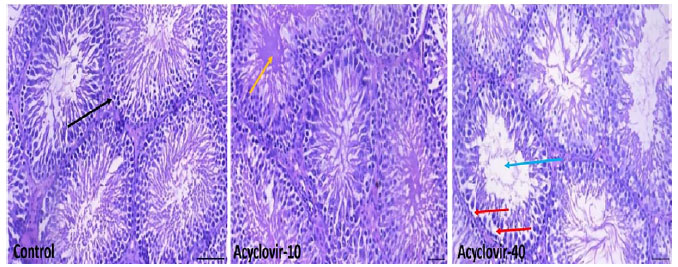
Figure 1. Testicular histoarchitecture of acyclovir treated rats (Magnification X 200, H/E staining, Scale Bar = 20µm). A. (Control) is normal testis showing numerous seminiferous tubules (black arrow) with germ cells at varying degree of maturation. B. (10 mg/kg acyclovir): there are edematous seminiferous tubules (yellow arrow). C. (40 mg/kg acyclovir): showed some seminiferous tubules with degenerated spermatogenic cells (red arrow), and scanty sperm cells in the lumen (blue arrow).