JBRA Assist. Reprod. 2025;29(4):817-822
CASE REPORT
doi: 10.5935/1518-0557.20250039
Hypogonadotropic Hypogonadism in a Central Obesity Patient with 46, XY, t(1; 10)(q42; q24)
1Department of Endocrinology, Union Hospital, Tongji Medical College, Huazhong University of Science and Technology, Wuhan 430022, China
2Hubei Provincial Clinical Research Center for Diabetes and Metabolic Disorders, Wuhan 430022, China
3Hukou County People’s Hospital, Jiujiang 332500, China
4Department of Cardiovascular Surgery, Union Hospital, Tongji Medical College, Huazhong University of Science and Technology, Wuhan 430022, China
†The authors contributed equally to this case report.
CONFLICTS OF INTEREST
The authors declare that the research was conducted without any commercial or financial relationships that could be seen as a potential conflict of interest.
ABSTRACT
Hypogonadotropic hypogonadism (HH) is characterized by absent or insufficient secretion of GnRH and/or pituitary gonadotropins, which consequently leads to testicular dysfunction. HH is commonly associated with obesity, particularly in specific genetic syndromes, and its etiology appears heterogeneous and remains far from fully understood. This report describes the case of a 31-year-old male with HH and central obesity, who has a karyotype of 46, XY, t(1; 10)(q42; q24). He demonstrated a good pituitary response to GnRH as well as a robust testicular reserve in both the GnRH stimulation test and the hCG stimulation test. Notably, he exhibited no lifestyle risk factors for obesity or comorbidities likely contributing to his condition. The SCD1 gene, located at 10q24.31, promotes the synthesis of fatty acids, and its abnormality may play a significant role in the pathogenesis of obesity. We propose that the translocation between chromosome 1q42 and chromosome 10q24 may disrupt the function of SCD1 at chromosome 10q24, resulting in the mutually reinforcing relationship between obesity and HH in this patient.
Keywords: Hypogonadism, Obesity, Chromosomal Abnormalities, Chromosomal Translocation
INTRODUCTION
Male hypogonadotropic hypogonadism (HH) is considered a consequence of congenital or acquired diseases that affect the hypothalamus and/or the pituitary gland. It is characterized by absent or inadequate secretion of gonadotropin-releasing hormone (GnRH) and/or pituitary gonadotropins, which consequently induces testicular dysfunction, impairing spermatogenesis and/or testosterone production (Fraietta et al., 2013). Numerous studies indicate that congenital hypogonadotropic hypogonadism (CHH) is clinically and genetically heterogeneous. More than 40 genes have been identified to play essential roles in the development, migration, and function of GnRH neurons, which are closely related to CHH (Butz et al., 2021).
In recent years, an increasing number of studies have shown that obesity is closely linked to lower free testosterone concentrations in male adolescents and adults. An extensive study conducted in the United States found that 40% of obese men had subnormal free testosterone levels (Dhindsa et al., 2010). Furthermore, it has been reported that the prevalence of HH among 160 obese men (mean age 58 years) is 36% in a study from the Netherlands (Dhindsa et al., 2018). Additionally, HH coincides with obesity in specific genetic syndromes, such as Prader-Willi syndrome, Bardet-Biedl syndrome, and Blakemore-Durmaz-Vasileiou syndrome, which are associated with complex disorders, including but not limited to growth hormone deficiency, cognitive impairment, neurodevelopmental delay, and rod-cone dystrophy (Bosch et al., 2021; Iughetti et al., 2019; Sloboda et al., 2022). The etiology of HH and obesity is not entirely clear. This article describes the case of a male with HH and obesity, with 46, XY, t(1; 10)(q42; q24) karyotype.
CASE REPORT
Case Presentation
The patient was a 31-year-old male, weighing 86 kg, with a height of 180 cm and an abdominal circumference of 90 cm. His pubic hair development was rated Tanner stage 4, while his penis and testicular development were rated Tanner stage 3. He had been obese since childhood without overfeeding and had experienced erectile dysfunction, diminished libido, and sparse pubic hair since puberty. His testosterone levels were low, and he used chorionic gonadotropin for three years, during which his secondary sexual characteristics developed and his erectile function improved. After discontinuing chorionic gonadotropin treatment, the symptoms of erectile dysfunction, diminished libido, and sparse pubic hair worsened. He denied any family history of obesity or abnormal sexual development but reported a history of hyperlipidemia and hyperuricemia for 5 years and osteoporosis for 2 years. Additionally, he underwent radiofrequency catheter ablation for supraventricular tachycardia at the age of 17.Further testing indicated that his testosterone level was 7.21 nmol/L (reference range 8.6-29.0 nmol/L), which was associated with anomalous luteinizing hormone (2.92 IU/L) and follicle-stimulating hormone (6.62 IU/L) levels, without any apparent abnormalities in adrenal and thyroid function. Therefore, he was diagnosed with obesity-related hypogonadotropic hypogonadism (Saboor Aftab et al., 2013), and treated with lifestyle interventions combined with orlistat at a dosage of 0.12 g three times daily. His body weight gradually decreased, and blood testosterone levels improved to 12.6 nmol/L after the first month and 18.6 nmol/L after three months.
Laboratory tests
The patient’s testosterone level was 7.21 nmol/L (Table 1), which is below the reference range. No significant abnormalities were noted in the complete blood count, routine urine analysis, serum electrolytes, cortisol and thyroid hormone levels, or liver and kidney function tests.
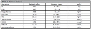
Table 1. Hormone analysis.
GnRH stimulation test
A GnRH stimulation test was performed between 8:00 AM and 10:00 AM after at least 8 hours of overnight fasting. Gonadorelin® (BBCA Pharmaceutical, Anhui, China) was administered intravenously at a dosage of 100 mg. Venous blood samples were collected at 0 (before the administration of Gonadorelin), and at 15, 30, 60, and 120 minutes after the injection to assess the levels of luteinizing hormone (LH) and follicle-stimulating hormone (FSH) (Mosbah et al., 2020). The results indicated peaks of LH and FSH of 56.39 IU/L and 20.86 IU/L, respectively (Figure 1).
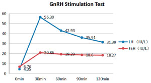
Figure 1. The GnRH stimulation test. LH and FSH levels peaked 30 minutes after the administration of Gonadorelin, reaching values of 56.39 IU/L and 20.86 IU/L, respectively.
Human chorionic gonadotropin (hCG) stimulation test
The hCG stimulation test was performed using an intramuscular injection of 2000 U hCG over three consecutive days, with blood samples collected before the first dose of hCG (0 minutes) and again after 24, 48, and 72 hours (Lucas-Herald et al., 2020). The peak testosterone level was 22.82 nmol/L following 72 hours of hCG injection (Figure 2).
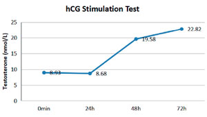
Figure 2. The hCG stimulation test. The testosterone level peaked at 72 hours after the hCG injection, reaching 22.82 nmol/L.
Oral glucose tolerance test
The oral glucose tolerance test was conducted using a 75-g glucose load after an overnight fast. Blood samples were collected before the test and at 30, 60, 120, and 180 minutes, with measurements taken for glucose and insulin levels (Wang et al., 2018). The results indicated that the plasma glucose level was 5.3 mmol/L before the administration of glucose. In comparison, the 2-hour post-load plasma glucose level was 10.2 mmol/L, surpassing the upper limit of normal plasma glucose (less than 7.8 mmol/L) after 2 hours, indicating that the patient had impaired glucose tolerance (Bartoli et al., 2011). His insulin level was 258.37 IU/mL at 30 minutes after glucose loading, peaking at 120 minutes (353.55 IU/mL), which was over 18 times higher than the baseline level (19.36 IU/mL); it remained at 53.93 IU/mL at 180 minutes, implying impairments in first-phase insulin secretion and increased late-phase insulin secretion (Figure 3).
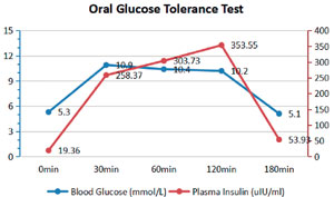
Figure 3. Oral glucose tolerance test. The 2-hour post-load plasma glucose was 10.2 mmol/L, and the peak insulin level was 353.55 uIU/ml, which occurred at the 2-hour post-load plasma glucose measurement.
Scrotal ultrasonography
Scrotal ultrasonography revealed the right testicle size to be 33.6×29.8×19.1 mm and the left testicle size to be 36.6×25.1×19.8 mm, which appeared within the normal reference range (Lotti et al., 2021). Each head of the epididymis had a cyst (1.5×1.2 mm on the right side, 1.4×1.3 mm on the left side) (Figure 4).
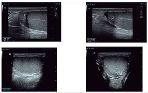
Figure 4. Scrotal ultrasonography. Both testicles were located in the scrotum. The right testicle measured 33.6×29.8×19.1 mm, while the left measured 36.6×25.1×19.8 mm. Each head of the epididymis contained a cyst (1.5×1.2 mm on the right side, 1.4×1.3 mm on the left side).
Chromosome analysis
G-band karyotyping analysis revealed that the patient’s karyotype was 46, XY, t(1;10)(q42;q24), indicating a translocation between chromosome 1q42 and chromosome 10q24 (Figure 5).
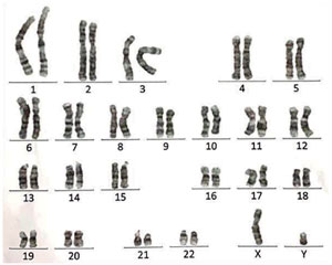
Figure 5. Karyotyping analysis. G-band karyotyping indicated that the patient’s karyotype is 46, XY, t(1;10)(q42;q24), suggesting a translocation between chromosome 1q42 and chromosome 10q24 in the patient.
Genetic sequencing
The patient’s exome and mitochondrial genome sequencing data were analyzed against the OMIM database (2021Q3). No significant pathogenic mutations were identified in association with the clinical phenotype.
DISCUSSION
This article describes the case of a 31-year-old male with central obesity and HH with 46, XY, t(1; 10)(q42; q24). The patient did not experience diseases likely to cause obesity, such as hypothyroidism, Cushing syndrome, and others. He did not show lifestyle risk factors for obesity, such as overeating. Notably, the patient suffered from hypogonadism, which is widely considered one of the significant causative factors in the etiology of obesity (Genchi et al., 2022). Hypogonadism may play a crucial role in the pathogenesis of obesity.
Our results indicated that the patient had no other conditions, such as Prader-Willi or Bardet-Biedl syndrome, that might cause or be associated with obesity. Interestingly, the results of the GnRH stimulation test revealed that the peak FSH was more than 12 times higher, and the peak LH was more than three times higher than the baseline levels (Figure 1), suggesting that the patient had a strong pituitary response to GnRH (Ab Rahim et al., 2020). Moreover, the results of the hCG stimulation test showed that the peak testosterone level was over two times higher than the baseline level (Figure 2), implying that the patient possessed a substantial testicular reserve (Bang et al., 2017). All these clinical features are closely related to obesity-related hypogonadism (Saboor Aftab et al., 2013). Additionally, it is widely acknowledged that HH and obesity create a vicious cycle, in which obesity can lead to HH, and HH, in turn, promotes obesity (Carrageta et al., 2019). Thus, we tentatively propose that the HH was likely attributed to obesity in this patient.
Karyotype analysis revealed a translocation between chromosomes 1q42 and 10q24. The stearoyl-CoA desaturase 1 (SCD1) gene, located at 10q24.31, promotes the synthesis of fatty acids and plays a crucial role in maintaining energy homeostasis. Dysfunction of SCD1 is a significant factor in obesity (Pan et al., 2021). Therefore, the translocation between chromosome 1q42 and chromosome 10q24 may result in an abnormal chromosome 10q24, subsequently leading to SCD1 dysfunction, which contributes to the development of obesity.
There is a lack of familial data to confirm the association between the chromosomal translocation of chromosomes 1q42 and 10q24, obesity, and HH. We were unable to identify an alteration in the SCD1 gene in the patient; however, the dysregulation of SCD1 likely mediated the obesity and HH stemming from the chromosomal translocation involving chromosomes 1q42 and 10q24. Additionally, we were unable to develop an animal model with the translocation to demonstrate the causative role of the chromosomal translocation described above in the pathogenesis of obesity and HH. Therefore, further family investigations and/or animal studies are necessary to verify the association among the chromosomal translocation, obesity, and HH.
Despite these limitations, we tentatively propose that the translocation between chromosome 1q42 and chromosome 10q24 is closely associated with obesity and HH, although further confirmatory research is needed. It seems reasonable to suggest that more attention should be given to chromosome abnormalities in obese patients with hypogonadism.
Acknowledgement
We are extremely grateful to the patient and his family for their involvement in this study.
Statement of Ethics
The study was conducted per the Declaration of Helsinki and was approved by the Ethics Committee of Huazhong University of Science and Technology (protocol code UHCT-IEC-SOP-016-03-01, dated July 24, 2024).Informed Consent StatementInformed written consent has been obtained from the patient for the publication of this article.
Data Availability Statement
The original contributions presented in the study are included in the article and supplementary material. Any further inquiries can be directed to the corresponding author.
REFERENCES
Ab Rahim SN, Omar J, Tuan Ismail TS. Gonadotropin-releasing hormone stimulation test and diagnostic cutoff in precocious puberty: a mini review. Ann Pediatr Endocrinol Metab. 2020;25:152-55. PMID: 32871650 DOI: 10.6065/apem.2040004.002 Medline
Bang AK, Nordkap L, Almstrup K, Priskorn L, Petersen JH, Rajpert-De Meyts E, Andersson AM, Juul A, Jørgensen N. Dynamic GnRH and hCG testing: establishment of new diagnostic reference levels. Eur J Endocrinol. 2017;176:379-91. PMID: 28077499 DOI: 10.1530/EJE-16-0912 Medline
Bartoli E, Fra GP, Carnevale Schianca GP. The oral glucose tolerance test (OGTT) revisited. Eur J Intern Med. 2011;22:8-12. PMID: 21238885 DOI: 10.1016/j.ejim.2010.07.008 Medline
Bosch E, Hebebrand M, Popp B, Penger T, Behring B, Cox H, Towner S, Kraus C, Wilson WG, Khan S, Krumbiegel M, Ekici AB, Uebe S, Trollmann R, Woelfle J, Reis A, Vasileiou G. BDV Syndrome: An Emerging Syndrome With Profound Obesity and Neurodevelopmental Delay Resembling Prader-Willi Syndrome. J Clin Endocrinol Metab. 2021;106:3413-27. PMID: 34383079 DOI: 10.1210/clinem/dgab592 Medline
Butz H, Nyírő G, Kurucz PA, Likó I, Patócs A. Molecular genetic diagnostics of hypogonadotropic hypogonadism: from panel design towards result interpretation in clinical practice. Hum Genet. 2021;140:113-34. PMID: 32222824 DOI: 10.1007/s00439-020-02148-0 Medline
Carrageta DF, Oliveira PF, Alves MG, Monteiro MP. Obesity and male hypogonadism: Tales of a vicious cycle. Obes Rev. 2019;20:1148-58. PMID: 31035310 DOI: 10.1111/obr.12863 Medline
Dhindsa S, Ghanim H, Batra M, Dandona P. Hypogonadotropic Hypogonadism in Men With Diabesity. Diabetes Care. 2018;41:1516-25. PMID: 29934480 DOI: 10.2337/dc17-2510 Medline
Dhindsa S, Miller MG, McWhirter CL, Mager DE, Ghanim H, Chaudhuri A, Dandona P. Testosterone concentrations in diabetic and nondiabetic obese men. Diabetes Care. 2010;33:1186-92. PMID: 20200299 DOI: 10.2337/dc09-1649 Medline
Fraietta R, Zylberstejn DS, Esteves SC. Hypogonadotropic hypogonadism revisited. Clinics. 2013;68:81-8. PMID: 23503957 DOI: 10.6061/clinics/2013(Sup01)09 Medline
Genchi VA, Rossi E, Lauriola C, D’Oria R, Palma G, Borrelli A, Caccioppoli C, Giorgino F, Cignarelli A. Adipose Tissue Dysfunction and Obesity-Related Male Hypogonadism. Int J Mol Sci. 2022;23:8194. PMID: 35897769 DOI: 10.3390/ijms23158194 Medline
Iughetti L, Vivi G, Balsamo A, Corrias A, Crinò A, Delvecchio M, Gargantini L, Greggio NA, Grugni G, Hladnik U, Pilotta A, Ragusa L, Salvatoni A, Wasniewska M, Weber G, Predieri B. Thyroid function in patients with Prader-Willi syndrome: an Italian multicenter study of 339 patients. J Pediatr Endocrinol Metab. 2019;32:159-65. PMID: 30703060 DOI: 10.1515/jpem-2018-0388 Medline
Lotti F, Bertolotto M, Maggi M. Historical trends for the standards in scrotal ultrasonography: What was, what is and what will be normal. Andrology. 2021;9:1331-55. PMID: 34089245 DOI: 10.1111/andr.13062 Medline
Lucas-Herald AK, Kyriakou A, Alimussina M, Guaragna-Filho G, Diver LA, McGowan R, Smith K, McNeilly JD, Ahmed SF. Serum Anti-Müllerian Hormone in the Prediction of Response to hCG Stimulation in Children With DSD. J Clin Endocrinol Metab. 2020;105:1608-16. PMID: 32016383 DOI: 10.1210/clinem/dgaa052 Medline
Mosbah H, Bouvattier C, Maione L, Trabado S, De Filippo G, Cartes A, Donzeau A, Chanson P, Brailly-Tabard S, Dwyer AA, Coutant R, Young J. GnRH stimulation testing and serum inhibin B in males: insufficient specificity for discriminating between congenital hypogonadotropic hypogonadism from constitutional delay of growth and puberty. Hum Reprod. 2020;35:2312-22. PMID: 32862222 DOI: 10.1093/humrep/deaa185 Medline
Pan G, Cavalli M, Wadelius C. Polymorphisms rs55710213 and rs56334587 regulate SCD1 expression by modulating HNF4A binding. Biochim Biophys Acta Gene Regul Mech. 2021;1864:194724. PMID: 34171462 DOI: 10.1016/j.bbagrm.2021.194724 Medline
Saboor Aftab SA, Kumar S, Barber TM. The role of obesity and type 2 diabetes mellitus in the development of male obesity-associated secondary hypogonadism. Clin Endocrinol. 2013;78:330-7. PMID: 23121123 DOI: 10.1111/cen.12092 Medline
Sloboda N, Lambert L, Ciorna V, Bruel AL, Tran Mau-Them F, Gomola V, Lemelle JL, Klein O, Camoin-Schweitzer MC, Magnavacca M, Legagneur C, Ezsto ML, Bonnet C, Philippe C, Leheup B. Atypical phenotype of a patient with Bardet-Biedl syndrome type 4. Mol Genet Genomic Med. 2022;10:e1869. PMID: 35318824 DOI: 10.1002/mgg3.1869 Medline
Wang X, Zhao X, Zhou R, Gu Y, Zhu X, Tang Z, Yuan X, Chen W, Zhang R, Qian C, Cui S. Delay in glucose peak time during the oral glucose tolerance test as an indicator of insulin resistance and insulin secretion in type 2 diabetes patients. J Diabetes Investig. 2018;9:1288-95. PMID: 29537733 DOI: 10.1111/jdi.12834 Medline