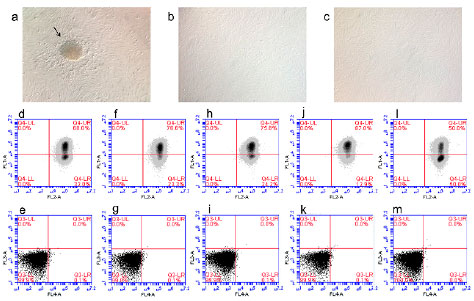
Figure 1. Characterization of EnMSC cultured in vitro. a-c: Morphology observed by a phase contrast microscope in 4x objective. a: Cells in passage 0 evidencing colony formation indicated by the black arrow. b and c: Cells in passage 1 and 2 respectively, without colony. d-m: Flow cytometry analysis with positive markers CD90 (FL-1) and CD105 (FL-2) and negative markers CD31 (FL-3) and CD45 (FL-4) of each volunteer. d-i: OC group. J-m: Non-OC group.