JBRA Assist. Reprod. 2026;30(1):120-131
ORIGINAL ARTICLE
doi: 10.5935/1518-0557.20250184
Acute stress induction downregulates RHOX-5 gene expression causing altered fertility indices in MALE Wistar rats
1Department of Medical Physiology, School of Medicine and Pharmacy, College of Medicine and Health Sciences, University of Rwanda, Butare, Rwanda
2Department of Physiology, Basic Medical Sciences, University of Global Health Equity, Butaro, Rwanda
CONFLICT OF INTEREST
The authors have no conflict of interest to declare.
ABSTRACT
Objective: Stress has been implicated in the onset and development of various diseases and conditions including cardiovascular diseases, respiratory diseases, obesity and infertility. The effects of stress on fertility were studied to elucidate possible mechanisms of stress-induced infertility.
Methods: Stress was induced using the flowerpot technique. Plasma concentration of fertility hormones, inflammatory and oxidative markers, sperm profile, histological studies and molecular studies were carried out in selected tissues from the study and control groups. Statistical analysis was carried out using the GraphPad Prism 8.0.1 version. Results were presented as Mean±SEM. The Student t-test was used to compare the means obtained; and p-values lower than 0.05 were considered statistically significant.
Results: Plasma concentration of testosterone, percentage of progressively motile sperm cells, percentage of normal sperm cells and relative expression of Rhox-5 gene were significantly decreased, while the C-reactive protein concentration was significantly increased in the stressed group when compared with the control group.
Conclusions: Stress reduces fertility through suppressed fertility hormone secretion and reduced genetic expression of the relevant genes, causing poor-quality spermatogenesis in male Wistar rats.
Keywords: stress, fertility hormones, infertility, Rhox-5 gene
INTRODUCTION
The prevalence of infertility amongst couples is becoming a cause for serious concern. Infertility has been linked with reproductive system disorders (Sabarre et al., 2013), age (Deyhoul et al., 2017), and hormonal disorders (Seth et al., 2013). Over the years, stress has been attributed to the onset or development of various diseases and conditions including cardiovascular diseases (Steptoe & Kivimäki, 2012), obesity (Scott et al., 2012), respiratory diseases (Aich et al., 2009), and infertility (Kalantaridou et al., 2010) amongst others. Stress has been linked to infertility in both males and females (Genazzani, 2005; Whirledge & Cidlowski, 2010; Dobson et al., 2012; Stárka & Duō¡ková, 2015), with little knowledge of the exact mechanisms involved. A study by Andersen et al. (2005) revealed that stress causes alteration in levels of reproductive hormones. Likewise, Whirledge & Cidlowski (2010), reported that modifications on the hypothalamic-pituitary-adrenal (HPA) axis, with the resultant changes in circulating levels of glucocorticoids in response to stress, leading to an inhibition in reproduction.
The reproductive system in both males and females is controlled by hormonal secretions from the hypothalamic-pituitary-gonadal (HPG) axis (Meccariello et al., 2014), with the secretion of gonadotropin-releasing hormone (GnRH) at the level of the hypothalamus; gonadotropins at the pituitary gland and testosterone from the testis, each hormone acting to stimulate or suppress the secretion of another in a classical feedback manner.
Stressful conditions result in the activation of the hypothalamic-pituitary-adrenal axis (HPA) - a key component of the stress system (Chrousos, 2009), as such, its integrity and precise regulation are essential for adapting to stressors. Increased levels of glucocorticoid, a principal component of the HPA axis, promotes gluconeogenesis and mobilization of amino acids, actions required to maintain circulating levels of glucose to mount a stress response (Whirledge & Cidlowski, 2010). It has also been extensively documented that elevated levels of glucocorticoids affect gonadal function at multiple levels of the HPG axis (Maeda & Tsukamura, 2006; Whirledge & Cidlowski, 2010). Decreased testosterone production, sperm production, maturation, erectile dysfunction, or impotence have been observed with stress (Daly et al., 2005; Whirledge & Cidlowski, 2010).
Infertility has been reported to cause stress (Chen et al., 2004; Ilacqua et al., 2018; Rooney & Domar, 2018), but the question of whether stress causes infertility has not been sufficiently answered. Various methods have been experimentally used to induce stress such as deprivation paradigms (Andersen et al., 2005), electric foot shocks (Cakır et al., 2010), forced swimming, and the restraint method (Jameel et al., 2014).
Rhox-5 and Cox-2 genes, both expressed in the Sertoli cells and Leydig cells respectively, have been observed to play active roles in gametogenesis and steroidogenesis. It has been reported that male mice lacking the Rhox-5 gene are sub-fertile, exhibiting increased germ cell apoptosis and a defect in sperm mobility (Hu et al., 2007). Inhibition of the stAR protein by the Cox-2 gene has also been reported to lead to suppression of steroidogenesis (Wang et al., 2005).
Hence, this study was aimed at examining if stress causes infertility and investigate the possible mechanisms of how stress modulates the morphology and function of the male reproductive systems in Wistar rats.
MATERIALS AND METHODS
Setting of the study
The study was carried out in the Department of Physiology Laboratory, University of Benin, Edo state, Nigeria. The animals were handled in accordance with the Guiding Principles for Research as recommended by the Declaration of Helsinki and the Guiding Principles for the Care and Use of Laboratory Animals. Consent and approval from the Research and Ethics Committee of the above University and department were received before the study.
Study design
Forty male and fifteen female Wistar rats (15 weeks old) weighing 250-280g were used for the study. They were housed in standard cages at room temperature, with 12 hours light/dark cycle. Before the study commenced, the animals were fed standard chow and water ad libitum and acclimatized for 2 weeks.
Stress induction
The rats were stressed using a customized form of the flowerpot technique as described by Heinrichs & Koob (2006).
-a waterproof pool is prepared and flooded with water at room temperature,
-a cylindrical pedestal with a height of about 1cm above the water level in the pool was fixed to the center of the floor of the pool,
-the animal is placed on the pedestal and is brought out of the pool to eat and rest for 1 hour each day.
Animal grouping
Forty male Wistar rats were sub-sectioned into 15 and 25 rats for sub-sections I and II, respectively. The animals in sub-section I were grouped into three (A, B, and C); with each group having 5 rats. The animals in groups A, B, and C were stressed using the flowerpot technique for 2, 3, and 5 days, respectively. The body weight of each rat was measured using an appropriate weighing balance, and blood samples were collected through the tail vein before and after stress induction for the evaluation of cortisol.
The rats in sub-section II were grouped into two. Five rats served as the control group, while 20 rats were stressed for 3 days and served as the stressed group. Fifteen animals from the stressed group were allowed to recover (recovery group) for 2, 4, and 6 weeks.
After stress induction and recovery, all the animals were sacrificed by cervical dislocation. Blood was drawn by direct cardiocentesis, centrifuged at 3500 RMP for 10 minutes and serum was collected for analysis of concentrations of follicle-stimulating hormone, luteinizing hormone, testosterone, anti-inflammatory, and antioxidant markers. Brain and testes were harvested for the pituitary gland and testicular histology. Testicular gene expression of Rhox-5 and Cox-2 were also investigated.
Fertility test
Fifteen unstressed female Wistar were grouped into three and allowed to mate with male rats in a ratio of 2:1 as follows;
Group A - Unstressed males (UM) + unstressed females (UF) (Control group).
Group B - Stressed males (SM) + unstressed females (UF)
Group C - Recovered + unstressed females.
The mating was confirmed by obtaining a vaginal smear through pipetting normal saline into the vagina and the mixture obtained was placed on a glass slide, covered with a cover slip, and viewed under the microscope. When mating occurred, sperm cells were visible in the vagina smear. After mating confirmation, the outcome of pregnancy was investigated i.e., pregnancy success rate, litter weight, litter numbers, and litter survival.
Hormone assays
The serum concentration of cortisol, follicle-stimulating hormone, luteinizing hormone, and testosterone were measured using an Enzyme-linked immunosorbent assay (ELISA) following the instructions on the manufacturer’s kit (Darwish, 2006). The Elisa kits used for the study were manufactured by Calbiotech Incorporation, United States.
Sperm analysis
Sperm motility and morphology were studied using the methods described by Ibeh et al. (2020).
Pituitary and testicular histology
The pituitary glands and testes were harvested and histological studies were done using standard methods of fixing, sectioning, and staining (Yang et al., 2006).
Inflammatory markers assay
C-reactive protein was measured using an Enzyme-linked immunosorbent assay (ELISA) following the instructions on the manufacturer’s kit (Darwish, 2006). The Elisa kits were manufactured by the Calbiotech Incorporation; the United States were used for the study. Fibrinogen concentration was measured using the gravimetric assay method (Ajayi et al., 2015).
Antioxidant assay
Catalase and superoxide dismutase activities were measured using spectrophotometry (Weydert & Cullen, 2010; Hadwan & Abed, 2015).
Molecular assay
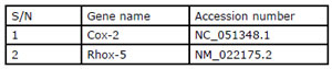
Specific gene expression analysis was done using the RT-PCR and gel electrophoresis technique (Mitchell & Iadarola, 2010). In the testes, Rhox-5 and Cox-2 genes were studied. The relative amount of cDNA (i.e., the intensities of the bands from agarose gel electrophoresis) was quantified through densitometry, using the ImageJ Software Version 5.0 and the gene expression was normalized with the GADPH gene as a housekeeping gene.

The accession numbers for the primers used are as follows:
Statistical Analysis
GraphPad Prism 8.0.1 (244) was used to analyze the data generated. Results are presented as Mean±SEM. Bar charts were plotted to show the results. Paired and unpaired student t-tests were used to compare the results in different groups and between various groups. P-values lower than 0.05 (p<0.05) were taken to be statistically significant.
RESULTS
Body weight and cortisol
Figure 1 shows a significant reduction (p<0.05) in the weight of male Wistar rats after stress for 2 days, 3 days, and 5 days. The serum concentration of cortisol was increased (p<0.05) after stress for 2 and 3 days but was not significantly different after 5 days of stress (Fig. 2).
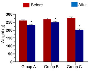
Figure 1. Weight of male Wistar rats before and after stress in Groups A, B, and C.
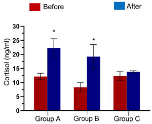
Figure 2. Serum Cortisol concentration of male Wistar rats before and after stress in Group A, B, and C. *p<0.05 is considered significant compared with the control.
Hormones
No difference was observed in the concentration of follicle-stimulating hormone and luteinizing hormone (Fig. 3). There was a significant reduction (p<0.05) in the concentration of testosterone (Fig. 3), while there were significant reductions in follicle stimulating hormone, luteinizing hormone, and testosterone at week 2 of recovery (Fig. 3) (p<0.05, respectively).
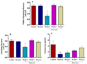
Figure 3. Serum follicle stimulating hormone, luteinizing hormone, and testosterone concentration in control, stressed and recovery groups of male Wistar rats. *p<0.05 is considered significant compared with the control.
Sperm Profile
Figure 4A shows no difference in total sperm count. There was a significant reduction (p<0.05) in the percentage of normal sperm cells (Fig. 4B), and sperm cells with progressive motility (Fig. 4C). Percentage of immotile sperm cells was unchanged (Fig. 4D).
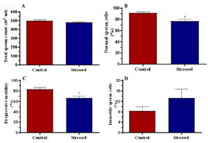
Figure 4. Total sperm count (A), percentage of normal sperm cells (B), progressive motility (C), and immotile sperm cells in control and stressed rats. *p<0.05 is considered significant compared with the control.
For sperm morphology, Table 1 shows the percentage of sperm cells in the control and stressed group which were headless, tailless, had big heads, short tails, double heads, head to head aggregation, tail to tail aggregation and body to body aggregation.
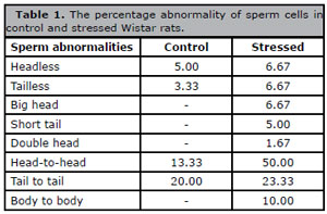
Table 1. The percentage abnormality of sperm cells in control and stressed Wistar rats.
Histology
Figure 5Pii shows that the pituitary gland of the stressed animals has decreased population and viability of acidophils (A) and basophils (B), compared with the control group (Fig. 5Pi); the chromophobes with inconspicuous nucleoli (C). The dilated and congested sinusoid (D) was observed in the stressed group when compared with the control. (Fig. 5Pi)
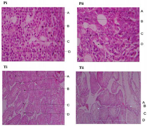
Figure 5. Photomicrograph of pituitary gland (Pi & ii) and testes (Ti & ii) of control and stressed male Wistar rats (H&E x 400).
Antioxidant markers
There was no difference in catalase activity (Fig. 6A) and superoxide dismutase activity (Fig. 6B).
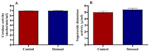
Figure 6. Catalase and superoxide dismutase activities in control and stressed male Wistar rats. *p<0.05 is considered significant compared with the control.
Anti-inflammatory markers
No difference was observed in fibrinogen concentration (Fig. 7A), but C-reactive protein was significantly (p<0.05) increased in the stressed group (Fig. 7B).
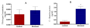
Figure 7. Fibrinogen and C-Reactive protein concentration in control and stressed male Wistar rats. *p<0.05 is considered significant compared with the control.
Pregnancy outcome
Table 2 shows a reduced pregnancy success rate, a significant reduction in litter numbers, an unchanged litter survival rate, and litter weight in group B. The table also shows no difference in pregnancy success rate, litter numbers, litter survival rate, and litter weight in Group C.
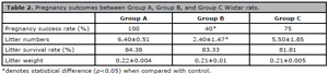
Table 2. Pregnancy outcomes between Group A, Group B, and Group C Wistar rats.
Molecular Assay
There was a significant reduction in the relative expression of the Rhox-5 gene (Fig. 8A). Figure 8B shows the unchanged relative expression of the Cox-2 gene.
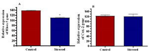
Figure 8. Relative expression of Rhox-5 gene (A) and Cox-2 gene (B) in control and stressed male Wistar rats. *p<0.05 is considered significant compared with the control.
DISCUSSION
This study employs the flowerpot technique of stress induction. This technique of stress induction works on various principles that have been observed to increase cortisol levels, hence inducing stress. They include rapid eye movement (REM) sleep deprivation (Andersen et al., 2005), restraint (Arakawa, 2020), food deprivation (Patchev & Patchev, 2006; Wunderink et al., 2012; Midwood et al., 2016), fear and anxiety (Adamec et al., 2005) and isolation (Heinrichs & Koob, 2006; Weiss et al., 2004).
Stress has been linked to infertility in both males and females (Genazzani, 2005; Whirledge & Cislowski, 2010; Dobson et al., 2012; Stárka & Duō¡ková, 2015), with little knowledge of the exact mechanisms involved. A study by Andersen et al. (2005) revealed that stress causes alterations in levels of reproductive hormones. Likewise, Whirledge & Cidlowski (2010), observed that modification of the hypothalamic-pituitary-adrenal (HPA) axis, with the resultant changes in circulating levels of glucocorticoids in response to stress, leads to an inhibition of reproduction. This study was aimed at elucidating the possible mechanisms by which stress modulates the morphologies and functions of male Wistar rats, hence causing infertility. Various parameters measured in this study include body weight, the concentration of cortisol to establish stress, concentrations of fertility hormones, inflammatory and oxidative biomarkers.
The results showed a significantly decreased weight of the animals after the 2nd, 3rd, and also 5th day of stress induction using the flowerpot technique. Acute stress has been associated with feeding suppression (anorexigenic effects) and reduced body weight gain (Halataei et al., 2011; Ranjbaran et al., 2013; Bagheri Nikoo et al., 2014; Rabasa & Dickson, 2016). Furthermore, the decrease in weight observed in this study could be attributed to the poor feeding of the animals since they were only fed for 1 hour daily, unlike before the experiment when they were fed ad libitum. Excessive gluconeogenesis and glycogenolysis characterize the neuroendocrine response to stress, leading to stress hyperglycemia (Dungan et al., 2009). During fasting, there is reduced glucose uptake to be stored in tissues, and there is increased glycogenolysis and gluconeogenesis to enrich the blood with glucose to sustain the tissues (Izumida et al., 2013), leading to a decrease in the body weight of the animal.
Serum cortisol concentrations were significantly increased in the groups stressed for 2 and 3 days using the flowerpot technique, supporting the observations of Cakır et al. (2010), who induced stress using electric foot shocks, and Jameel et al. (2014) who induced stress using forced swim and restraint methods of stress induction. Increased concentration of cortisol from the adrenal gland is normally prompted by an adrenocorticotropic hormone from the anterior pituitary glands; whose secretion is triggered by the release of Corticotropin-releasing hormone from the hypothalamus in response to stress (Gallo-Payet et al., 2017; Vaeroy et al., 2019). The results also show that for the animals stressed for 5 days, the serum concentration of cortisol remained statistically unchanged, suggesting that by the fifth day of stress induction, the animals have acclimatized to the flowerpot technique, i.e., the animals have developed resistance to the stressor.
Significantly decreased serum concentration of testosterone was observed in the stressed group, supporting the observation of Daly et al. (2005) and Brownlee et al. (2005) that a negative relationship exists between cortisol and testosterone. In earlier years, a direct inhibitory effect of high doses of glucocorticoids upon testicular Leydig cell function in rats has been established, resulting in a decrease in the production of testosterone (Brownlee et al., 2005). This occurs via glucocorticoid-induced apoptosis (Hu et al., 2008). Brownlee et al. (2005) also observed that pharmacologically increased levels of cortisol have a significant negative effect on circulating testosterone. With reduced testosterone concentration in the stressed group, there would be reduced sex drive (libido), and reduced or poor spermatogenesis as testosterone is essential for both.
The gonadotropins were statistically unchanged after stress induction. This finding opposes the observation of Xiao et al. (2002), Li et al. (2004) and Whirledge & Cidlowski (2010) that stress decreases circulating levels of gonadotropins. This is possibly because of the short stress duration in the present study, as time is required for the effect of stress to reflect on the circulating levels of the hormones. This is confirmed by the significantly reduced serum concentrations of the gonadotropins at 2 weeks after stress-induction, followed by a rise to the same level as the control by the end of the fourth week.
Hormonal levels of testosterone and gonadotropins returned to baseline at the fourth week of rest after stress, around the same period, mating could occur when an estrus female was present. This confirms the importance of testosterone in the initiation of sexual acts (Shulman & Spritzer, 2014).
The results of the study also showed no change in total sperm count following stress. However, the qualities of the sperm cells were altered, specifically, the percentage of normal sperm cells and progressive sperm motility were reduced significantly in the stressed group. These findings are in line with various observations that stress suppresses spermatogenesis resulting in decreased sperm motility and viability (Juárez-Rojas et al., 2017; Nargund, 2015). For fertilization to occur after mating, efficient passage of spermatozoa through the cervical mucus is required to meet up with the ovum and this depends on rapid progressive motility (Vogiatzi et al., 2022). While some sperm cells may move around the cervix till and outlive their lifespan, only those with the ability to move forward in a straight line can travel through the cervical mucus to fertilize the egg. The high percentage of sperm cells with progressive motility, among other factors, shows an increased probability of conception in the presence of a viable egg (Buck Louis et al., 2014).
This study also reported a higher number of headless and tailless sperm cells in the stressed group. Headless sperm cells cannot fertilize the eggs due to the absence of proteolytic enzymes and genetic materials required for fertilization, which are all contained in the head. Likewise, tailless sperm cells cannot fertilize the eggs since they cannot swim to the eggs without their flagella-like tails. Sperm cells with broken midpieces would die off easily since the midpiece houses the mitochondria which is the powerhouse of the cells. Also, a high percentage of head-to-head or tail-to-tail aggregation would result in a lower probability of fertilization since these aggregations would slow down the movement of the sperm cells toward the eggs i.e., impedes progressive motility (Du Plessis et al., 2013). An increased percentage of sperm cells with normal morphology shows an increased probability of conception, while a reduced percentage shows a reduced probability of conception (Slama et al., 2002).
The alteration in sperm parameters could be attributed to the indirect effect of cortisol on testicular tissues via reduced steroidogenesis, causing changes in Sertoli cells and the blood-testis barrier, and leading to the arrest of spermatogenesis (Nargund, 2015).
The stressed groups showed tubules containing degenerating spermatogenic cells, Sertoli cells, and Leydig cell atrophy as well as mild to severe spermatogenic arrest. The effect of stress on the testes appears to be degeneration of the cellular components including spermatocytes, Sertoli cells, Leydig cells as well as interstitial connective tissue. Spermatogenic arrest as seen in the stressed group is an interruption of sperm cell maturation, which of course impairs the production of mature sperm cells and overtime results in oligospermia (low sperm count) and azoospermia (semen containing no sperm), which are common causes of infertility in males. Since testosterone is one of the most important hormones responsible for both initiation and maintenance of spermatogenesis (Smith & Walker, 2014), a decrease in the serum concentration of testosterone as obtained in this study could account for the spermatogenic arrest observed.
Histopathological changes as shown in the present study clearly demonstrate that acute restraint stress causes marked degeneration of Sertoli cells. These data are in accordance with findings by Rai et al. (2003) and Aziz et al. (2013). The observed reduction in the serum testosterone of acute restrain stressed rats may be responsible for the disintegration of Sertoli cells. Immobilization stress and heat stress in Wistar rats have been reported to damage Sertoli cells through a decline in androgen (Yazawa et al., 1999), oxidative stress (Vallés et al., 2014), activation of pro-inflammatory cytokines, and damage of blood-testis-barrier (Xu et al., 2015). Nevertheless, the present study reports unchanged oxidative markers by stress, accompanied by increased levels of C-reactive protein.
Histology of the pituitary gland from the group subjected to stress showed disorganized micro architecture comprising a decreased population of acidophils, basophils, and chromophobes. The remaining cells showed variable morphological features, while some appeared normal, others appeared shrunken, and some have pyknotic nuclei. The nuclei of the chromophobes contain inconspicuous nucleoli, which is a sign of a reduction in protein storage. The sinusoids are markedly dilated and congested, taking up spaces originally occupied by parenchymal cells. The glands of the animals subjected to stress have decreased population of neuroendocrine cells, some of which are not viable because of structural derangements. These features give an impression of depletion in protein stores and gonadotropin-producing cells, which will over time reflect as reduced levels of gonadotropins (Ganapathy & Tadi, 2022).
The increased concentration of C-reactive protein observed in the stressed group supports the findings of Brunner et al. (1996), Kittel et al. (2002), Miller et al. (2005), Hamer et al. (2006), Nijm et al. (2007), Coussons-Read et al. (2007) and Ranjit et al. (2007). These authors found associations between acute and chronic stress with elevated C-reactive protein and serum fibrinogen concentrations.
Stress is known to activate the sympathetic nervous system in addition to the activation of the HPA axis, resulting in the release of catecholamines and glucocorticoids (Miller & O’Callaghan, 2002). According to Black & Garbutt (2002), the chronic secretion of these hormones may result in endothelial distension and initiate the acute phase inflammatory response involving the release of cytokines and acute phase reactant proteins (C - reactive protein and fibrinogen), though this study observed an unchanged fibrinogen concentration with stress.
Catalase and superoxide dismutase (SOD) activities were unchanged between the stressed and control groups. According to Birben et al. (2012) and Sharifi-Rad et al. (2018), stressful conditions cause an overwhelming of the body’s antioxidant systems by reactive oxygen species (ROS), causing oxidative stress. Decreased catalase and SOD activity in association with stress has been reported in the past (Zaidi et al., 2003; Zafir & Banu, 2009; Priya & Reddy, 2012), but those studies involved chronic stress, unlike the present study which involves acute stress, indicating that the duration of stress determines the stress response.
Significantly reduced pregnancy success rates and litter numbers were observed in the stressed group. There is paucity of data on the effect of paternal stress on pregnancy outcome, though the same has been established for maternal stress (Dunkel Schetter & Tanner, 2012; Traylor et al., 2020). The results obtained from this study could be linked to the poor quality of spermatozoa observed. With fewer fertile and viable sperm cells in the stressed group, only a few successful fertilizations could occur, accounting for the reduced pregnancy success rates. Litter weight and litter survival (beyond 1 week) seem to be unaffected by paternal stress, showing that the litter’s development and ability to survive in the external world were like those of the control group. Also, pregnancy outcome after recovery was like that observed in the control group, confirming that complete recovery occurs in males after the stressor is removed and a period of rest is allowed.
The relative expressions of Rhox-5 and Cox-2 genes to the housekeeping gene, glyceraldehyde 3-phosphate dehydrogenase (GADPH) were studied. The significantly reduced expression of the Rhox-5 gene (p<0.05) observed in the stressed group could be linked to the poor quality of spermatogenesis observed since the Rhox-5 gene facilitates spermatogenesis (Maclean et al., 2005; Hu et al., 2010; Welborn et al., 2015). There are earlier reports of rodents lacking the Rhox-5 gene being sub-fertile, exhibiting increased germ-cell apoptosis and defects in sperm motility (Maclean et al., 2005; Hu et al., 2007). This reduced expression of the Rhox-5 gene can be linked to the reduced pregnancy success rate and litter numbers observed in the groups with the stressed males.
Cox-2 gene is a down-regulator of testosterone secretion, and its expression is inversely related to the concentration of testosterone (Wang et al., 2005). An increased expression of the Cox-2 gene would lead to a decreased secretion of testosterone and vice versa. The results show no significant difference in the relative expression of the Cox-2 gene between the control and stressed groups. This signifies that the reduced concentration of testosterone observed earlier in the stressed males was not due to down-regulation at the molecular level, but instead because of the atrophic Leydig cells observed in the histology of the testes following stress.
ACKNOWLEDGEMENTS
The authors wish to acknowledge the inputs of the staff from the Physiology Laboratory, UNIBEN at the time of this project, especially Mr. Wisdom Silas and Mr. Oge Aloamaka. The scholarly input from Dr. Oyeyemi Wahab in the preparation of this manuscript is also appreciated.
REFERENCES
Adamec RE, Blundell J, Burton P. Neural circuit changes mediating lasting brain and behavioral response to predator stress. Neurosci Biobehav Rev. 2005;29:1225-41. PMID: 16099042 DOI: 10.1016/j.neubiorev.2005.05.007 Medline
Aich P, Potter AA, Griebel PJ. Modern approaches to understanding stress disease susceptibility: A review with special emphasis on respiratory disease. Int J Gen Med. 2009;2:19-32. PMID: 20360883 DOI: 10.2147/IJGM.S4843 Medline
Andersen ML, Martins PJ, D’Almeida V, Bignotto M, Tufik S. Endocrinological and catecholaminergic alterations during sleep deprivation and recovery in male rats. J Sleep Res. 2005;14:83-90. PMID: 15743338 DOI: 10.1111/j.1365-2869.2004.00428.x Medline
Arakawa H. Restraint stress activates defensive behaviors in male rats depending on age and housing condition. Physiol Behav. 2020;224:113073. PMID: 32659391 DOI: 10.1016/j.physbeh.2020.113073 Medline
Aziz NM, Ragy MM, Gayyed MF. Effect of acute immobilization stress with or without a heme oxygenase inducer on testicular structure and function in male albino rats. J Basic Clin Physiol Pharmacol. 2013;24:255-62. PMID: 23509214 DOI: 10.1515/jbcpp-2012-0066 Medline
Birben E, Sahiner UM, Sackesen C, Erzurum S, Kalayci O. Oxidative stress and antioxidant defense. World Allergy Organ J. 2012;5:9-19. PMID: 23268465 DOI: 10.1097/WOX.0b013e3182439613 Medline
Black PH, Garbutt LD. Stress, inflammation and cardiovascular disease. Stress, inflammation and cardiovascular disease. J Psychosom Res. 2002;52:1-23. PMID: 11801260 DOI: 10.1016/S0022-3999(01)00302-6 Medline
Brownlee KK, Moore AW, Hackney AC. Relationship between circulating cortisol and testosterone: influence of physical exercise. J Sports Sci Med. 2005;4:76-83. PMID: 24431964 Medline
Brunner E, Davey Smith G, Marmot M, Canner R, Beksinska M, O’Brien J. Childhood social circumstances and psychosocial and behavioral factors as determinants of plasma fibrinogen. Lancet. 1996;347:1008-13. PMID: 8606563 DOI: 10.1016/S0140-6736(96)90147-6 Medline
Buck Louis GM, Sundaram R, Schisterman EF, Sweeney A, Lynch CD, Kim S, Maisog JM, Gore-Langton R, Eisenberg ML, Chen Z. Semen quality and time to pregnancy: the Longitudinal Investigation of Fertility and the Environment Study. Fertil Steril. 2014;101:453-62. PMID: 24239161 DOI: 10.1016/j.fertnstert.2013.10.022 Medline
Cakır B, Kasımay O, Kolgazi M, Ersoy Y, Ercan F, Yeğen BC. Stress-induced multiple organ damage in rats is ameliorated by the antioxidant and anxiolytic effects of regular exercise. Cell Biochem Funct. 2010;28:469-79. PMID: 20803705 DOI: 10.1002/cbf.1679 Medline
Chen TH, Chang SP, Tsai CF, Juang KD. Prevalence of depressive and anxiety disorders in an assisted reproductive technique clinic. Hum Reprod. 2004;19:2313-8. PMID: 15242992 DOI: 10.1093/humrep/deh414 Medline
Chrousos GP. Stress and disorders of the stress system. Nat Rev Endocrinol. 2009;5:374-81. PMID: 19488073 DOI: 10.1038/nrendo.2009.106 Medline
Coussons-Read ME, Okun ML, Nettles CD. Psychosocial stress increases inflammatory markers and alters cytokine production across pregnancy. Brain Behav Immun. 2007;21:343-50. PMID: 17029703 DOI: 10.1016/j.bbi.2006.08.006 Medline
Daly W, Seegers CA, Rubin DA, Dobridge JD, Hackney AC. Relationship between stress hormones and testosterone with prolonged endurance exercise. Eur J Appl Physiol. 2005;93:375-80. PMID: 15618989 DOI: 10.1007/s00421-004-1223-1 Medline
Darwish IA. Immunoassay methods and their applications in pharmaceutical analysis. Basic methodology and recent advances. Int J Biomed Sci. 2006;2:217-35. PMID: 23674985 DOI: 10.59566/IJBS.2006.2217 Medline
Dobson H, Fergani C, Routly JE, Smith RF. Effects of stress on reproduction in ewes. Anim Reprod Sci. 2012;130:135-40. PMID: 22325927 DOI: 10.1016/j.anireprosci.2012.01.006 Medline
Du Plessis SS, Gokul S, Agarwal A. Semen hyperviscosity: causes, consequences and cures. Front Biosci. 2013;5:224-31. PMID: 23276984 DOI: 10.2741/e610 Medline
Dungan KM, Braithwaite SS, Preiser JC. Lancet. 2009;373:1798-807. PMID: 19465235 DOI: 10.1016/S0140-6736(09)60553-5 Medline
Dunkel Schetter C, Tanner L. Anxiety, depression and stress in pregnancy: implications for mothers, children, research, and practice. Curr Opin Psychiatry. 2012;25:141-8. PMID: 22262028 DOI: 10.1097/YCO.0b013e3283503680 Medline
Gallo-Payet N, Martinez A, Lacroix A. ACTH action in the adrenal cortex: from molecular biology to pathophysiology. Front Endocrinol. 2017;8:101. PMID: 28659864 DOI: 10.3389/fendo.2017.00101 Medline
Genazzani AD. Neuroendocrine aspects of amenorrhea related to stress. Pediatr Endocrinol Rev. 2005;2:661-8. PMID: 16208279 Medline
Hadwan MH, Abed HN. Data supporting the spectrophotometric method for the estimation of catalase activity. Data Brief. 2015;6:194-9. PMID: 26862558 DOI: 10.1016/j.dib.2015.12.012 Medline
Halataei BA, Khosravi M, Arbabian S, Sahraei H, Golmanesh L, Zardooz H, Jalili C, Ghoshooni H. Saffron (Crocus sativus) aqueous extract and its constituent crocin reduces stress-induced anorexia in mice. Phytother Res. 2011;25:1833-8. PMID: 21503997 DOI: 10.1002/ptr.3495 Medline
Hamer M, Gibson EL, Vuononvirta R, Williams E, Steptoe A. Inflammatory and hemostatic responses to repeated mental stress: individual stability and habituation over time. Brain Behav Immun. 2006;20:456-9. PMID: 16488574 DOI: 10.1016/j.bbi.2006.01.001 Medline
Heinrichs SC, Koob GF. Application of experimental stressors in laboratory rodents. Curr Protoc Neurosci. 2006;Chapter 8:Unit8.4. PMID: 18428648 DOI: 10.1002/0471142301.ns0804s34 Medline
Hu Z, MacLean JA, Bhardwaj A, Wilkinson MF. Regulation and function of the Rhox-5 homeobox gene. Ann N Y Acad Sci. 2007;1120:72-83. PMID: 18184911 DOI: 10.1196/annals.1411.011 Medline
Hu GX, Lian QQ, Lin H, Latif SA, Morris DJ, Hardy MP, Ge RS. Rapid mechanisms of glucocorticoid signaling in the Leydig cell. Steroids. 2008;73:1018-24. PMID: 18281069 DOI: 10.1016/j.steroids.2007.12.020 Medline
Hu Z, Dandekar D, O’Shaughnessy PJ, De Gendt K, Verhoeven G, Wilkinson MF. Androgen-induced Rhox homeobox genes modulate the expression of AR-regulated genes. Mol Endocrinol. 2010;24:60-75. PMID: 19901196 DOI: 10.1210/me.2009-0303 Medline
Ilacqua A, Izzo G, Emerenziani GP, Baldari C, Aversa A. Lifestyle and fertility: the influence of stress and quality of life on male fertility. Reprod Biol Endocrinol. 2018;16:115. PMID: 30474562 DOI: 10.1186/s12958-018-0436-9 Medline
Jameel MK, Joshi AR, Dawane J, Padwal M, Joshi A, Pandit VA, Melinkeri R. Effect of various physical stress models on serum cortisol level in wistar rats. J Clin Diagn Res. 2014;8:181-3. PMID: 24783129 DOI: 10.7860/JCDR/2014/7210.4116 Medline
Juárez-Rojas L, Vigueras-Villaseñor RM, Casillas F, Retana-Márquez S. Gradual decrease in spermatogenesis caused by chronic stress. Acta Histochem. 2017;119:284-91. PMID: 28236448 DOI: 10.1016/j.acthis.2017.02.004 Medline
Kalantaridou SN, Zoumakis E, Makrigiannakis A, Lavasidis LG, Vrekoussis T, Chrousos GP. Corticotropin-releasing hormone, stress and human reproduction: an update. J Reprod Immunol. 2010;85:33-9. PMID: 20412987 DOI: 10.1016/j.jri.2010.02.005 Medline
Kittel F, Leynen F, Stam M, Dramaix M, de Smet P, Mak R, De Backer G, Kornitzer M. Job conditions and fibriniogen in 14226 Belgium workers: the Belstress study. Eur Heart J. 2002;23:1841-8. PMID: 12445532 DOI: 10.1053/euhj.2002.3258 Medline
Li XF, Edward J, Mitchell JC, Shao B, Bowes JE, Coen CW, Lightman SL, O’Byrne KT. Differential effects of repeated restraint stress on pulsatile luteinizing hormone secretion in female Fischer, Lewis and Wistar rats. J Neuroendocrinol. 2004;16:620-7. PMID: 15214865 DOI: 10.1111/j.1365-2826.2004.01209.x Medline
Maeda K, Tsukamura H. The Impact of Stress on Reproduction: Are Glucocorticoids Inhibitory or Protective to Gonadotropin Secretion? Endocrinology. 2006;147:1085-6. PMID: 16481480 DOI: 10.1210/en.2005-1523 Medline
Maclean JA 2nd, Chen MA, Wayne CM, Bruce SR, Rao M, Meistrich ML, Macleod C, Wilkinson MF. Rhox: a new homeobox gene cluster. Cell. 2005;120:369-82. PMID: 15707895 DOI: 10.1016/j.cell.2004.12.022 Medline
Midwood JD, Larsen MH, Aarestrup K, Cooke SJ. Stress and food deprivation: linking physiological state to migration success in a teleost fish. J Exp Biol. 2016;219:3712-8. PMID: 27618858 DOI: 10.1242/jeb.140665 Medline
Miller DB, O’Callaghan JP. Neuroendocrine aspects of the response to stress. Metabolism. 2002;51:5-10. PMID: 12040534 DOI: 10.1053/meta.2002.33184 Medline
Miller GE, Rohleder N, Stetler C, Kirschbaum C. Clinical depression and regulation of the inflammatory response during acute stress. Psychosom Med. 2005;67:679-87. PMID: 16204423 DOI: 10.1097/01.psy.0000174172.82428.ce Medline
Mitchell K, Iadarola MJ. RT-PCR analysis of pain genes: use of gel-based RT-PCR for studying induced and tissue-enriched gene expression. Methods Mol Biol. 2010;617:279-95. PMID: 20336429 DOI: 10.1007/978-1-60327-323-7_21 Medline
Nargund VH. Effects of psychological stress on male fertility. Nat Rev Urol. 2015;12:373-82. PMID: 26057063 DOI: 10.1038/nrurol.2015.112 Medline
Nijm J, Kristenson M, Olsson AG, Jonasson L. Impaired cortisol response to acute stressors in patients with coronary disease. Implications for inflammatory activity. J Intern Med. 2007;262:375-84. PMID: 17697159 DOI: 10.1111/j.1365-2796.2007.01817.x Medline
Patchev VK, Patchev AV. Experimental models of stress. Dialogues Clin Neurosci. 2006;8:417-32. PMID: 17290800 DOI: 10.31887/DCNS.2006.8.4/vpatchev Medline
Priya PH, Reddy PS. Effect of restraint stress on lead-induced male reproductive toxicity in rats. J Exp Zool A Ecol Genet Physiol. 2012;317:455-65. PMID: 22753343 DOI: 10.1002/jez.1738 Medline
Ranjit N, Diez-Roux AV, Shea S, Cushman M, Seeman T, Jackson SA, Ni H. Psychosocial factors and inflammation in the multi-ethnic study of atherosclerosis. Arch Intern Med. 2007;167:174-81. PMID: 17242319 DOI: 10.1001/archinte.167.2.174 Medline
Ranjbaran M, Mirzaei P, Lotfi F, Behzadi S, Sahraei H. Reduction of metabolic signs of acute stress in male mice by Papaver rhoaes hydro-alcoholic extract. Pak J Biol Sci. 2013;16:1016-21. PMID: 24502164 DOI: 10.3923/pjbs.2013.1016.1021 Medline
Rooney KL, Domar AD. The relationship between stress and infertility. Dialogues Clin Neurosci. 2018;20:41-7. PMID: 29946210 DOI: 10.31887/DCNS.2018.20.1/klrooney Medline
Sabarre KA, Khan Z, Whitten AN, Remes O, Phillips KP. A qualitative study of Ottawa university students’ awareness, knowledge and perceptions of infertility, infertility risk factors and assisted reproductive technologies (ART). Reprod Health. 2013;10:41. PMID: 23962162 DOI: 10.1186/1742-4755-10-41 Medline
Scott KA, Melhorn SJ, Sakai RR. Effects of Chronic Social Stress on Obesity. Curr Obes Rep. 2012;1:16-25. PMID: 22943039 DOI: 10.1007/s13679-011-0006-3 Medline
Seth B, Arora S, Singh R. Association of obesity with hormonal imbalance in infertility: a cross-sectional study in north Indian women. Indian J Clin Biochem. 2013;28:342-7. PMID: 24426235 DOI: 10.1007/s12291-013-0301-8 Medline
Sharifi-Rad J, Sharifi-Rad M, Salehi B, Iriti M, Roointan A, Mnayer D, Soltani-Nejad A, Afshari A. In vitro and in vivo assessment of free radical scavenging and antioxidant activities of Veronica persica Poir. Cell Mol Biol (Noisy-le-grand). 2018;64:57-64. PMID: 29981684 DOI: 10.14715/cmb/2018.64.8.9 Medline
Shulman LM, Spritzer MD. Changes in the sexual behavior and testosterone levels of male rats in response to daily interactions with estrus females. Physiol Behav. 2014;133:8-13. PMID: 24813700 DOI: 10.1016/j.physbeh.2014.05.001 Medline
Slama R, Eustache F, Ducot B, Jensen TK, Jørgensen N, Horte A, Irvine S, Suominen J, Andersen AG, Auger J, Vierula M, Toppari J, Andersen AN, Keiding N, Skakkebaek NE, Spira A, Jouannet P. Time to pregnancy and semen parameters: a cross-sectional study among fertile couples from four European cities. Hum Reprod. 2002;17:503-15. PMID: 11821304 DOI: 10.1093/humrep/17.2.503 Medline
Smith LB, Walker WH. The regulation of spermatogenesis by androgens. Semin Cell Dev Biol. 2014;30:2-13. PMID: 24598768 DOI: 10.1016/j.semcdb.2014.02.012 Medline
Stárka L, Dušková M. Functional hypothalamic amenorrhea. Vnitr Lek. 2015;61:882-5. PMID: 26486482 Medline
Steptoe A, Kivimäki M. Stress and cardiovascular disease. Nat Rev Cardiol. 2012;9:360-70. PMID: 22473079 DOI: 10.1038/nrcardio.2012.45 Medline
Traylor CS, Johnson JD, Kimmel MC, Manuck TA. Effects of psychological stress on adverse pregnancy outcomes and nonpharmacologic approaches for reduction: an expert review. Am J Obstet Gynecol MFM. 2020;2:100229. PMID: 32995736 DOI: 10.1016/j.ajogmf.2020.100229 Medline
Vaeroy H, Schneider F, Fetissov SO. Neurobiology of Aggressive Behavior-Role of Autoantibodies Reactive With Stress-Related Peptide Hormones. Front Psychiatry. 2019;10:872. PMID: 31866881 DOI: 10.3389/fpsyt.2019.00872 Medline
Vallés AS, Aveldaño MI, Furland NE. Altered lipid homeostasis in Sertoli cells stressed by mild hyperthermia. PLoS One. 2014;9:e91127. PMID: 24690895 DOI: 10.1371/journal.pone.0091127 Medline
Vogiatzi P, Pouliakis A, Sakellariou M, Athanasiou A, Athanasiou A, Colaghis A, Finelli R, Loutradis D, Henkel R, Agarwal A. Male Age and Progressive Sperm Motility Are Critical Factors Affecting Embryological and Clinical Outcomes in Oocyte Donor ICSI Cycles. Reprod Sci. 2022;29:883-95. PMID: 34782988 DOI: 10.1007/s43032-021-00801-1 Medline
Wang X, Shen CL, Dyson MT, Eimerl S, Orly J, Hutson JC, Stocco DM Cyclooxygenase-2 regulation of the age-related decline in testosterone biosynthesis. Endocrinology. 2005;146:4202-8. PMID: 16002525 DOI: 10.1210/en.2005-0298 Medline
Weiss IC, Pryce CR, Jongen-Rêlo AL, Nanz-Bahr NI, Feldon J. Effect of social isolation on stress-related behavioural and neuroendocrine state in the rat. Behav Brain Res. 2004;152:279-95. PMID: 15196796 DOI: 10.1016/j.bbr.2003.10.015 Medline
Welborn JP, Davis MG, Ebers SD, Stodden GR, Hayashi K, Cheatwood JL, Rao MK, MacLean JA 2nd. Rhox8 Ablation in the Sertoli Cells Using a Tissue-Specific RNAi Approach Results in Impaired Male Fertility in Mice. Biol Reprod. 2015;93:8. PMID: 25972016 DOI: 10.1095/biolreprod.114.124834 Medline
Weydert CJ, Cullen JJ. Measurement of superoxide dismutase, catalase and glutathione peroxidase in cultured cells and tissue. Nat Protoc. 2010;5:51-66. PMID: 20057381 DOI: 10.1038/nprot.2009.197 Medline
Whirledge S, Cidlowski JA. Glucocorticoids, stress, and fertility. Minerva Endocrinol. 2010;35:109-25. PMID: 20595939 Medline
Wunderink YS, Martínez-Rodríguez G, Yúfera M, Montero IM, Flik G, Mancera JM, Klaren PH. Food deprivation induces chronic stress and affects thyroid hormone metabolism in Senegalese sole (Solea senegalensis) post-larvae. Comp Biochem Physiol A Mol Integr Physiol. 2012;162:317-22. PMID: 22516685 DOI: 10.1016/j.cbpa.2012.03.023 Medline
Xiao E, Xia-Zhang L, Ferin M. Inadequate luteal function is the initial clinical cyclic defect in a 12-day stress model that includes a psychogenic component in the Rhesus monkey. J Clin Endocrinol Metab. 2002;87:2232-7. PMID: 11994369 DOI: 10.1210/jcem.87.5.8500 Medline
Xu B, Chen M, Ji X, Yao M, Mao Z, Zhou K, Xia Y, Han X, Tang W. Metabolomic profiles reveal key metabolic changes in heat stress-treated mouse Sertoli cells. Toxicol In Vitro. 2015;29:1745-52. PMID: 26165742 DOI: 10.1016/j.tiv.2015.07.009 Medline
Yang ZW, Kong LS, Guo Y, Yin JQ, Mills N. Histological changes of the testis and epididymis in adult rats as a result of Leydig cell destruction after ethane dimethane sulfonate treatment: a morphometric study. Asian J Androl. 2006;8:289-99. PMID: 16625278 DOI: 10.1111/j.1745-7262.2006.00140.x Medline
Yazawa H, Sasagawa I, Ishigooka M, Nakada T. Effect of immobilization stress on testicular germ cell apoptosis in rats. Hum Reprod. 1999;14:1806-10. PMID: 10402394 DOI: 10.1093/humrep/14.7.1806 Medline
Zafir A, Banu N. Modulation of in vivo oxidative status by exogenous corticosterone and restraint stress in rats. Stress. 2009;12:167-77. PMID: 18850490 DOI: 10.1080/10253890802234168 Medline
Zaidi SM, Al-Qirim TM, Hoda N, Banu N. Modulation of restraint stress induced oxidative changes in rats by antioxidant vitamins. J Nutr Biochem. 2003;14:633-6. PMID: 14629894 DOI: 10.1016/S0955-2863(03)00117-7 Medline