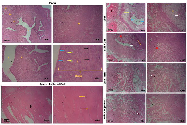
Figure 2. Shows the histology of the uterus and peritoneal wall of the control groups (H&E; x40, x100, x400) and the endometriotic tissue of the experimental groups (H&E; x40, x100). M - myometrium; P - perimetrium; E - endometrium; L - _lumen; F - Fasciculi; Long yellow arrows – _nuclei of muscle cells; Blue arrows – _surface epithelium of the endometrium; Short yellow arrows – _endometrial glands; Black arrows – _nuclei of endometrial stroma cells. White arrows – _hemorrhage/artery; *represents endometrial cyst; Red arrowhead - fibro-adipose tissue.