JBRA Assist. Reprod. 2026;30(2):353-363
REVIEW
doi: 10.5935/1518-0557.20250177
In vitro maturation 2.0: a new era for an underdog in ART
1Nilo Frantz Medicina Reprodutiva, Porto Alegre, Brazil
2Departamento de Ciências Morfológicas, Instituto de Ciências Básicas da Saúde, Universidade Federal do Rio Grande do Sul, Porto Alegre, Brazil
CONFLICT OF INTERESTS
The authors declare no competing interest
ABSTRACT
In vitro maturation (IVM) of human oocytes, once a pioneering concept predating conventional IVF, has long remained an underutilized technique in assisted reproduction. Despite early promise, clinical adoption of IVM has been limited due to lower embryo developmental competence and live birth rates compared to IVF. However, recent advances in the understanding of oocyte physiology, including cumulus-oocyte communication, the regulatory roles of cAMP/cGMP signaling pathways, and endocrine modulation of meiotic resumption, have reignited interest in optimizing IVM protocols. Innovations such as biphasic Capacitation (CAPA) IVM systems and the use of ovarian somatic support cells (OSCs) derived from induced pluripotent stem cells (iPSCs) aim to replicate the dynamic follicular environment more accurately and enhance oocyte competence. Clinical studies suggest that, while IVM still results in modestly lower cumulative live birth rates compared to conventional IVF, it offers significant advantages for selected patient populations, particularly women with polycystic ovary syndrome (PCOS), high ovarian reserve, or those requiring fertility preservation. Importantly, current evidence supports the genetic and epigenetic safety of IVM-derived offspring. As technical refinements continue and professional education expands, IVM is poised to fulfill its potential as a safer, less invasive, and more accessible alternative within the landscape of assisted reproductive technologies.
Keywords: In Vitro Maturation, meiotic maturation, cytoplasmic maturation, patient-friendly
INTRODUCTION
In Vitro Maturation (IVM) is an alternative Assisted Reproductive Technology (ART), traditionally defined as the maturation of immature oocytes in vitro from the germinal vesicle (GV, prophase I) to the metaphase II (MII) stage (Fig. 1A). These oocytes are typically retrieved from mid-sized antral follicles in unstimulated ovaries. Over time, IVM protocols have evolved to include minimal stimulation with follicle-stimulating hormone (FSH) and/or human chorionic gonadotropin (hCG). In recognition of its clinical value, the American Society for Reproductive Medicine (ASRM) declared IVM a non-experimental technique in 2021 (Practice Committees of the ASRM, 2021), especially applicable to women with polycystic ovaries (PCO), polycystic ovary syndrome (PCOS), and those requiring fertility preservation, such as cancer patients.
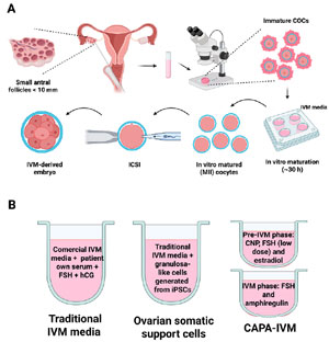
Figure 1. In vitro maturation of human oocytes. A) Workflow for the IVM treatment. B) Media composition for different IVM systems. Created in BioRender. Roos Kulmann, M. I. (2025) https://BioRender.com/31kaba0.
While commonly associated with these populations, IVM may also be suitable for normo-ovulatory patients with high ovarian reserve. Although initially perceived as a strategy to avoid ovarian hyperstimulation syndrome (OHSS), a complication now largely mitigated by the use of GnRH antagonists (Mills & Dahan, 2022), IVM offers additional benefits. It represents a more patient-friendly alternative to conventional in vitro fertilization (IVF), which requires controlled ovarian stimulation with high-dose gonadotropins to collect multiple mature (MII) oocytes. By reducing or eliminating the need for ovarian stimulation, IVM can lower treatment costs (Braam et al., 2021), minimize the number of clinic visits and ultrasound monitoring, and reduce medication-related side effects (Marchante et al., 2024), factors that often contribute to treatment discontinuation.
The concept of IVM was first demonstrated in rabbits (Pincus & Enzmann, 1935) and subsequently in other mammalian species, including humans (Edwards, 1965). In the late 1960s, Robert Edwards, widely regarded as a pioneer in reproductive medicine, initially explored IVM as a treatment for infertility (Edwards et al., 1969), before shifting his focus toward developing IVF. Reflecting on the field trajectory decades later, Edwards wrote in a 2007 publication: “With hindsight, should we have developed IVM rather than ovarian stimulation in 1969?” (Edwards, 2007).
Although IVF went on to become a mainstream technique, IVM remained on the sidelines for decades. The first live birth following IVM was reported in 1991 (Cha et al., 1991), and the first successful IVM pregnancy in a PCO patient was documented in 1994 (Trounson et al., 1994). In Latin America, the first IVM birth occurred in 2008 (Frantz et al., 2008). Despite its early origins, even predating IVF, IVM clinical development has been remarkably slow. To date, only an estimated 5,000-6,000 babies have been born worldwide using IVM (Das & Son, 2023), compared to millions born through IVF.
This limited adoption may be attributed to several factors, including lower embryo developmental potential in early IVM protocols and the technical complexity of the procedure. Additionally, the development of commercially available IVM systems did not keep pace with recent scientific discoveries on the physiology of oocyte maturation in vivo. As a result, IVM has consistently demonstrated lower success rates compared to conventional IVF (Vuong et al., 2020) and remains largely confined to a few specialized centers worldwide.
Recent scientific advances and the development of innovative culture systems have reignited interest in IVM, offering promising opportunities to broaden access to ART and enhance clinical outcomes. Key innovations include biphasic IVM systems designed to preserve oocyte-cumulus communication (Sanchez et al., 2019), the addition of granulosa-like cells (Piechota et al., 2023) or follicular fluid-derived extracellular vesicles (EVs) (Makieva et al., 2025) to better support oocyte maturation in vitro, and microfluidic chips (Zargari et al., 2016). Could this new era of IVM represent the next breakthrough in ART, positioned as a gentler alternative between intrauterine insemination (IUI) and conventional IVF? This review explores this possibility by focusing on molecular mechanisms underlying physiological oocyte maturation, current IVM clinical protocols, their safety, and whether IVM still lags behind standard IVF treatment in terms of clinical efficacy.
PHYSIOLOGY OF OOCYTE MATURATION
Oocyte maturation is a finely regulated physiological process that begins during fetal development and spans decades, culminating in the resumption of meiosis just prior to ovulation (Jamnongjit & Hammes, 2005). The initiation of meiosis during fetal life results in the formation of primordial follicles, wherein oocytes become arrested in prophase I, also known as the GV stage, surrounded by a single layer of flattened pre-granulosa cells (as reviewed in Telfer et al., 2023). This arrest is maintained until the follicle is recruited for growth and maturation, a process that remains tightly coordinated by the somatic compartment of the follicle.
The maintenance of meiotic arrest in fully grown oocytes is primarily governed by high levels of intracellular cyclic adenosine monophosphate (cAMP) (Cho et al., 1974), which inhibits the maturation-promoting factor (MPF) via activation of cAMP-dependent protein kinase A (PKA) (Shitsukawa et al., 2001). Two sources contribute to intra-oocyte cAMP levels: (1) endogenous production within the oocyte (Mehlmann et al., 2002), and (2) transzonal flux of cAMP from surrounding cumulus cells (CCs) under the influence of FSH (Anderson & Albertini, 1976). Critically, cyclic guanosine monophosphate (cGMP), synthesized in granulosa and CCs under the regulation of C-type natriuretic peptide (CNP), diffuses through gap junctions to the oocyte and inhibits phosphodiesterase 3A (PDE3A) (Zhang et al., 2010a; Tsuji et al., 2012), thereby preventing cAMP hydrolysis and reinforcing meiotic arrest (Norris et al., 2009). Therefore, maintaining the communication with CCs is crucial for oocyte meiotic arrest.
FSH plays a central role in promoting folliculogenesis and oocyte growth by acting on its receptor, expressed in granulosa cells from early/mid antral stage (Casarini et al., 2022) and possibly in the oocyte (Méduri et al., 2002). FSH drives granulosa cell proliferation, steroidogenesis, and antrum formation, eventually leading to the differentiation in mural granulosa cells (MGCs) and CCs (Eppig, 2001). The latter remain in intimate communication with the oocyte via transzonal projections (TZPs) and gap junctions, facilitating the bidirectional exchange of signals and metabolites (Kidder & Mhawi, 2002). During the growth phase, the oocyte increases in diameter and accumulates essential organelles, mRNAs, and proteins, collectively referred to as cytoplasmic maturation, largely supported by the surrounding CCs. Notably, the oocyte actively regulates CC function through the secretion of oocyte-secreted factors (OSFs), including growth differentiation factor 9 (GDF9) and bone morphogenetic protein 15 (BMP15) to modulate CC proliferation, metabolic activity, inhibition of premature luteinization, and responsiveness to meiotic inducers (Gilchrist, 2011).
Meiotic resumption is triggered by the preovulatory surge of luteinizing hormone (LH), which does not act directly on the oocyte or CCs, both of which normally lack LH receptors before ovulation (Diaz et al., 2007), but instead induces the expression of epidermal growth factor (EGF)-like peptides (amphiregulin, epiregulin, and betacellulin) in MGCs (Park et al., 2004). These peptides activate EGF receptors on CCs, a receptor system whose functional maturation depends on FSH signaling in concert with OSFs (Fan et al., 2009). The resulting signaling cascade likely leads to cumulus expansion, retraction of TZPs, and gap junction breakdown, culminating in a rapid decrease in intra-oocyte cGMP. This decline lifts the inhibition on PDE3A, leading to cAMP degradation, MPF activation, and germinal vesicle breakdown (GVBD) (Gilchrist, 2011).
The physiological coordination of meiotic arrest and resumption is lost when the oocyte is removed from its follicular niche, as occurs in conventional IVM protocols (Gilchrist & Smitz, 2023), resulting in spontaneous and asynchronous nuclear maturation. This unphysiological resumption of meiosis is hypothesized to compromise developmental competence by bypassing essential cytoplasmic and nuclear maturation events.
Understanding the molecular orchestration of oocyte maturation in vivo provides critical insights for designing culture systems that can faithfully mimic these conditions.
A CLINICAL PERSPECTIVE ON IVM
Patient selection
IVM was initially proposed for patients with PCOS or PCO-like conditions who face an elevated risk of developing OHSS during controlled ovarian stimulation. However, with the widespread use of GnRH antagonist triggers, the risk of OHSS has significantly reduced. As a result, the scope of IVM has broadened to include other patient groups, such as egg donors (Holzer et al., 2007), women with ovarian resistance to FSH (Galvão et al., 2018) and those requiring fertility preservation due to gonadotoxic treatments like cancer therapies (Cao & Chian, 2009; Huang et al., 2010; Chian et al., 2013) (Practice Committees of the ASRM, 2021).
A major advantage of IVM lies in its potential to reduce treatment costs compared to IVF cycles. Cost-effectiveness analyses suggest that IVM can decrease expenses related to gonadotropin use, office visits, and ultrasound examinations, leading to an estimated 34% reduction in overall treatment costs (Braam et al., 2021). As a result, the implementation of an IVM program could significantly expand access to ART for patients who might otherwise be unable to afford them, particularly in areas where IVF costs are high and not covered by insurance. Additionally, many patients may choose IVM to avoid the physical and emotional side effects associated with the high doses of gonadotropins, making IVM a more comfortable and less stressful alternative. In fact, women undergoing minimal gonadotropin priming during IVM have reported experiencing fewer side effects, such as pain, nausea, bleeding, and breast swelling, compared to those undergoing conventional controlled ovarian stimulation for IVF (Marchante et al., 2024). This improved patient experience adds to the appeal of IVM as a less invasive and more patient-friendly option.
IVM definitions
There are varying definitions and approaches to IVM. A more traditional or “purist” view of IVM involves the retrieval of COCs containing GV oocytes without any prior gonadotropin stimulation (Gilchrist & Smitz, 2023). However, some groups have suggested that using gonadotropin priming, administering FSH, hCG, or both, could improve pregnancy rates in IVM treatments (Fadini et al., 2009).
In standard IVF cycles, some of the collected oocytes are immature, typically at the GV or MI stages. Despite losing communication with CCs after denudation, these oocytes often undergo spontaneous maturation in vitro without the need for specialized IVM media or stimulation, a process referred to as Rescue IVM (Coticchio et al., 2025). Clinically, these oocytes can serve as surplus, potentially enhancing cumulative pregnancy rates, especially in patients with poor prognosis (Liu et al., 2020; Coticchio et al., 2025). It is important to distinguish this form of rescue maturation from the conventional IVM approach, which typically involves minimal or no gonadotropin stimulation.
FSH and hCG priming
Human follicles measuring 2-6 mm in diameter are known to express high levels of FSH receptors, and when activated by FSH, this promotes follicular growth and estradiol production (Jeppesen et al., 2012; Kristensen et al., 2018; Gilchrist & Smitz, 2023). Various regimens have been suggested in the literature, including cumulative doses of 450 to 600 IU of FSH administered over 2 to 5 days, typically starting on day 2 or 3 of the menstrual cycle (Wynn et al., 1998; De Vos et al., 2011). The effectiveness of FSH priming without hCG in IVM cycles remains a topic of debate. Junk et al. (2003) reported improved maturation rates with FSH priming but no significant impact on embryonic development. Since FSH priming does not trigger meiotic resumption in oocytes in vivo, immature compact COCs are collected during oocyte retrieval (Das & Son, 2023).
Interestingly, granulosa cells from antral follicles smaller than 6 mm do not usually express LH receptors, making the benefits of hCG priming questionable (Jeppesen et al., 2012; Gilchrist & Smitz, 2023). In practice, hCG priming may primarily promote the maturation of dominant follicles within the cohort, leading to a mixture of asynchronous mature and immature oocytes at collection, which complicates the strict definition of IVM. Similar to FSH priming, studies have demonstrated that hCG priming alone can increase oocyte maturation rates but does not necessarily improve pregnancy outcomes (Chian et al., 2000; Junk et al., 2003; Zheng et al., 2012; Lin et al., 2020).
The debate extends to the combined use of FSH and hCG priming. Fadini et al. (2009) showed that combining FSH and hCG priming significantly increased pregnancy rates (29.9%) compared to unstimulated cycles (15.3%), FSH priming alone (17.3%), or hCG priming alone (7.6%). However, Lin et al. (2003) reported that adding FSH priming to hCG priming did not result in any noticeable improvements in pregnancy rates. Therefore, further research is needed to determine whether gonadotropin priming can provide additional benefits to patients undergoing IVM treatment.
Oocyte retrieval
There is no consensus regarding the ideal timing for oocyte retrieval in IVM cycles. However, most studies suggest selecting a lead follicle diameter of up to 10 mm, based on findings by Cobo et al. (1999), which demonstrated that oocyte retrieval at this stage resulted in a higher number of recovered oocytes and improved blastocyst rates compared to retrieval when the lead follicle exceeded 10 mm. Larger follicles may induce atresia in sibling oocytes, potentially compromising their quality and development. Typically, the oocyte retrieval is scheduled 42-46 h after the last FSH injection in FSH-primed cycles (De Vos et al., 2011; Gilchrist et al., 2024), or 36-38 hours after administration in hCG-primed cycles (Zheng et al., 2012).
Compared to IVF, oocyte retrieval in IVM presents additional complexities due to the smaller follicular size (typically 2 to 6 mm), lack of CCs expansion and tighter adherence of immature oocytes to the follicle wall, requiring a more delicate and technically demanding aspiration process (Practice Committees of the ASRM, 2021). The retrieval procedure is more time-consuming, often taking twice as long as conventional IVF, as precise handling is necessary to optimize oocyte recovery while minimizing follicular collapse.
The choice of aspiration needle plays a crucial role in IVM success. While a single-lumen 17-gauge needle commonly used in IVF can be utilized, many centers opt for finer-gauge (19-21G) double-lumen needles (Wynn et al., 1998; Yan et al., 2021). These finer needles enhance precision when aspirating smaller follicles and reduce trauma to the follicular environment. Additionally, aspiration pressure must be carefully controlled to preserve COCs. Lower pressures, typically between 80 and 120 mmHg, are recommended to minimize shear stress and prevent dissociation of CCs from the oocyte, thereby maintaining its developmental competence in subsequent in vitro maturation steps (Hashimoto et al., 2007).
IVM IN THE LABORATORY
The IVM media
Early clinical findings revealed that oocytes matured in vitro did not exhibit comparable developmental competence to oocytes matured in vivo (Chian, 2004). This discrepancy has sparked global research efforts to optimize IVM. However, progress has been exceedingly slow. Commercially available systems have failed to keep pace with recent scientific advances, largely due to the limited interest of IVF clinics in adopting this alternative ART. As a result, a fully effective IVM protocol that accurately replicates in vivo maturation remains elusive.
Currently available IVM systems are relatively basic, typically relying on the addition of FSH and hCG to induce maturation (Fig. 1B). However, as previously discussed, the LH surge does not usually act directly on CCs or the oocyte. Instead, it initiates a signaling cascade leading to the expression and secretion of EGF-like peptides, such as amphiregulin, epiregulin, and betacellulin in MGCs. These peptides are promising additions to IVM media, as they promote meiotic resumption in a more physiological manner. In a murine IVM model, EGF-p supplementation led to improved blastocyst formation rates and embryo quality compared to FSH or EGF alone (Richani et al., 2013).
EGF-p exert their effects through activation of EGF receptor, whose expression is supported by FSH, GDF9 and BMP15. Supplementation of GDF9 and BMP15 has also been shown to enhance blastocyst and live birth rates in bovine (Hussein et al., 2006) and murine (Yeo et al., 2007) IVM models, respectively. In parallel, emerging IVM strategies highlight the importance of maintaining cumulus-oocyte communication to support cytoplasmic maturation, avoiding premature TZP retraction and meiosis resumption. In this sense, CNP plays a critical role in maintaining intra-oocyte cAMP levels and prophase I arrest (Zhang et al., 2010a), while the expression of its receptor in CCs is supported by estradiol (Zhang et al., 2011), a hormone that has been shown to enhance oocyte competence in multiple IVM models (Zheng, 2003; Maksura et al., 2021).
Collectively, these findings suggest that an improved baseline IVM medium should incorporate EGF-like peptides, FSH, GDF9, BMP15, and estradiol to better replicate in vivo signaling and support oocyte competence.
New IVM systems
In an effort to overcome the suboptimal outcomes typically observed in IVM cycles, new IVM systems have been developed. Capacitation IVM (CAPA-IVM) is an emerging approach designed to preserve oocyte-cumulus communication by preventing premature maturation and TZP retraction, both of which are critical for cytoplasmic maturation and enhanced developmental potential (Romero et al., 2016; Sanchez et al., 2019). Spontaneous oocyte maturation occurs primarily due to a decline in intracellular cAMP. While cAMP is sustained through endogenous oocyte production and FSH-stimulated synthesis in CCs, it also depends on cGMP, which inhibits PDE3 and prevents cAMP degradation (Degerman et al., 1997).
As previously mentioned, cGMP production in CCs is stimulated by CNP secreted by MGCs (Zhang et al., 2010a). Following IVM COC retrieval, this source of CNP is lost, leading to a drop in cGMP levels and a consequent decline in cAMP within the oocyte, triggering premature meiotic resumption (Degerman et al., 1997). Capacitation IVM (CAPA-IVM) mitigates this issue by introducing a pre-IVM phase with exogenous CNP (Gilchrist et al., 2024). The rationale for this supplementation is to sustain cGMP levels, stabilize intra-oocyte cAMP, and delay nuclear maturation, thereby allowing additional time for cytoplasmic maturation (Sanchez et al., 2019).
CAPA-IVM has demonstrated superior efficiency compared to commercially available IVM systems, leading to higher oocyte maturation rates, increased yield of good-quality embryos, and improved clinical pregnancy outcomes (Gilchrist et al., 2024). The first randomized controlled trial (RCT) comparing CAPA-IVM with conventional IVF found that CAPA-IVM was only slightly inferior in terms of Day-3 embryo quality and cumulative pregnancy rates (Vuong et al., 2020). Importantly, studies assessing the safety of CAPA-IVM have shown no significant differences in DNA methylation (Saenz-de-Juano et al., 2019) and health of children (Vuong et al., 2022).
Another innovative approach to IVM involves co-culturing cumulus-oocyte complexes (COCs) with ovarian somatic support cells (OSCs) generated from human-induced pluripotent stem cells (iPSCs). These OSCs, differentiated through the expression of specific transcription factors, exhibit granulosa-like characteristics (FOXL2+, AMHR2+, NR2F2+) (Pierson Smela et al., 2023) and may restore critical signaling from mural granulosa cells (MGCs) within the IVM culture. Upon FSH stimulation, OSCs produce growth factors and steroids, potentially recreating key elements of the follicular microenvironment (Pierson Smela et al., 2023).
As previously discussed, the preovulatory LH surge stimulates MGCs to produce EGF-like peptides, amphiregulin, epiregulin, and betacellulin, which activate EGF receptors in both an autocrine manner (within MGCs) and a paracrine manner (in CCs) (Park et al., 2004). This cascade triggers meiotic resumption via the ERK1/2 pathway. While not yet fully characterized, OSCs may also express CNP, helping sustain intra-oocyte cAMP levels and maintaining cumulus-oocyte communication.
Unlike conventional static culture media, OSCs dynamically respond to co-culture signals, potentially fine-tuning their gene expression in response to cues from CCs and the oocyte (Paulsen et al., 2024). Notably, OSC-assisted IVM has been shown to generate oocytes with transcriptomic profiles more closely resembling those of in vivo-matured oocytes (Paulsen et al., 2024). Furthermore, this approach has resulted in higher MII maturation and euploid blastocyst formation rates in comparison to commercially available IVM systems (Piechota et al., 2023). Future clinical data on pregnancy outcomes will determine whether OSC-IVM significantly enhances success rates in IVM cycles.
Although additional laboratory procedures in IVM cycles are well established, the technique complexity may require a larger and more experienced team of embryologists. Oocyte retrieval from antral follicles is technically challenging due to the smaller size of COCs, increased blood content in follicular fluid, and the need for additional sieves to aid in oocyte capture (Practice Committees of the ASRM, 2021). Moreover, hCG priming can cause oocyte asynchrony, leading to a mixture of maturation stages at the time of collection (Gilchrist & Smitz, 2023). In that case, COC morphology can help infer oocyte maturity: expanded COCs should be denuded and MII oocytes inseminated immediately, while unexpanded ones are directed to IVM culture. This results in staggered insemination times for oocytes from the same patient, often at inconvenient hours, thereby increasing the overall workload in the laboratory.
IVM VERSUS IVF: HOW FAR ARE WE?
Multiple studies have investigated whether IVM offers comparable efficiency to IVF, particularly in women with PCO/PCOS. Initial research began with case-control studies and later progressed to RCTs. Child (2002) conducted the first case-control study comparing unstimulated IVM with IVF in age-matched PCOS patients. The study reported clinical pregnancy and live birth rates per oocyte retrieval of 26.2% and 15.9%, respectively, in IVM cycles, compared to 38.3% and 26.2% in IVF cycles (Table 1). Although these differences were not statistically significant, there was a trend towards lower success rates with IVM. Furthermore, 11.2% of patients undergoing IVF developed moderate or severe OHSS, while none of the IVM patients experienced this complication. Similarly, a retrospective case-control study by Gremeau et al. (2012) found lower clinical pregnancy (50.5% vs. 19.6%) and live birth rates (44.3% vs. 16.5%) with IVM, along with a significantly reduced risk of OHSS (8.2% vs. 0%).
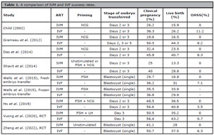
Table 1. A comparison of IVM and IVF success rates.
Both of these initial studies utilized fresh transfers of multiple IVM cleavage-stage embryos (averaging 3.2 and 1.9 embryos, respectively) to compensate for their reduced developmental competence. This approach led to multiple pregnancy rates as high as 40% (Child, 2002; Gremeau et al., 2012). These findings were further supported by additional retrospective studies involving cleavage-stage embryo transfers, which reported multiple pregnancy rates exceeding 30% (Das et al., 2014; Ho et al., 2019). However, these rates were comparable to those in the IVF control groups, suggesting that the high incidence of multiple pregnancies was more related to the transfer strategy employed rather than being specific to the IVM technology itself.
The shift in reproductive medicine toward transferring fewer embryos with higher developmental potential also extended to IVM. Walls et al. (2015) were the first to compare IVM and IVF outcomes after the transfer of a single blastocyst. In their study, which involved priming IVM cycles with FSH, they reported similar blastocyst formation rates (45% vs. 46%), usable blastocyst rates (38% vs. 40%), and a comparable proportion of cycles with complete failure of blastocyst development (16% vs. 14%) between IVM and IVF. However, IVM cycles yielded fewer total blastocysts (3.0 vs. 4.6), and the fresh transfer of a single blastocyst led to significantly lower live birth rate in IVM (18.8% vs. 31%).
Interestingly, a subset of IVM cycles involved freezing all blastocysts due to inadequate endometrial conditions for fresh transfer. When these frozen embryos were later transferred, similar live birth rates were observed between IVM and IVF (29.9% vs. 33.9%) (Walls et al., 2015), suggesting that poor endometrial conditions may contribute, at least in part, to the lower success rates observed with IVM. In a subsequent study, Zheng et al. (2022) conducted the first RCT comparing IVM and IVF after a freeze-all strategy and single blastocyst transfer, this time without any priming in IVM cycles. Their design yielded markedly different outcomes between IVM and IVF, with IVM showing a lower rate of vitrified blastocysts (27.2% vs. 46.5%), a higher proportion of cycles with no available blastocysts (36.7% vs. 3.8%) and reduced live birth rates (28% vs. 37.5%). This highlights the potential role of FSH priming in improving the developmental competence of IVM embryos.
With the advent of embryo freezing, each oocyte retrieval cycle has the potential to result in multiple embryo transfers, making the cumulative live birth rate arguably the most clinically meaningful measure of success. This is particularly relevant for IVM, which tends to result in a lower number of good-quality embryos compared to IVF (Das et al., 2014; Ho et al., 2019). In light of this, several studies have consistently reported inferior cumulative live birth rates for IVM compared to IVF, with differences ranging from 10.5% to 18.6%, even when employing newer protocols like CAPA-IVM (Walls et al., 2015; Ho et al., 2019; Vuong et al., 2020; Zheng et al., 2022).
Overall, few studies have examined the differential success rates between IVM and IVF over the past 20 years, and those that do exist are quite heterogeneous regarding hormonal priming, laboratory protocols, and the stage of embryos transferred. Even fewer studies are RCTs. The available evidence suggests that IVM continues to result in lower-quality embryos and reduced live birth rates compared to IVF, although the cumulative difference may be as small as 10.5%. Whether a reduction in success rates of approximately 10% is an acceptable trade-off for the benefits of IVM, such as reduced costs, shorter treatment times, and avoidance of the unpleasant side effects associated with gonadotropins, remains an important topic for further discussion.
IVM SAFETY
The consistently lower embryo quality observed in IVM combined with the additional culture steps required in the laboratory raises concerns about whether genetic and epigenetic factors in IVM embryos, while still compatible with live birth, could potentially impact the long-term health of children born through this technique.
Human oocytes undergo significant remodeling of their methylation and genomic imprinting patterns during oocyte growth (Yan et al., 2021), which may be hypothetically compromised by disruption of this process in vitro, affecting the epigenetic safety of the offspring. However, studies have shown that IVM does not significantly alter methylation patterns in imprinted genes when compared to oocytes matured in vivo (Kuhtz et al., 2014). Furthermore, blastocysts derived from IVM treatment in PCOS patients did not show any significant differences in methylation patterns or the expression of key epigenetic regulators compared to blastocysts from IVF-treated PCOS patients (Saenz-de-Juano et al., 2019). Finally, bisulfite sequencing analysis of chorionic villus and cord-blood samples from children conceived through IVM revealed no significant differences in the methylation of key developmentally relevant genomic loci (Pliushch et al., 2015). Overall, the available data suggests that the epigenetic stability of the offspring is not adversely affected by IVM.
Another primary concern relates to the chromosomal integrity of IVM embryos. Although most aneuploidies do not result in live births, certain chromosomal abnormalities can still manifest in viable offspring (Capalbo et al., 2022). To investigate this issue, Zhang et al. (2010b) analyzed chromosomal abnormality rates in cleavage-stage embryos derived from both IVM and IVF using FISH on chromosomes 13, 15, 16, 18, 21, 22, X, and Y. The results revealed no significant difference in chromosomal abnormality rates between IVM and IVF embryos (58.7% vs. 57.4%, respectively). However, the study also found that the incidence of chromosomal abnormalities increased with the time required for oocyte maturation. Specifically, IVM embryos derived from oocytes that took 48 hours to reach the MII stage exhibited a higher frequency of abnormalities compared to those that matured within 24 hours. This suggests that the timing of oocyte maturation is a critical factor to consider when selecting IVM embryos for transfer. Similarly, Requena et al. (2009) reported aneuploidy rates of 60% in cleavage-stage embryos from IVM cycles compared to 33% in IVF cycles, although this difference was not statistically significant.
Later studies have employed advanced genetic methodologies, such as comparative genomic hybridization (aCGH) and next-generation sequencing (NGS), to analyze aneuploidies across all chromosomes in IVM-derived cleavage-stage embryos, rather than focusing on a specific set of chromosomes. These technologies enable the detection of a broader range of aneuploidies, previously undetectable with FISH analysis (ESHRE PGT-SR/PGT-A Working Group et al., 2020). Notably, these studies reported comparable incidence of aneuploidies between IVM and IFV embryos, with rates ranging from 28% to 55.6% (Spits et al., 2015; Li et al., 2021).
When assessing the health of IVM offspring, several studies have consistently shown no significant differences in perinatal and obstetric outcomes compared to IVF (Cha et al., 2005; Mikkelsen, 2005; Shu-Chi et al., 2006; Söderström-Anttila et al., 2006; Buckett et al., 2007; Fadini et al., 2012; Roesner et al., 2017; Mostinckx et al., 2019; Yu et al., 2019; Belva et al., 2020). These studies, with follow-up periods extending up to 2 and 7.5 years, evaluated key parameters such as birth weight, gestational age at delivery, sex ratio, developmental milestones (assessed via Bayley Scales), Apgar scores, pregnancy complications, congenital abnormalities, psychomotor and neuropsychological development, and karyotype. Notably, Foix-L’Hélias et al. (2014) reported an increase in average birth weight, length, and head circumference among female infants conceived via IVM compared to those conceived via IVF. It is noteworthy that some studies properly matched IVM patients with PCOS to IVF patients with PCOS (Cha et al., 2005; Mostinckx et al., 2019; Yu et al., 2019; Belva et al., 2020), while others did not account for the specific PCOS background (Shu-Chi et al., 2006; Söderström-Anttila et al., 2006; Buckett et al., 2007; Foix-L’Hélias et al., 2014; Roesner et al., 2017), which could be an influencing factor. However, a recent meta-analysis, as well as findings from the first RCT comparing IVM and IVF, confirmed no significant differences in perinatal outcomes between the two techniques, reaffirming the safety of IVM (Vuong et al., 2020; Strowitzki et al., 2021).
Overall, the current evidence supports the notion that IVM is a safe treatment, showing similar rates of methylation at epigenetically relevant loci, chromosomal abnormalities in embryos, and obstetric/perinatal outcomes when compared to IVF. However, it is important to recognize that the available research is still limited and highly heterogeneous, particularly concerning the IVM protocols used and the study populations involved. To enhance the understanding of IVM safety, future research should: i) go beyond analyzing DNA methylation and consider other epigenetic factors such as histone modifications and chromatin remodeling; ii) assess chromosomal integrity using state-of-the-art procedures, as trophectoderm biopsy from blastocysts; and iii) focus specifically on PCOS patients, allowing for a more accurate comparison of ART procedures without the confounding effects of underlying infertility conditions.
CONCLUSIONS AND PERSPECTIVES
Despite being one of the earliest ART explored, IVM has historically remained on the sidelines of mainstream IVF, with few significant innovations over the last five decades. However, recent scientific advances in the understanding of oocyte physiology have revitalized efforts to optimize IVM protocols. Innovations such as biphasic CAPA-IVM systems and the use of OSCs derived from iPSCs demonstrate that mimicking the native follicular microenvironment can significantly enhance oocyte competence and clinical outcomes.
Nevertheless, IVM still faces notable challenges. Despite improvements, embryo developmental potential and cumulative live birth rates remain slightly lower compared to standard IVF. Additionally, the technical demands of oocyte retrieval and laboratory culture require specialized expertise and resources, limiting its widespread adoption. While current data support the genetic and epigenetic safety of IVM offspring, longer-term and larger-cohort studies are needed to fully validate these findings.
Future innovative strategies may involve the integration of biphasic maturation systems, OSCs, microfluidic platforms, and dynamic three-dimensional (3D) culture models to simulate the dynamic follicular environment more accurately. Additionally, training IVF professionals and educating both staff and patients on the unique benefits of IVM could be key steps toward broader acceptance and implementation of this technique.
Ultimately, IVM holds the potential to expand access to ART by offering a safer, more affordable, and less invasive treatment option, particularly for patients with high ovarian reserve, PCOS, or those requiring fertility preservation. As research continues to bridge the gap between laboratory culture and physiological conditions, IVM may yet fulfill its early promise as a mainstream alternative in reproductive medicine.
REFERENCES
Anderson E, Albertini DF. Gap junctions between the oocyte and companion follicle cells in the mammalian ovary. J Cell Biol. 1976;71:680-6. PMID: 825522 DOI: 10.1083/jcb.71.2.680 Medline
Belva F, Roelants M, Vermaning S, Desmyttere S, De Schepper J, Bonduelle M, Tournaye H, Hes F, De Vos M. Growth and other health outcomes of 2-year-old singletons born after IVM versus controlled ovarian stimulation in mothers with polycystic ovary syndrome. Hum Reprod Open. 2020;2020:hoz043. PMID: 32055712 DOI: 10.1093/hropen/hoz043 Medline
Braam SC, Ho VNA, Pham TD, Mol BW, van Wely M, Vuong LN. In-vitro maturation versus IVF: a cost-effectiveness analysis. Reprod Biomed Online. 2021;42:143-9. PMID: 33132059 DOI: 10.1016/j.rbmo.2020.09.022 Medline
Buckett WM, Chian RC, Holzer H, Dean N, Usher R, Tan SL. Obstetric outcomes and congenital abnormalities after in vitro maturation, in vitro fertilization, and intracytoplasmic sperm injection. Obstet Gynecol. 2007;110:885-91. PMID: 17906024 DOI: 10.1097/01.AOG.0000284627.38540.80 Medline
Cao YX, Chian RC. Fertility Preservation with Immature and in Vitro Matured Oocytes. Semin Reprod Med. 2009;27:456-64. PMID: 19806514 DOI: 10.1055/s-0029-1241055 Medline
Capalbo A, Poli M, Jalas C, Forman EJ, Treff NR. On the reproductive capabilities of aneuploid human preimplantation embryos. Am J Hum Genet. 2022;109:1572-81. PMID: 36055209 DOI: 10.1016/j.ajhg.2022.07.009 Medline
Casarini L, Paradiso E, Lazzaretti C, D’Alessandro S, Roy N, Mascolo E, Zaręba K, García-Gasca A, Simoni M. Regulation of antral follicular growth by an interplay between gonadotropins and their receptors. J Assist Reprod Genet. 2022;39:893-904. PMID: 35292926 DOI: 10.1007/s10815-022-02456-6 Medline
Cha KY, Chung HM, Lee DR, Kwon H, Chung MK, Park LS, Choi DH, Yoon TK. Obstetric outcome of patients with polycystic ovary syndrome treated by in vitro maturation and in vitro fertilization-embryo transfer. Fertil Steril. 2005;83:1461-5. PMID: 15866585 DOI: 10.1016/j.fertnstert.2004.11.044 Medline
Cha KY, Koo JJ, Ko JJ, Choi DH, Han SY, Yoon TK. Pregnancy after in vitro fertilization of human follicular oocytes collected from nonstimulated cycles, their culture in vitro and their transfer in a donor oocyte program. Fertil Steril. 1991;55:109-13. PMID: 1986950 DOI: 10.1016/S0015-0282(16)54068-0 Medline
Chian RC. In-vitro maturation of immature oocytes for infertile women with PCOS. Reprod Biomed Online. 2004;8:547-52. PMID: 15151718 DOI: 10.1016/S1472-6483(10)61101-7 Medline
Chian RC, Buckett WM, Tulandi T, Tan SL. Prospective randomized study of human chorionic gonadotrophin priming before immature oocyte retrieval from unstimulated women with polycystic ovarian syndrome. Hum Reprod. 2000;15:165-70. PMID: 10611207 DOI: 10.1093/humrep/15.1.165 Medline
Chian RC, Uzelac PS, Nargund G. In vitro maturation of human immature oocytes for fertility preservation. Fertil Steril. 2013;99:1173-81. PMID: 23433515 DOI: 10.1016/j.fertnstert.2013.01.141 Medline
Child T. A comparison of in vitro maturation and in vitro fertilization for women with polycystic ovaries. Obstet Gynecol. 2002;100:665-70. PMID: 12383531 DOI: 10.1016/s0029-7844(02)02193-2 Medline
Cho WK, Stern S, Biggers JD. Inhibitory effect of dibutyryl cAMP on mouse oocyte maturation in vitro. J Exp Zool. 1974;187:383-6. PMID: 4362350 DOI: 10.1002/jez.1401870307 Medline
Cobo AC, Requena A, Neuspiller F, Aragon s M, Mercader A, Navarro J, Simón C, Remohí J, Pellicer A. Maturation in vitro of human oocytes from unstimulated cycles: selection of the optimal day for ovum retrieval based on follicular size. Hum Reprod. 1999;14:1864-8. PMID: 10402406 DOI: 10.1093/humrep/14.7.1864 Medline
Coticchio G, Cimadomo D, De Vos M, Ebner T, Esbert M, Escribá MJ, Gilchrist RB, Rienzi L. To rescue or not to rescue immature oocytes: prospects and challenges. Fertil Steril. 2025;123:749-58. PMID: 40058555 DOI: 10.1016/j.fertnstert.2025.02.039 Medline
Das M, Son WY. In vitro maturation (IVM) of human immature oocytes: is it still relevant? Reprod Biol Endocrinol. 2023;21:110. PMID: 37993914 DOI: 10.1186/s12958-023-01162-x Medline
Das M, Son WY, Buckett W, Tulandi T, Holzer H. In-vitro maturation versus IVF with GnRH antagonist for women with polycystic ovary syndrome: treatment outcome and rates of ovarian hyperstimulation syndrome. Reprod Biomed Online. 2014;29:545-51. PMID: 25262236 DOI: 10.1016/j.rbmo.2014.07.019 Medline
De Vos M, Ortega-Hrepich C, Albuz FK, Guzman L, Polyzos NP, Smitz J, Devroey P. Clinical outcome of non-hCG-primed oocyte in vitro maturation treatment in patients with polycystic ovaries and polycystic ovary syndrome. Fertil Steril. 2011;96:860-4. PMID: 21868003 DOI: 10.1016/j.fertnstert.2011.07.1108 Medline
Degerman E, Belfrage P, Manganiello VC. Structure, Localization, and Regulation of cGMP-inhibited Phosphodiesterase (PDE3). J Biol Chem. 1997;272:6823-6. PMID: 9102399 DOI: 10.1074/jbc.272.11.6823 Medline
Diaz FJ, Wigglesworth K, Eppig JJ. Oocytes determine cumulus cell lineage in mouse ovarian follicles. J Cell Sci. 2007;120:1330-40. PMID: 17389684 DOI: 10.1242/jcs.000968 Medline
Edwards RG. IVF, IVM, natural cycle IVF, minimal stimulation IVF - time for a rethink. Reprod Biomed Online. 2007;15:106-19. PMID: 17623547 DOI: 10.1016/s1472-6483(10)60699-2 Medline
Edwards RG. Maturation in vitro of mouse, sheep, cow, pig, rhesus monkey and human ovarian oocytes. Nature. 1965;208:349-51. PMID: 4957259 DOI: 10.1038/208349a0 Medline
Edwards RG, Bavister BD, Steptoe PC. Early Stages of Fertilization in vitro of Human Oocytes Matured in vitro. Nature. 1969;221:632-5. PMID: 4886881 DOI: 10.1038/221632a0 Medline
Eppig JJ. Oocyte control of ovarian follicular development and function in mammals. Reproduction. 2001;122:829-38. PMID: 11732978 DOI: 10.1530/rep.0.1220829 Medline
ESHRE PGT-SR/PGT-A Working Group; Coonen E, Rubio C, Christopikou D, Dimitriadou E, Gontar J, Goossens V, Maurer M, Spinella F, Vermeulen N, De Rycke M. ESHRE PGT Consortium good practice recommendations for the detection of structural and numerical chromosomal aberrations†. Hum Reprod Open. 2020;2020:hoaa017. PMID: 32500102 DOI: 10.1093/hropen/hoaa017 Medline
Fadini R, Dal Canto M, Renzini MM, Brambillasca F, Comi R, Fumagalli D, Lain M, Merola M, Milani R, De Ponti E. Effect of different gonadotrophin priming on IVM of oocytes from women with normal ovaries: a prospective randomized study. Reprod Biomed Online. 2009;19:343-51. PMID: 19778479 DOI: 10.1016/S1472-6483(10)60168-X Medline
Fadini R, Mignini Renzini M, Guarnieri T, Dal Canto M, De Ponti E, Sutcliffe A, Shevlin M, Comi R, Coticchio G. Comparison of the obstetric and perinatal outcomes of children conceived from in vitro or in vivo matured oocytes in in vitro maturation treatments with births from conventional ICSI cycles. Hum Reprod. 2012;27:3601-8. PMID: 23042796 DOI: 10.1093/humrep/des359 Medline
Fan HY, Liu Z, Shimada M, Sterneck E, Johnson PF, Hedrick SM, Richards JS. MAPK3/1 (ERK1/2) in ovarian granulosa cells are essential for female fertility. Science. 2009;324:938-41. PMID: 19443782 DOI: 10.1126/science.1171396 Medline
Foix-L’Hélias L, Grynberg M, Ducot B, Frydman N, Kerbrat V, Bouyer J, Labrune P. Growth development of French children born after in vitro maturation. PLoS One. 2014;9:e89713. PMID: 24586979 DOI: 10.1371/journal.pone.0089713 Medline
Galvão A, Segers I, Smitz J, Tournaye H, De Vos M. In vitro maturation (IVM) of oocytes in patients with resistant ovary syndrome and in patients with repeated deficient oocyte maturation. J Assist Reprod Genet. 2018;35:2161-71. PMID: 30238176 DOI: 10.1007/s10815-018-1317-z Medline
Gilchrist RB. Recent insights into oocyte-follicle cell interactions provide opportunities for the development of new approaches to in vitro maturation. Reprod Fertil Dev. 2011;23:23-31. PMID: 21366977 DOI: 10.1071/RD10225 Medline
Gilchrist RB, Ho TM, De Vos M, Sanchez F, Romero S, Ledger WL, Anckaert E, Vuong LN, Smitz J. A fresh start for IVM: capacitating the oocyte for development using pre-IVM. Hum Reprod Update. 2024;30:3-25. PMID: 37639630 DOI: 10.1093/humupd/dmad023 Medline
Gilchrist RB, Smitz J. Oocyte in vitro maturation: physiological basis and application to clinical practice. Fertil Steril. 2023;119:524-39. PMID: 36804961 DOI: 10.1016/j.fertnstert.2023.02.010 Medline
Gremeau AS, Andreadis N, Fatum M, Craig J, Turner K, Mcveigh E, Child T. In vitro maturation or in vitro fertilization for women with polycystic ovaries? A case-control study of 194 treatment cycles. Fertil Steril. 2012;98:355-60. PMID: 22658347 DOI: 10.1016/j.fertnstert.2012.04.046 Medline
Hashimoto S, Fukuda A, Murata Y, Kikkawa M, Oku H, Kanaya H, Sonoda M, Sugihara K, Murata T, Nagata F, Nakaoaka Y, Morimoto Y. Effect of aspiration vacuum on the developmental competence of immature human oocytes retrieved using a 20-gauge needle. Reprod Biomed Online. 2007;14:444-9. PMID: 17425825 DOI: 10.1016/S1472-6483(10)60891-7 Medline
Ho VNA, Braam SC, Pham TD, Mol BW, Vuong LN. The effectiveness and safety of in vitro maturation of oocytes versus in vitro fertilization in women with a high antral follicle count. Hum Reprod. 2019;34:1055-64. PMID: 31111879 DOI: 10.1093/humrep/dez060 Medline
Holzer H, Scharf E, Chian RC, Demirtas E, Buckett W, Tan SL. In vitro maturation of oocytes collected from unstimulated ovaries for oocyte donation. Fertil Steril. 2007;88:62-7. PMID: 17336971 DOI: 10.1016/j.fertnstert.2006.11.087 Medline
Huang JY, Chian RC, Gilbert L, Fleiszer D, Holzer H, Dermitas E, Elizur SE, Gidoni Y, Levin D, Son WY, Tan SL. Retrieval of immature oocytes from unstimulated ovaries followed by in vitro maturation and vitrification: A novel strategy of fertility preservation for breast cancer patients. Am J Surg. 2010;200:177-83. PMID: 20637351 DOI: 10.1016/j.amjsurg.2009.04.004 Medline
Hussein TS, Thompson JG, Gilchrist RB. Oocyte-secreted factors enhance oocyte developmental competence. Dev Biol. 2006;296:514-21. PMID: 16854407 DOI: 10.1016/j.ydbio.2006.06.026 Medline
Jamnongjit M, Hammes SR. Oocyte maturation: the coming of age of a germ cell. Semin Reprod Med. 2005;23:234-41. PMID: 16059829 DOI: 10.1055/s-2005-872451 Medline
Jeppesen JV, Kristensen SG, Nielsen ME, Humaidan P, Dal Canto M, Fadini R, Schmidt KT, Ernst E, Yding Andersen C. LH-receptor gene expression in human granulosa and cumulus cells from antral and preovulatory follicles. J Clin Endocrinol Metab. 2012;97:E1524-31. PMID: 22659248 DOI: 10.1210/jc.2012-1427 Medline
Junk SM, Dharmarajan A, Yovich JL. FSH priming improves oocyte maturation, but priming with FSH or hCG has no effect on subsequent embryonic development in an in vitro maturation program. Theriogenology. 2003;59:1741-9. PMID: 12566148 DOI: 10.1016/S0093-691X(02)01234-7 Medline
Kidder G, Mhawi A. Gap junctions and ovarian folliculogenesis. Reproduction. 2002;123:613-20. PMID: 12006089 DOI: 10.1530/rep.0.1230613 Medline
Kristensen SG, Mamsen LS, Jeppesen JV, Bøtkjær JA, Pors SE, Borgbo T, Ernst E, Macklon KT, Andersen CY. Hallmarks of Human Small Antral Follicle Development: Implications for Regulation of Ovarian Steroidogenesis and Selection of the Dominant Follicle. Front Endocrinol (Lausanne). 2018;8:376. PMID: 29375481 DOI: 10.3389/fendo.2017.00376 Medline
Kuhtz J, Romero S, De Vos M, Smitz J, Haaf T, Anckaert E. Human in vitro oocyte maturation is not associated with increased imprinting error rates at LIT1, SNRPN, PEG3 and GTL2. Hum Reprod. 2014;29:1995-2005. PMID: 24963167 DOI: 10.1093/humrep/deu155 Medline
Li J, Chen J, Sun T, Zhang S, Jiao T, Chian RC, Li Y, Xu Y. Chromosome aneuploidy analysis in embryos derived from in vivo and in vitro matured human oocytes. J Transl Med. 2021;19:416. PMID: 34625066 DOI: 10.1186/s12967-021-03080-1 Medline
Lin Y, Zheng X, Ma C, Li X, Zhang X, Yang P, Xu J, Zhu J. Human Chorionic Gonadotropin Priming Does Not Improve Pregnancy Outcomes of PCOS-IVM Cycles. Front Endocrinol (Lausanne). 2020;11:279. PMID: 32425891 DOI: 10.3389/fendo.2020.00279 Medline
Lin YH, Hwang JL, Huang LW, Mu SC, Seow KM, Chung J, Hsieh BC, Huang SC, Chen CY, Chen PH. Combination of FSH priming and hCG priming for in-vitro maturation of human oocytes. Hum Reprod. 2003;18:1632-6. PMID: 12871873 DOI: 10.1093/humrep/deg335 Medline
Liu Y, Jiang H, Du X, Huang J, Wang X, Hu Y, Ni F, Liu C. Contribution of rescue in-vitro maturation versus double ovarian stimulation in ovarian stimulation cycles of poor-prognosis women. Reprod Biomed Online. 2020;40:511-7. PMID: 32199798 DOI: 10.1016/j.rbmo.2019.12.012 Medline
Maksura H, Akon N, Islam MN, Akter I, Modak AK, Khatun A, Alam MH, Hashem MA, Amin MR, Moniruzzaman M. Effects of estradiol on in vitro maturation of buffalo and goat oocytes. Reprod Med Biol. 2021;20:62-70. PMID: 33488284 DOI: 10.1002/rmb2.12350 Medline
Marchante M, Barrachina F, Piechota S, Fernandez-González M, Giovannini A, Smith T, Kats S, Paulsen B, González E, Calvente V, Silvan A, Abittan B, Klein J, Klatsky P, Ordonez D, Kramme CC. Donor side effects experienced under minimal controlled ovarian stimulation with in vitro maturation vs. conventional controlled ovarian stimulation for in vitro fertilization treatment. FS Sci. 2024;5:242-51. PMID: 38838957 DOI: 10.1016/j.xfss.2024.05.002 Medline
Méduri G, Charnaux N, Driancourt MA, Combettes L, Granet P, Vannier B, Loosfelt H, Milgrom E. Follicle-Stimulating Hormone Receptors in Oocytes? J Clin Endocrinol Metab. 2002;87:2266-76. PMID: 11994374 DOI: 10.1210/jcem.87.5.8502 Medline
Mehlmann LM, Jones TLZ, Jaffe LA. Meiotic arrest in the mouse follicle maintained by a Gs protein in the oocyte. Science. 2002;297:1343-5. PMID: 12193786 DOI: 10.1126/science.1073978 Medline
Mikkelsen AL. Strategies in human in-vitro maturation and their clinical outcome. Reprod Biomed Online. 2005;10:593-9. PMID: 15949216 DOI: 10.1016/S1472-6483(10)61666-5 Medline
Mills G, Dahan MH. Gonadotropin releasing hormone (GnRH) antagonist administration to decrease the risk of ovarian hyperstimulation syndrome in GNRH agonist cycles triggered with human chorionic gonadotropin. Arch Gynecol Obstet. 2022;306:1731-7. PMID: 35932297 DOI: 10.1007/s00404-022-06717-8 Medline
Mostinckx L, Segers I, Belva F, Buyl R, Santos-Ribeiro S, Blockeel C, Smitz J, Anckaert E, Tournaye H, De Vos M. Obstetric and neonatal outcome of ART in patients with polycystic ovary syndrome: IVM of oocytes versus controlled ovarian stimulation. Hum Reprod. 2019;34:1595-607. PMID: 31347678 DOI: 10.1093/humrep/dez086 Medline
Norris RP, Ratzan WJ, Freudzon M, Mehlmann LM, Krall J, Movsesian MA, Wang H, Ke H, Nikolaev VO, Jaffe LA. Cyclic GMP from the surrounding somatic cells regulates cyclic AMP and meiosis in the mouse oocyte. Development. 2009;136:1869-78. PMID: 19429786 DOI: 10.1242/dev.035238 Medline
Park JY, Su YQ, Ariga M, Law E, Jin SLC, Conti M. EGF-like growth factors as mediators of LH action in the ovulatory follicle. Science. 2004;303:682-4. PMID: 14726596 DOI: 10.1126/science.1092463 Medline
Paulsen B, Piechota S, Barrachina F, Giovannini A, Kats S, Potts KS, Rockwell G, Marchante M, Estevez SL, Noblett AD, Figueroa AB, Aschenberger C, Kelk DA, Forti M, Marcinyshyn S, Wiemer K, Sanchez M, Belchin P, Lee JA, Buyuk E, et al. Rescue in vitro maturation using ovarian support cells of human oocytes from conventional stimulation cycles yields oocytes with improved nuclear maturation and transcriptomic resemblance to in vivo matured oocytes. J Assist Reprod Genet. 2024;41:2021-36. PMID: 38814543 DOI: 10.1007/s10815-024-03143-4 Medline
Pierson Smela MD, Kramme CC, Fortuna PRJ, Adams JL, Su R, Dong E, Kobayashi M, Brixi G, Kavirayuni VS, Tysinger E, Kohman RE, Shioda T, Chatterjee P, Church GM. Directed differentiation of human iPSCs to functional ovarian granulosa-like cells via transcription factor overexpression. Elife. 2023;12:e83291. PMID: 36803359 DOI: 10.7554/eLife.83291 Medline
Pincus G, Enzmann EV. The comparative behavior of mammalian eggs in vivo and in vitro: I. The activation of ovarian eggs. J Exp Med. 1935;62:665-75. PMID: 19870440 DOI: 10.1084/jem.62.5.665 Medline
Pliushch G, Schneider E, Schneider T, El Hajj N, Rösner S, Strowitzki T, Haaf T. In vitro maturation of oocytes is not associated with altered deoxyribonucleic acid methylation patterns in children from in vitro fertilization or intracytoplasmic sperm injection. Fertil Steril. 2015;103:720-7.e1. PMID: 25572872 DOI: 10.1016/j.fertnstert.2014.12.096 Medline
Practice Committees of the American Society for Reproductive Medicine, the Society of Reproductive Biologists and Technologists, and the Society for Assisted Reproductive Technology. Electronic address: jgoldstein@asrm.org In vitro maturation: a committee opinion. Fertil Steril. 2021;115:298-304. PMID: 33358333 DOI: 10.1016/j.fertnstert.2020.11.018 Medline
Requena A, Bronet F, Guillén A, Agudo D, Bou C, García-Velasco JA. The impact of in-vitro maturation of oocytes on aneuploidy rate. Reprod Biomed Online 2009;18:777-83. PMID: 19490781 DOI: 10.1016/S1472-6483(10)60026-0 Medline
Richani D, Ritter LJ, Thompson JG, Gilchrist RB. Mode of oocyte maturation affects EGF-like peptide function and oocyte competence. Mol Hum Reprod. 2013;19:500-9. PMID: 23594928 DOI: 10.1093/molehr/gat028 Medline
Roesner S, Von Wolff M, Elsaesser M, Roesner K, Reuner G, Pietz J, Bruckner T, Strowitzki T. Two-year development of children conceived by IVM: a prospective controlled single-blinded study. Hum Reprod. 2017;32:1341-50. PMID: 28387798 DOI: 10.1093/humrep/dex068 Medline
Romero S, Sanchez F, Lolicato F, Van Ranst H, Smitz J. Immature Oocytes from Unprimed Juvenile Mice Become a Valuable Source for Embryo Production When Using C-Type Natriuretic Peptide as Essential Component of Culture Medium. Biol Reprod. 2016;95:64. PMID: 27488026 DOI: 10.1095/biolreprod.116.139808 Medline
Saenz-de-Juano MD, Ivanova E, Romero S, Lolicato F, Sánchez F, Van Ranst H, Krueger F, Segonds-Pichon A, De Vos M, Andrews S, Smitz J, Kelsey G, Anckaert E. DNA methylation and mRNA expression of imprinted genes in blastocysts derived from an improved in vitro maturation method for oocytes from small antral follicles in polycystic ovary syndrome patients. Hum Reprod. 2019;34:1640-9. PMID: 31398248 DOI: 10.1093/humrep/dez121 Medline
Sanchez F, Le AH, Ho VNA, Romero S, Van Ranst H, De Vos M, Gilchrist RB, Ho TM, Vuong LN, Smitz J. Biphasic in vitro maturation (CAPA-IVM) specifically improves the developmental capacity of oocytes from small antral follicles. J Assist Reprod Genet. 2019;36:2135-44. PMID: 31399916 DOI: 10.1007/s10815-019-01551-5 Medline
Shavit T, Ellenbogen A, Michaeli M, Kartchovsky E, Ruzov O, Shalom-Paz E. In-vitro maturation of oocytes vs in-vitro fertilization with a gonadotropin-releasing hormone antagonist for women with polycystic ovarian syndrome: can superiority be defined? Eur J Obstet Gynecol Reprod Biol. 2014 Aug;179:46-50. doi: 10.1016/j.ejogrb.2014.05.013. Epub 2014 Jun 2. PMID: 24965979. Medline
Shitsukawa K, Andersen CB, Richard FJ, Horner AK, Wiersma A, Van Duin M, Conti M. Cloning and Characterization of the Cyclic Guanosine Monophosphate-Inhibited Phosphodiesterase PDE3A Expressed in Mouse Oocyte1. Biol Reprod. 2001;65:188-96. PMID: 11420239 DOI: 10.1095/biolreprod65.1.188 Medline
Shu-Chi M, Jiann-Loung H, Yu-Hung L, Tseng-Chen S, Ming-I L, Tsu-Fuh Y. Growth and development of children conceived by in-vitro maturation of human oocytes. Early Hum Dev. 2006;82:677-82. PMID: 16690233 DOI: 10.1016/j.earlhumdev.2006.01.012 Medline
Söderström-Anttila V, Salokorpi T, Pihlaja M, Serenius-Sirve S, Suikkari AM. Obstetric and perinatal outcome and preliminary results of development of children born after in vitro maturation of oocytes. Hum Reprod. 2006;21:1508-13. PMID: 16449308 DOI: 10.1093/humrep/dei503 Medline
Spits C, Guzman L, Mertzanidou A, Jacobs K, Ortega-Hrepich C, Gilchrist RB, Thompson JG, De Vos M, Smitz J, Sermon K. Chromosome constitution of human embryos generated after in vitro maturation including 3-isobutyl-1-methylxanthine in the oocyte collection medium. Hum Reprod. 2015;30:653-63. PMID: 25475586 DOI: 10.1093/humrep/deu329 Medline
Strowitzki T, Bruckner T, Roesner S. Maternal and neonatal outcome and children’s development after medically assisted reproduction with in-vitro matured oocytes-a systematic review and meta-analysis. Hum Reprod Update. 2021;27:460-73. PMID: 33377477 DOI: 10.1093/humupd/dmaa056 Medline
Telfer EE, Grosbois J, Odey YL, Rosario R, Anderson RA. Making a good egg: human oocyte health, aging, and in vitro development. Physiol Rev. 2023;103:2623-77. PMID: 37171807 DOI: 10.1152/physrev.00032.2022 Medline
Trounson A, Wood C, Kausche A. In vitro maturation and the fertilization and developmental competence of oocytes recovered from untreated polycystic ovarian patients. Fertil Steril. 1994;62:353-62. PMID: 8034085 DOI: 10.1016/S0015-0282(16)56891-5 Medline
Tsuji T, Kiyosu C, Akiyama K, Kunieda T. CNP/NPR2 signaling maintains oocyte meiotic arrest in early antral follicles and is suppressed by EGFR-mediated signaling in preovulatory follicles. Mol Reprod Dev. 2012;79:795-802. PMID: 22987720 DOI: 10.1002/mrd.22114 Medline
Vuong LN, Ho VNA, Ho TM, Dang VQ, Phung TH, Giang NH, Le AH, Pham TD, Wang R, Smitz J, Gilchrist RB, Norman RJ, Mol BW. In-vitro maturation of oocytes versus conventional IVF in women with infertility and a high antral follicle count: a randomized non-inferiority controlled trial. Hum Reprod. 2020;35:2537-47. PMID: 32974672 DOI: 10.1093/humrep/deaa240 Medline
Vuong LN, Nguyen MHN, Nguyen NA, Ly TT, Tran VTT, Nguyen NT, Hoang HLT, Le XTH, Pham TD, Smitz JEJ, Mol BW, Norman RJ, Ho TM. Development of children born from IVM versus IVF: 2-year follow-up of a randomized controlled trial. Hum Reprod. 2022;37:1871-9. PMID: 35595193 DOI: 10.1093/humrep/deac115 Medline
Walls ML, Hunter T, Ryan JP, Keelan JA, Nathan E, Hart RJ. In vitro maturation as an alternative to standard in vitro fertilization for patients diagnosed with polycystic ovaries: a comparative analysis of fresh, frozen and cumulative cycle outcomes. Hum Reprod. 2015;30:88-96. PMID: 25355587 DOI: 10.1093/humrep/deu248 Medline
Wynn P, Picton HM, Krapez JA, Rutherford AJ, Balen AH, Gosden RG. Pretreatment with follicle stimulating hormone promotes the numbers of human oocytes reaching metaphase II by in-vitro maturation. Hum Reprod. 1998;13:3132-8. PMID: 9853870 DOI: 10.1093/humrep/13.11.3132 Medline
Yan R, Gu C, You D, Huang Z, Qian J, Yang Q, Cheng X, Zhang L, Wang H, Wang P, Guo F. Decoding dynamic epigenetic landscapes in human oocytes using single-cell multi-omics sequencing. Cell Stem Cell. 2021;28:1641-56.e7. PMID: 33957080 DOI: 10.1016/j.stem.2021.04.012 Medline
Yeo CX, Gilchrist RB, Thompson JG, Lane M. Exogenous growth differentiation factor 9 in oocyte maturation media enhances subsequent embryo development and fetal viability in mice. Hum Reprod. 2007;23:67-73. PMID: 17933754 DOI: 10.1093/humrep/dem140 Medline
Yu EJ, Yoon TK, Lee WS, Park EA, Heo JY, Ko YK, Kim J. Obstetrical, neonatal, and long-term outcomes of children conceived from in vitro matured oocytes. Fertil Steril. 2019;112:691-9. PMID: 31371040 DOI: 10.1016/j.fertnstert.2019.05.034 Medline
Zhang M, Su YQ, Sugiura K, Wigglesworth K, Xia G, Eppig JJ. Estradiol Promotes and Maintains Cumulus Cell Expression of Natriuretic Peptide Receptor 2 (NPR2) and Meiotic Arrest in Mouse Oocytes In Vitro. Endocrinology. 2011;152:4377-85. PMID: 21914782 DOI: 10.1210/en.2011-1118 Medline
Zhang M, Su YQ, Sugiura K, Xia G, Eppig JJ. Granulosa Cell Ligand NPPC and Its Receptor NPR2 Maintain Meiotic Arrest in Mouse Oocytes. Science. 2010a;330:366-9. PMID: 20947764 DOI: 10.1126/science.1193573 Medline
Zhang XY, Ata B, Son WY, Buckett WM, Tan SL, Ao A. Chromosome abnormality rates in human embryos obtained from in-vitro maturation and IVF treatment cycles. Reprod Biomed Online. 2010b;21:552-9. PMID: 20800547 DOI: 10.1016/j.rbmo.2010.05.002 Medline
Zheng P. 17Beta-estradiol and progesterone improve in-vitro cytoplasmic maturation of oocytes from unstimulated prepubertal and adult rhesus monkeys. Hum Reprod. 2003;18:2137-44. PMID: 14507835 DOI: 10.1093/humrep/deg410 Medline
Zheng X, Wang L, Zhen X, Lian Y, Liu P, Qiao J. Effect of hCG priming on embryonic development of immature oocytes collected from unstimulated women with polycystic ovarian syndrome. Reprod Biol Endocrinol. 2012;10:40. PMID: 22621829 PMID: 22621829 DOI: 10.1186/1477-7827-10-40 Medline