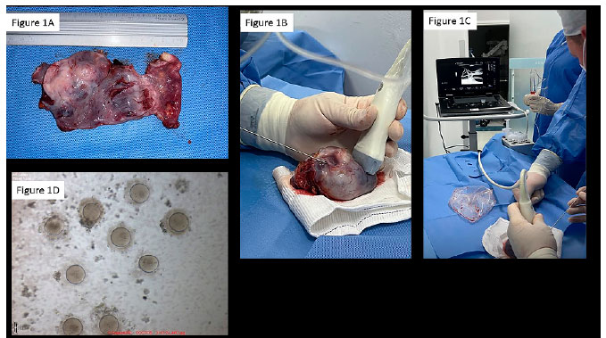
Figure 1. A. Right ovary after ex vivo follicular aspiration. B. Ex vivo ultrasound-guided aspiration of the right ovary. C. Ex vivo ultrasound-guided aspiration of the right ovary - follicular fluid visible on the ultrasound screen. D. Nine of the ten mature oocytes after denudation.