JBRA Assisted Reproduction 2026;30(3):609-627
REVIEW
doi: 10.5935/1518-0557.20260014
Chemotherapy-Induced Infertility: Mechanisms, Risk Factors, and Fertility Preservation Strategies in Cancer Patients
1Plant Design Department, Pars Abnoos Sanat Consulting Engineering Company, Shiraz, Iran
2Department of Reproductive Biology, School of Advanced Medical Sciences and Technologies, Shiraz University of Medical Sciences, Shiraz, Iran
3Stem Cells Technology Research Center, Shiraz University of Medical Sciences, Shiraz, Iran
4Infertility Research Center, Shiraz University of Medical Sciences, Shiraz, Iran
5Department of Biochemistry, School of Medicine, Shiraz University of Medical Sciences, Shiraz, Iran
6Department of Natural Sciences, West Kazakhstan Marat Ospanov Medical University, Aktobe 030012, Kazakhstan
COMPETING INTERESTS
The authors declare that no conflict of interest would prejudice the impartiality of this scientific work.
ABSTRACT
Chemotherapy remains a cornerstone of cancer treatment, effectively targeting rapidly dividing cancer cells, yet its systemic effects often extend to healthy reproductive cells, resulting in temporary or permanent infertility. This review explores the mechanisms by which chemother-apy impairs fertility in both men and women, focusing on the damage to ovarian follicles and spermatogenesis. Key factors influencing fertility outcomes include the type and dosage of chemotherapy drugs, patient age, and pre-existing reproductive health. Alkylating agents, such as cyclophosphamide, and platinum-based drugs, like cisplatin, pose the highest risk of gonad-otoxicity, while other agents exhibit milder, often reversible effects. Fertility preservation op-tions such as oocyte and embryo freezing for women, sperm banking for men, and emerging techniques like ovarian and testicular tissue cryopreservation offer hope for maintaining re-productive potential. The psychological burden of infertility and the promising yet complex role of nanoparticles in reproductive medicine are also discussed. By synthesizing current research, this review underscores the need for personalized fertility counseling and advanced preserva-tion strategies to balance cancer treatment efficacy with reproductive goals.
Keywords: chemotherapy, infertility, fertility preservation, gonadotoxicity, reproductive health
INTRODUCTION
Chemotherapy is a potent and systemic treatment used to target rapidly dividing cancer cells, but it can inadvertently affect healthy cells in the body, such as cells in the reproductive system (Mustapha et al., 2021). Both men and women have reproductive cells that are sensitive to chemotherapy, and the damage caused by these drugs can lead to temporary or permanent infertility. This unintended consequence is due to chemotherapy’s mechanism of action, which targets not just cancer cells but any other cells, including those responsible for oocyte and sperm production (Papathanasiou et al., 2024). The risk of infertility depends on several factors, including the specific drugs used, the dosage, the patient’s age, and their pre-existing fertility health (Papathanasiou et al., 2024).
In women, chemotherapy primarily affects the ovaries, which contain a finite number of oocytes from birth (Papathanasiou et al., 2024). These oocytes do not regenerate, so any damage to them is permanent. Chemotherapy drugs, particularly alkylating agents such as cyclophosphamide, can cause significant ovarian toxicity by damaging the DNA in oocytes or triggering cell death (Markowska et al., 2024). As a result, chemotherapy can reduce the ovarian reserve, leading to diminished fertility or even premature ovarian failure (POF), which is an early onset of menopause (Horan, 2022). Women undergoing chemotherapy may experience temporary amenorrhea, but in some cases, this can become permanent. The extent of damage is often age-dependent; younger women tend to have a better chance of recovering fertility after treatment, whereas older women, who have fewer oocytes, are at a greater risk of permanent infertility (Horan, 2022; Sabetian et al., 2022a).
In men, chemotherapy can severely impact spermatogenesis, the process of sperm production in the testes (Delessard et al., 2020; Yaghutian Nezhad et al., 2021). Spermatogonia are rapidly dividing and thus vulnerable to chemotherapy’s effects. Chemotherapy can reduce or disrupt sperm production, leading to conditions like oligospermia or azoospermia (Javed, 2020). Depending on the type of drug and dosage, this infertility may be temporary. Even if sperm production resumes, chemotherapy can cause genetic damage to the DNA within sperm, which raises concerns about the health of future offspring (Meistrich, 2020). As with women, the age of the patient plays a significant role; younger men are more likely to recover sperm production after treatment, while older men may face a higher risk of irreversible infertility (Jensen et al., 2022).
The type of chemotherapy drug used plays a significant role in determining fertility outcomes. Alkylating agents, such as cyclophosphamide and busulfan, are particularly damaging to reproductive cells (Bhardwaj et al., 2023; Bahmyari et al., 2024). Platinum-based drugs, like cisplatin, are also highly gonadotoxic and can cause severe ovarian or testicular damage (Tian En et al., 2020). Other chemotherapy drugs, such as taxanes and anthracyclines, pose a moderate risk to fertility but may still cause significant reproductive harm, especially when used in high doses or combined with other treatments (Poorvu et al., 2019). The total dose and length of chemotherapy also influence the likelihood of infertility, with higher doses and longer treatments posing a greater risk (Hales et al., 2005; Markowska et al., 2024).
Ultimately, the effects of chemotherapy on fertility can vary greatly depending on individual circumstances. Age, the type of cancer, the specific chemotherapy drugs, and the patient’s overall reproductive health all play a role in determining whether fertility will be affected temporarily or permanently (Li et al., 2024). While chemotherapy is a crucial tool in the fight against cancer, its potential to cause infertility can have profound psychological and emotional impacts on patients, especially those of reproductive age (Goldman et al., 2015). Therefore, patients need to discuss their fertility concerns with their healthcare providers before beginning treatment to explore possible preservation options.
Table 1 provides a comprehensive snapshot of the fertility effects associated with twelve widely used chemotherapy drugs-Cyclophosphamide, Doxorubicin, Paclitaxel, Cisplatin, Methotrexate, Fluorouracil (5-FU), Vincristine, Etoposide, Bortezomib, Carboplatin, Ifosfamide, Bleomycin, and Gemcitabine-alongside Bumetanide, a non-chemotherapy diuretic, as of February 27, 2025. The data reveals a spectrum of reproductive impacts, ranging from severe, often permanent infertility risks posed by alkylating agents like Cyclophosphamide and Ifosfamide, to milder, potentially reversible effects from drugs like Gemcitabine and Bleomycin. Platinum-based agents such as Cisplatin and Carboplatin show significant but varying degrees of gonadotoxicity, while targeted therapies like Bortezomib and less toxic agents like Vincristine and Fluorouracil demonstrate minimal direct impact on fertility (Abdel-Latif et al., 2022; American Cancer Society and American Society of Clinical Oncology (ASCO), 2025). Bumetanide stands apart with no evidence of fertility effects, underscoring its distinct role outside oncology (NHS - National Health Service, 2022). High-risk drugs necessitate proactive fertility preservation strategies, such as oocyte or sperm banking, particularly for younger patients, while those with milder profiles may allow for natural recovery post-treatment (Li et al., 2024). However, gaps in research-especially for newer agents like Bortezomib and less-studied drugs like Gemcitabine-suggest a need for further investigation to fully elucidate their long-term effects, particularly in women. This table serves as a valuable resource for clinicians and patients navigating treatment decisions, emphasizing the importance of individualized counseling to balance cancer management with reproductive goals.
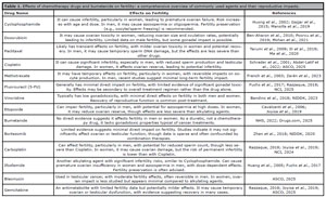
Table 1. Effects of chemotherapy drugs and bumetanide on fertility: a comprehensive overview of commonly used agents and their reproductive impacts.
Recent updates in clinical practice guidelines from the American Society of Clinical Oncology underscore the importance of discussing and offering fertility preservation options to all patients of reproductive age at the time of cancer diagnosis (Su et al., 2025). All guidelines endorse established methods such as embryo, oocyte, and ovarian tissue cryopreservation, as well as ovarian transposition (ESHRE Guideline Group on Female Fertility Preservation et al., 2020; Kieu et al., 2022). For females, options include embryo cryopreservation, oocyte freezing, and ovarian tissue freezing. Hormonal suppression and ovarian shielding during radiation may also help reduce damage. However, these methods often require time-sensitive procedures, which can be challenging when urgent cancer treatment is needed. For males, sperm banking is the most common and effective method, while testicular sperm extraction or testicular tissue freezing may be options for those unable to produce a sample (Ní Dhonnabháin et al., 2022; Pathak et al., 2023; Katzir et al., 2024). The Cancer Council Australia emphasizes the importance of early discussions between patients and healthcare providers to explore fertility preservation before treatment begins. A multidisciplinary approach involving oncologists, fertility specialists, and counselors ensures patients receive comprehensive care, including psychological and financial support (Kieu et al., 2022).
This review integrates such up-to-date guideline recommendations and pooled clinical evidence to provide a robust framework for assessing fertility risks and preservation strategies in men and women undergoing gonadotoxic treatments. Also includes preclinical research using animal models to provide a comprehensive understanding of chemotherapy-induced gonadotoxicity and strategies for fertility preservation.
We searched PubMed, Scopus, Web of Science, and Embase using keywords such as ‘chemotherapy AND fertility preservation,’ ‘gonadotoxicity,’ ‘premature ovarian failure,’ and ‘oocyte cryopreservation.’ The search covered publications from January 2015 to June 2025. Only English-language articles were included.
CHEMOTHERAPY’S EFFECTS ON FEMALE FERTILITY
Female fertility is intricately tied to the health and number of ovarian follicles containing immature oocytes (Telfer et al., 2023). From birth, a woman has a fixed number of oocytes that are gradually lost throughout her life, with no possibility of regeneration (Moghadam et al., 2022). These oocytes are highly sensitive to environmental factors, including chemotherapy, which can cause both temporary and permanent damage (Sonigo et al., 2019). Chemotherapy targets rapidly dividing cells, a hallmark of cancer, but this also makes healthy reproductive cells vulnerable to damage (Alimbetov et al., 2018). The extent of chemotherapy’s impact on fertility depends on the individual’s ovarian reserve before treatment (Blumenfeld, 2012).
Ovarian Follicles and Their Role in Female Fertility
Throughout a woman’s reproductive life, a small percentage of follicles are selected to mature and eventually release oocytes in a process known as ovulation (Johnson, 2015). However, most follicles never reach maturity and undergo a natural process called atresia, where they degrade and are reabsorbed by the body (Corriero et al., 2021). This gradual depletion of follicles is a normal part of the aging process, but when accelerated by external factors such as chemotherapy, it can significantly impair fertility (Yan et al., 2022). Each ovarian follicle contains an oocyte surrounded by support cells that help regulate its development and maturation (Clarke, 2018). The health of these oocytes is critical for reproductive success, and damage to them can lead to infertility or diminished fertility. Chemotherapy, particularly certain classes of drugs, can interfere with this process by damaging the DNA within oocytes or inducing apoptosis, reducing viable oocytes over time (Winship et al., 2018).
Chemotherapy and Its Mechanisms of Ovarian Damage
Chemotherapy drugs exert their effects by interfering with cell division and inducing DNA damage (Sun et al., 2021). While this is crucial for killing cancer cells, it also impacts other rapidly dividing cells, including those in the ovaries. Among the most harmful chemotherapy agents for fertility are alkylating agents, which include drugs like cyclophosphamide, ifosfamide, and busulfan (Kapoor et al., 2025). These drugs work by directly binding to the DNA in cells, creating cross-links and breaking DNA strands, which prevents cells from dividing and ultimately leads to cell death (Bergamo et al., 2018). In ovarian follicles, this can destroy oocytes, causing a significant reduction in the ovarian reserve (Monniaux, 2018). The ovaries are particularly susceptible to chemotherapy-induced gonadotoxicity because they contain cells that are in various stages of division and development. Preantral and antral follicles, which are the follicles closest to releasing mature oocytes, are especially vulnerable to damage (Das et al., 2023). These follicles require a precise hormonal environment and intact DNA to complete the maturation process, and chemotherapy can disrupt both, either by directly damaging the oocyte’s DNA or by impairing the support cells and hormone production needed for development (Winship et al., 2018). One major risk posed by toxic elements is oxidative stress, which also occurs when chemotherapy drugs generate free radicals that can damage cellular structures, including DNA, proteins, and lipids (Cauli, 2021; Talaie et al., 2025). Environmental factors such as air pollution, UV radiation, and chemical toxins induce oxidative stress by generating excess reactive oxygen species, which overwhelm antioxidant defenses and damage cellular components like lipids, proteins, and DNA (Alaee, 2018; Banagar et al., 2018; Alaee et al., 2024a; Khodabandeh et al., 2025). Antioxidants neutralize reactive oxygen species (ROS) and protect cells from oxidative damage by donating electrons, scavenging free radicals, and enhancing endogenous defense systems (Patil et al., 2015; Flora et al., 2021; Neisy et al., 2025; Zahedi et al., 2025).
Oocytes, being long-lived cells, are particularly prone to accumulating damage from oxidative stress. This can lead to DNA mutations or other types of molecular damage that not only reduce the chances of successful fertilization but may also raise concerns about the quality and health of any offspring (Musson et al., 2022).
Long-Term Consequences: Premature Ovarian Failure and Diminished Ovarian Reserve
The most serious consequence of chemotherapy on female fertility is the potential for premature ovarian failure (POF), also known as primary ovarian insufficiency (POI) (Hao et al., 2019). POF occurs when the ovaries stop functioning properly before the age of 40, leading to early menopause and a loss of fertility. The onset of POF is marked by the cessation of menstruation (amenorrhea), low levels of estrogen, and high levels of follicle-stimulating hormone (FSH), which indicate that the ovaries are no longer producing oocytes. POF can be a direct result of extensive damage to the ovarian reserve caused by chemotherapy. Since women are born with a limited number of oocytes, once these are depleted, the ovaries cannot regenerate them (Monniaux, 2018). The risk of POF is particularly high in women who receive high doses of alkylating agents or undergo prolonged chemotherapy regimens (Cui et al., 2018). Women who experience POF may have no remaining viable oocytes, making natural conception impossible and requiring alternative methods like donor oocytes or adoption to achieve parenthood.
In cases where chemotherapy does not result in complete ovarian failure, it can still lead to diminished ovarian reserve (DOR) (Spears et al., 2019). This condition is characterized by a reduction in the number of viable oocytes, which can make conception more difficult and reduce the chances of successful in vitro fertilization if fertility treatment is pursued (Parsanezhad et al., 2017; Al Abdali & Gowri, 2021; Ashourzadeh et al., 2024). Women with DOR often face shorter reproductive windows and may experience challenges with oocyte quality, which further complicates efforts to conceive (Swan & Colino, 2022).
Hormonal Disruption and Its Impact on Reproduction
In addition to directly damaging oocytes, chemotherapy can disrupt the hormonal environment required for normal ovarian function (Xiong et al., 2021). Ovarian follicles rely on the balanced production of several key hormones, including FSH and luteinizing hormone (LH), which stimulate the growth and maturation of oocytes (Salih & Jaafar, 2013; Ma et al., 2023). Chemotherapy can interfere with the hypothalamic-pituitary-ovarian axis, disrupting the normal feedback loops that regulate these hormones (Markowska et al., 2024). This disruption can lead to irregular menstrual cycles, anovulation, and ultimately, infertility (Song & Li, 2021). Hormonal imbalances can also contribute to temporary amenorrhea, folliculogenesis arrest, and disruption of oocyte maturation (Alaee et al., 2024b; Singh et al., 2024). In some women, chemotherapy-induced hormonal disruptions may persist, reducing their chances of conceiving naturally even if menstruation resumes (Markowska et al., 2024).
Factors Influencing Fertility Outcomes Post-Chemotherapy
Several factors determine whether chemotherapy will cause temporary or permanent infertility. One of the most significant factors is the age of the patient at the time of treatment (Blumenfeld, 2002). Younger women generally have a larger ovarian reserve, which offers some protection against complete ovarian failure (Richardson et al., 2014). They are more likely to recover ovarian function after chemotherapy, though this is not guaranteed. Older women, particularly those approaching menopause, have fewer remaining oocytes, making them more vulnerable to permanent infertility (Qiao et al., 2014). The risk of long-term fertility issues increases significantly for women over 35 who undergo chemotherapy (Blumenfeld, 2012). The type of chemotherapy drug used is another critical factor. Alkylating agents are among the most gonadotoxic, causing the greatest damage to the ovaries (Bhardwaj et al., 2023). Other chemotherapy drugs, such as platinum-based compounds (e.g., cisplatin) and taxanes (e.g., paclitaxel), also pose a risk but may cause less severe damage depending on the dosage and duration of treatment (Rottenberg et al., 2021). The overall dosage and length of chemotherapy also influence fertility outcomes (van den Berg et al., 2021). Higher doses of chemotherapy, as well as prolonged treatment regimens, increase the likelihood of permanent ovarian damage (Mauri et al., 2020). Additionally, combination therapies, which involve using multiple chemotherapy agents, are more likely to cause irreversible harm to the ovaries. Figure 1 shows the gonadotoxic effects of chemotherapy drug on female fertility.
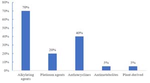
Figure 1. Comparative gonadotoxic effects of major chemotherapy drug classes on female fertility (data have been extracted from Mulder et al. (2021), Dumenigo et al. (2024), Himpe et al. (2023), and Santaballa et al. (2022).
CHEMOTHERAPY’S EFFECTS ON MALE FERTILITY
Chemotherapy carries significant risks for male fertility (Di Tucci et al., 2022). The primary target of chemotherapy in men is the process of spermatogenesis, which occurs in the testes and involves sperm production from precursor germ cells (Lopes et al., 2021). These germ cells are among the most rapidly dividing cells in the body, making them particularly susceptible to the damaging effects of any toxicants (Mohammadi et al., 2023). The drugs used in chemotherapy can lead to a reduction in sperm count, known as oligospermia, or even a complete absence of sperm in the semen, referred to as azoospermia (Mendes et al., 2022). Additionally, some chemotherapy agents can damage the DNA within sperm, raising concerns about fertility and the potential for genetic abnormalities in future offspring (Meistrich, 2020).
Mechanism of Action
The mechanism of chemotherapy affecting male fertility revolves around its impact on the rapidly dividing germ cells responsible for sperm production (Duffin et al., 2024). Chemotherapy drugs, especially those classified as alkylating agents, can inflict substantial damage on these cells, leading to impaired spermatogenesis (Sriram et al., 2024). By disrupting the normal cell division process, these drugs can interfere with the production of healthy sperm, causing various fertility issues. This damage is compounded by the fact that sperm production is a continuous process; therefore, any disruption can have immediate and long-lasting effects on a man’s fertility. The effects may not only result in reduced sperm counts but may also lead to sperm with compromised motility and morphology, further diminishing the chances of successful conception (Ben-Aharon et al., 2010).
Impact of Specific Chemotherapy Agents
The specific chemotherapy agents used play a critical role in determining the degree of fertility impairment in men (Figure 2) (Delessard et al., 2020). Alkylating agents, such as cyclophosphamide and chlorambucil, are known for their high gonadotoxic potential (Sriram et al., 2024). These drugs can cause long-term or even permanent infertility by damaging the testicular germ cells directly (Lorenzi et al., 2021). The likelihood of developing infertility increases with the cumulative dose and duration of treatment. Platinum compounds, including cisplatin, also pose a significant risk to male fertility (Tian En et al., 2020). These drugs can harm the spermatogenic cells, leading to both temporary and, in some cases, permanent infertility (Schrader et al., 2001). Men treated with these agents may experience varying degrees of fertility impairment based on their response to treatment. Other classes of chemotherapy drugs, such as antimetabolites and microtubule inhibitors (e.g., vinblastine and paclitaxel), have a moderate risk of inducing infertility (Markowska et al., 2024). While many men may experience temporary infertility during and after treatment, some may regain normal sperm production within months. However, the timeline for recovery can vary significantly among individuals, with some men taking years to return to baseline sperm counts, if they recover at all.
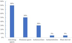
Figure 2. Comparative gonadotoxic effects of major chemotherapy drug classes on male fertility (the data have been extracted from Himpe et al., 2023; Reynolds & McKenzie, 2023).
Other Reproductive Effects of Chemotherapy in Men
Beyond directly impacting sperm production, chemotherapy can induce other reproductive issues in men (Mendes et al., 2022). One notable concern is hormonal disruption, particularly regarding the production of testosterone. Chemotherapy may affect the hormonal signaling pathways that regulate testosterone synthesis, potentially impacting sexual function, libido, and overall reproductive health (Babaniyi et al., 2024). While hormonal disruptions are less common than the direct effects on sperm, they can still contribute to fertility challenges and affect a man’s quality of life. Additionally, testicular damage can occur as a result of chemotherapy (Amiri et al., 2021). The structural integrity of the testicles is crucial for proper sperm production, and any damage to the testes can further impair fertility. The cumulative effect of chemotherapy on both sperm production and testicular health underscores the importance of monitoring reproductive health in men undergoing cancer treatment (McHugh et al., 2024).
SHORT-TERM VS. LONG-TERM EFFECTS
The effects of chemotherapy on male fertility can be categorized into short-term and long-term outcomes (Yumura et al., 2023). In the short term, many men experience a temporary reduction in sperm count, motility, or quality during chemotherapy (Yumura et al., 2023). This transient infertility can be distressing, especially for men who are planning to start families. While sperm production often resumes after the completion of chemotherapy, the timeline for recovery is unpredictable (Schlegel et al., 2021). In some cases, it can take several months to years for sperm counts to return to normal levels, and not all men will fully recover their fertility. In contrast, the long-term effects of chemotherapy may include permanent infertility, especially in men who receive high doses of gonadotoxic drugs (Duffin et al., 2024). Older men are particularly vulnerable; their reproductive function may already be declining due to age, and chemotherapy may exacerbate this decline, leading to an inability to produce viable sperm after treatment (Nguyen-Powanda & Robaire, 2020). The risk of long-term infertility necessitates careful consideration and planning for men undergoing chemotherapy who wish to preserve their reproductive options (Di Tucci et al., 2022). Table 2 highlights that chemotherapy can cause temporary fertility issues, like amenorrhea in women and low sperm counts in men, which often improve in younger patients. However, long-term effects, such as permanent infertility from ovarian failure or azoospermia, are more likely with high doses of drugs like cyclophosphamide, especially in older individuals. These differences emphasize the need for early fertility planning to address both short-term setbacks and lasting impacts.
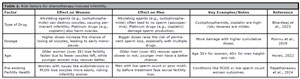
Table 2. Risk factors for chemotherapy-induced infertility.
Table 3 summarizes the shortand long-term effects of chemotherapy on male and female fertility, while Figure 3 illustrates the underlying mechanism of chemotherapy-induced infertility.

Table 3. Short-term vs. long-term effects of chemotherapy on fertility.
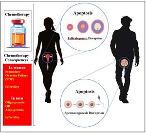
Figure 3. Schematic representation of folliculogenesis and spermatogenesis disruption after chemotherapy.
FACTORS INFLUENCING FERTILITY LOSS IN CHEMOTHERAPY
Chemotherapy-induced infertility is influenced by several critical factors, each playing a significant role in determining the extent of reproductive damage and the likelihood of fertility recovery after treatment (Bhardwaj et al., 2023). These factors include the type and dosage of chemotherapy drugs used, the age of the patient, and the individual’s pre-existing fertility health. Understanding these factors is essential for assessing the risk of infertility and considering fertility preservation options for cancer patients undergoing chemotherapy. Table 4 illustrates that chemotherapy-induced infertility hinges on drug type, dosage, age, and pre-existing fertility health. Alkylating agents and platinum compounds, especially at high doses, pose the greatest threat to reproductive cells in both women and men, often causing irreversible damage. Older patients face higher risks due to naturally declining fertility, while those with prior reproductive issues are more vulnerable. These factors highlight the need for tailored treatment plans and early fertility discussions to mitigate risks and preserve future reproductive options.
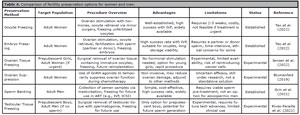
Table 4. Comparison of fertility preservation options for women and men.
Type and Dose of Chemotherapy
One of the most influential factors in chemotherapy-induced fertility loss is the type and dosage of the chemotherapy drugs administered (Di Tucci et al., 2022). Different chemotherapy agents vary in their level of gonadotoxicity, or their ability to damage reproductive cells. Alkylating agents, such as cyclophosphamide, ifosfamide, and busulfan, are among the most harmful to both male and female reproductive cells (Olowe et al., 2024). These drugs work by binding to and cross-linking DNA, which disrupts cell division and leads to cell death. In reproductive cells, this process can result in the destruction of oocytes in women and sperm-producing germ cells in men, often leading to irreversible infertility. Cisplatin, a platinum-based compound, also poses a high risk of fertility loss. While it is effective in targeting cancer cells, cisplatin is highly toxic to germ cells, potentially causing long-term or permanent infertility, especially when administered at high doses (Katanić Stanković et al., 2023). The cumulative dose of chemotherapy is a crucial factor; the higher the dose and the longer the treatment duration, the greater the risk of significant reproductive damage (van den Boogaard et al., 2022). For some patients, even moderate doses of gonadotoxic drugs can lead to a reduction in ovarian reserve or sperm count, making it essential to tailor chemotherapy regimens carefully for patients concerned about fertility (Table 2).
Age of the Patient
The age of the patient is another critical determinant of fertility loss during chemotherapy, particularly for women (Lee et al., 2011). Female fertility is closely linked to the size and health of the ovarian reserve, which consists of a finite number of oocytes present from birth (Gleicher et al., 2011). As women age, their ovarian reserve naturally declines, with the quantity and quality of oocytes diminishing over time. Older women, especially those approaching menopause, are at higher risk of permanent infertility following chemotherapy, as they may have fewer viable oocytes remaining before treatment even begins (Sparidaens et al., 2022). The ovaries of younger women tend to be more resilient and may have a greater capacity to recover after chemotherapy, although this is not guaranteed. In men, the impact of age on chemotherapy-induced infertility is somewhat less pronounced but still important. Older men may experience a slower or incomplete recovery of sperm production following chemotherapy due to age-related declines in reproductive function (de Almeida et al., 2024). As a result, men over the age of 40 undergoing chemotherapy may face a higher risk of permanent fertility loss than younger men. Both male and female patients should consider their age when assessing the potential impact of chemotherapy on their long-term fertility.
Pre-existing Fertility Health
A patient’s pre-existing fertility health is a significant factor in determining their susceptibility to chemotherapy-induced infertility (Mauri et al., 2020). Individuals with underlying fertility issues, such as endometriosis, polycystic ovarian syndrome (PCOS), or low sperm count, may be more vulnerable to the damaging effects of chemotherapy (Brassard et al., 2008; Shiravani et al., 2024). For women with conditions like endometriosis, where the health of the ovarian reserve may already be compromised, chemotherapy can further reduce the number of viable oocytes, increasing the likelihood of premature ovarian failure or diminished ovarian reserve (Mustapha et al., 2021). Similarly, men with suboptimal sperm parameters, such as low sperm count or poor sperm motility, may experience a more profound impact on their fertility, as chemotherapy could exacerbate these issues (Blumenfeld, 2012). Patients with compromised reproductive health should be particularly cautious when undergoing chemotherapy and may benefit from pre-treatment fertility assessments (Di Tucci et al., 2022). These evaluations can help determine baseline fertility and guide decisions about fertility preservation strategies, such as sperm banking for men or oocyte/embryo freezing for women. By understanding their unique reproductive risks, patients can make informed choices about safeguarding their fertility before beginning cancer treatment. The factors influencing fertility loss during chemotherapy are multifaceted and include the type and dose of chemotherapy drugs, the age of the patient, and their pre-existing fertility health. Alkylating agents and platinum compounds pose the highest risk of reproductive damage, particularly at high doses (Tian En et al., 2020; Sriram et al., 2024). Recognizing these factors is essential for assessing fertility risks and exploring preservation options to protect future reproductive potential.
FERTILITY PRESERVATION OPTIONS FOR INDIVIDUALS UNDERGOING CHEMOTHERAPY
Fertility preservation strategies are available for patients who wish to have biological children after chemotherapy.
For patients undergoing chemotherapy who are concerned about their future reproductive potential, there are several medical interventions available to help preserve fertility. These options vary depending on the patient’s sex, age, and specific circumstances, such as whether they are preor post-pubertal. Fertility preservation methods are critical in helping patients retain the possibility of having biological children after cancer treatment (Klipstein et al., 2020). Table 4 shows that fertility preservation options vary widely for women and men facing chemotherapy. These options highlight the importance of personalized planning to protect reproductive potential before cancer treatment begins.
Fertility Preservation Options for Women Undergoing Chemotherapy
Fortunately, several fertility preservation strategies are available for women undergoing chemotherapy (Razeti et al., 2023). The effectiveness and suitability of each depend on factors like age, ovarian reserve, and the type of chemotherapy being administered. Oocyte cryopreservation is one of the most common methods (Cho et al., 2020). This involves stimulating the ovaries with hormones to produce multiple oocytes, which are then retrieved and frozen for future use. This technique allows women to preserve their oocytes before undergoing chemotherapy, providing an opportunity to conceive through IVF later (11). Another option is embryo freezing, which is similar to oocyte freezing but involves fertilizing the retrieved oocytes with sperm to create embryos before freezing them (Cho et al., 2020). This approach is typically chosen by women who have a partner or wish to use donor sperm. Ovarian tissue cryopreservation is an emerging experimental technique where ovarian tissue is removed, frozen, and reimplanted after treatment to restore ovarian function (Eijkenboom et al., 2022). This method is particularly useful for prepubescent girls who cannot undergo oocyte retrieval but still wish to preserve fertility. A significant concern with ovarian tissue transplantation is the potential reintroduction of malignant cells. To mitigate this risk, alternative approaches have been developed. Following ovarian tissue cryopreservation, follicles can be isolated and cultured in vitro, enabling their maturation without the need for tissue retransplantation. This technique eliminates the risk of cancer cell transfer while preserving fertility potential (Telfer et al., 2023). In vitro maturation of oocytes from unstimulated or minimally stimulated ovaries offers another promising approach, particularly for patients who cannot undergo conventional ovarian stimulation.
New frontier approaches, including artificial ovary constructs using biomaterials, 3D scaffolds, and ovary-on-a-chip systems, are being explored to restore ovarian function and growth of follicles ex vivo (Dadashzadeh et al., 2021).
Tissue engineering strategies using decellularized ovarian scaffolds are being developed to create a supportive microenvironment for follicular development while eliminating cancer cells (Alaee et al., 2021; 2024b). These acellular matrices, when repopulated with patient-derived follicles or ovarian cells, may restore endocrine function and fertility without the risk of cancer recurrence (Dadashzadeh et al., 2021). Other scaffold-based approaches incorporating biomaterials like alginate or fibrin aim to mimic the ovarian extracellular matrix to support follicle survival and growth (Dadashzadeh et al., 2021). These emerging technologies represent important alternatives to traditional transplantation of cryopreserved ovarian tissue, particularly for patients with blood-borne malignancies where the risk of malignant cell transfer remains a significant concern (Kuchakzadeh et al., 2024).
For some women, the use of GnRH agonists (gonadotropin-releasing hormone agonists) during chemotherapy may help protect the ovaries by temporarily suppressing ovarian function (Lee & Choi, 2021). By shutting down the ovaries and reducing hormonal stimulation, GnRH agonists aim to shield oocytes from chemotherapy’s damaging effects. However, the effectiveness of this approach is still under investigation and may not provide complete protection against fertility loss (Lee & Choi, 2021).
Fertility Preservation Options for Men
Men undergoing chemotherapy also have a range of fertility preservation options, with sperm banking being the most common and well-established technique (Jensen et al., 2022).
The most common fertility preservation method for men is sperm banking, which involves collecting and freezing sperm samples before starting chemotherapy (Liu et al., 2021). The sperm can be stored for extended periods and used later in assisted reproductive technologies, such as in vitro fertilization or intracytoplasmic sperm injection (Moungala, 2023). Sperm cryopreservation is a straightforward and effective option for men, with success rates for conception remaining high even after long-term storage (Akbarzadeh-Jahromi et al., 2022; Moungala, 2022). For men who can produce sperm, this method is highly recommended before undergoing chemotherapy, as sperm production may be impaired or lost permanently after treatment (Sharma & Sharma, 2020).
For prepubescent boys who have not yet begun producing sperm, traditional sperm banking is not an option. In such cases, an experimental technique known as testicular tissue freezing may be considered (Saulnier et al., 2021). This procedure involves surgically removing a small portion of testicular tissue that contains sperm precursor cells, which are immature cells that have the potential to develop into mature sperm eventually (Mitchell & Ives, 2024). The tissue is then frozen and stored for future use, with the hope that advances in reproductive technology will allow these precursor cells to be used to restore fertility or generate viable sperm in the future. While this method is still in the experimental stages, it offers hope for young boys who would otherwise face a high risk of infertility due to cancer treatment (Mitchell & Ives, 2024).
Successful testicular tissue reconstruction for the reintroduction of cryopreserved spermatogonial stem cells is a critical step towards restoring fertility (Forouzandegan et al., 2025). This process typically involves the enzymatic digestion of a sterile testicular cell suspension, which is then co-cultured with supportive somatic cells and often integrated into a three-dimensional biomaterial scaffold, such as decellularized testis, agarose, or Matrigel, to reform a tissue-like structure (Del Vento et al., 2018). Following in vitro culture to allow for cell aggregation and reorganization, this reconstructed tissue is subsequently grafted, often subcutaneously or under the tunica albuginea, into an immunodeficient mouse host (Del Vento et al., 2018). This in vivo environment provides the necessary physiological signals and vascularization to support the survival, engraftment, and crucially, the initiation of spermatogenesis from the transplanted spermatogonial stem cells, thereby validating the functional potential of the reconstructed testicular niche (Izadi et al., 2025).
Improving the success of fertility preservation via sperm and oocyte cryopreservation requires a multi-pronged approach that addresses both logistical and biological challenges. A primary strategy is the urgent timeline optimization between diagnosis and the start of therapy to harvest gametes at the highest possible quality (Ozimic et al., 2023). For post-pubertal males, this means multiple sperm collections if time permits, while for females, random-start ovarian stimulation protocols have revolutionized the process by allowing immediate initiation of cycle synchronization, shaving weeks off the timeline (Walker et al., 2022). Biologically, the administration of antioxidants is a key intervention to counteract the oxidative stress induced by both the disease state and cancer treatments themselves. Supplementation with compounds like Coenzyme Q10, Melatonin, and Vitamin E can help protect mitochondrial function in oocytes and reduce DNA fragmentation in sperm, thereby improving post-thaw survival and developmental competence (Karimfar et al., 2015; Cao et al., 2022; Zhang et al., 2024).
POST-CHEMOTHERAPY FERTILITY ASSESSMENT AND TREATMENT
After chemotherapy, patients who wish to conceive may undergo fertility assessments to determine whether natural conception is possible. In cases where infertility is diagnosed, assisted reproductive technologies such as in vitro fertilization, sperm retrieval, or donor oocytes/sperm may be explored.
PSYCHOLOGICAL AND EMOTIONAL IMPACT OF INFERTILITY
Infertility resulting from chemotherapy can have profound psychological and emotional consequences, particularly for young cancer survivors who have aspirations for future parenthood (Banser, 2022). Getting a cancer diagnosis can make people scared and unsure. Finding out they might not be able to have kids can make them feel even more sad, lost, and hopeless. Many people feel angry, sad, or worried when they realize they might not be able to have kids. For young people who beat cancer, not being able to have kids can affect more than just their feelings-it can also change things with their partner, family, and friends. The stress of trying to have a baby, along with the tough physical and emotional effects of cancer treatment, can put pressure on close relationships. Partners might not get the emotional side of not having kids, which can leave the person feeling alone. Doctors and nurses are really important in helping with these emotional struggles. They need to talk to patients about ways to save fertility and the chance of not having kids before starting chemo. These talks can help patients make smart choices about having kids later. Also, giving patients counseling or support groups can help them deal with their sadness and worries. Support groups can be particularly beneficial as they create a safe space for individuals to share their experiences and feelings with others who are facing similar challenges. These connections can help combat feelings of isolation and foster a sense of community. Mental health professionals trained in reproductive health can also offer therapeutic interventions tailored to the unique experiences of cancer survivors, helping them process their emotions and navigate the complexities of infertility (Banser, 2022; Dong et al., 2023; Ensafdaran et al., 2024). Overall, recognizing the psychological and emotional impact of infertility is vital in providing comprehensive care to cancer survivors. By addressing these issues, healthcare providers can help individuals regain a sense of control and hope for the future, whether that involves exploring alternative paths to parenthood or finding peace with their situation.
Low-income patients, rural residents, and those without health insurance often face significant barriers to timely fertility preservation, including high out-of-pocket costs, lack of nearby specialized clinics, and the need to travel long distances to access reproductive services (Peipert et al., 2023). Studies from multiple countries have reported that patients from socioeconomically disadvantaged backgrounds are less likely to be referred to fertility specialists compared with those from higher-income groups (Flanagan et al., 2022). Geographic disparities also play a major role, as rural or remote communities may have limited or no knowledge and access to reproductive endocrinologists or cryopreservation facilities, leading to delayed or missed opportunities for fertility preservation before cancer treatment (Letourneau et al., 2012; Alaee et al., 2019). In addition, cultural norms, language differences, and lack of awareness among patients and healthcare providers can further reduce the likelihood of timely referral and intervention (Letourneau et al., 2012). Variations in national health policies and insurance coverage add another layer of inequality, with some healthcare systems fully covering fertility preservation costs, while others provide no reimbursement, making such (Gadson et al., 2024).
NANOPARTICLES’ EFFECTS ON HUMAN FERTILITY
The advent of nanoparticle-based cancer therapies represents a monumental leap forward in oncology, offering the potential for highly targeted drug delivery that maximizes tumor destruction while minimizing the systemic side effects that have long plagued conventional treatments like chemotherapy and radiation (Abbasi, 2017; Taghipour et al., 2017). By engineering these microscopic carriers to seek out and penetrate cancerous cells specifically, physicians can administer powerful cytotoxic agents with unprecedented precision (Abdollahii et al., 2020). This targeted approach spares much of the body’s healthy tissues from exposure, thereby reducing the severity of common side effects such as nausea, nerve damage, and extreme fatigue (Andone et al., 2023). The promise of nanoparticles lies in their ability to make cancer treatment more effective and more tolerable, potentially turning certain cancers into manageable chronic conditions (Andone et al., 2023).
However, this significant progress introduces a complex and serious secondary concern: the potential risk of infertility (Falchi et al., 2018; Ilani et al., 2018). While nanoparticles are designed for specificity, their small size and unique pharmacokinetic properties raise questions about unintended biodistribution. Studies have shown that some nanoparticles, particularly those with poorly optimized formulations, may impair spermatogenesis by causing oxidative stress and DNA damage (Ilani & Alaee, 2017; Falchi et al., 2018). Nanoparticles can cross biological barriers, such as the blood-testis barrier, and accumulate in reproductive tissues, which can be harmful. Studies have shown that some nanoparticles, particularly those with poorly optimized formulations, may impair spermatogenesis by causing oxidative stress and DNA damage (Falchi et al., 2018). This can lead to oligospermia, azoospermia, or abnormal sperm morphology, ultimately reducing male fertility (Sabetian et al., 2022b). In women, the impact of nanoparticles on ovarian function and oocyte quality is less well understood, but concerns have been raised about their potential to disrupt the ovarian environment or harm oocytes. While nanoparticles offer groundbreaking potential in cancer treatment and even in diagnosing and treating infertility, their use must be carefully optimized to avoid harming reproductive health.
Researchers are working on refining nanoparticle formulations, incorporating antioxidants, vitamins, or other protective agents to reduce toxicity (Sabetian et al., 2021).
This improved detection helps in the early diagnosis of both male and female infertility, leading to more timely and effective treatments. In therapeutic applications, nanoparticles can be designed to deliver targeted drugs to tissues (Taghipour et al., 2017). For example, gold, zinc, silver, and cerium nanoparticles are under investigation for their ability to enhance sperm quality or protect oocytes from oxidative damage during assisted reproductive technologies, but some of them have adverse effects (Davis et al., 2024).
Consequently, the development and deployment of these innovative treatments must be paired with proactive fertility preservation strategies. For patients of reproductive age, this means oncologists must integrate discussions about infertility risks into the informed consent process before starting treatment. The scientific community is also obligated to conduct rigorous long-term toxicology studies focused specifically on the gonadal impact of various nanoparticle formulations. Ultimately, the goal is to harness the life-saving power of nanotechnology without sacrificing the possibility of future life, ensuring that beating cancer does not come at the cost of a patient’s ability to build a family.
CONCLUSION
Chemotherapy is a vital tool in combating cancer, yet its impact on fertility represents a significant challenge for patients of reproductive age. This review highlights how chemotherapy, particularly through alkylating agent compounds, disrupts ovarian function and spermatogenesis, often leading to diminished fertility or permanent infertility. The risk is modulated by factors such as drug type, dosage, patient age, and baseline fertility, with younger patients showing greater potential for recovery. Advances in fertility preservation, including oocyte, embryo, and sperm cryopreservation, alongside experimental techniques like ovarian and testicular tissue freezing, provide viable options for safeguarding reproductive potential. However, the emotional toll of infertility underscores the importance of pre-treatment counseling and psychological support. Ultimately, a multidisciplinary approach-integrating oncology, reproductive medicine, and mental health support-is essential to address the complex interplay between cancer treatment and fertility, empowering patients to make informed decisions about their reproductive futures.
AUTHORS’ CONTRIBUTION
All authors of this study have a complete contribution for data collection, data analyses and manuscript writing.
Acknowledgments
We would like to express our sincere gratitude to the Department of Natural Sciences at West Kazakhstan Marat Ospanov Medical University for their invaluable support in the development of this paper. Special thanks to the faculty and staff for their continued guidance, encouragement, and assistance throughout this research. Their dedication to fostering academic growth and advancing scientific understanding has played a crucial role in the successful completion of this study.
Ethical issue
Authors are aware of, and comply with, best practice in publication ethics specifically concerning authorship (avoidance of guest authorship), dual submission, manipulation of figures, competing interests, and compliance with policies on research ethics. Authors adhere to publication requirements that submitted work is original and has not been published elsewhere in any language.
Declaration of generative AI and AI-assisted technologies in the writing process
During the preparation of this work the authors used ChatGPT in order to enhance the manuscript writing. After using this tool, the authors reviewed and edited the content as needed and take full responsibility for the content of the published article.
REFERENCES
Abbasi M. Nanoparticles as a Promising Innovative Treatment Towards Infertility. J Infertil Reprod Bio. 2017;5:1-4.Abdel-Latif R, Fathy M, Anwar HA, Naseem M, Dandekar T, Othman EM. Cisplatin-Induced Re-productive Toxicity and Oxidative Stress: Ameliorative Effect of Kinetin. Antioxidants. 2022;11:863. PMID: 35624727 DOI: 10.3390/antiox11050863 Medline
Akbarzadeh-Jahromi M, Jafari F, Parsanezhad ME, Alaee S. Evaluation of supplementation of cryopreservation medium with gallic acid as an antioxidant in quality of post-thaw human spermatozoa. Andrologia. 2022;54:e14571. PMID: 36054457 DOI: 10.1111/and.14571 Medline
Alaee S. Air Pollution and Infertility - A letter to Editor. J Environ Treat Tech. 2018;6:72-3.
Alaee S, Asadollahpour R, Hosseinzadeh Colagar A, Talaei-Khozani T. The decellularized ovary as a potential scaffold for maturation of preantral ovarian follicles of prepubertal mice. Syst Biol Reprod Med. 2021;67:413-27. PMID: 34445905 DOI: 10.1080/19396368.2021.1968542 Medline
Alaee S, Khodabandeh Z, Dara M, Hosseini E, Sharma M. Curcumin mitigates acrylamide-induced ovarian antioxidant disruption and apoptosis in female Balb/c mice: A comprehensive study on gene and protein expressions. Food Sci Nutr. 2024a;12:4160-72. PMID: 38873462 DOI: 10.1002/fsn3.4076 Medline
Alaee S, Zal F, Razban V, Talaei-Khozani T, Shokri S, Khodabandeh Z. PRP Influences Maturation and Fertilisation of Immature Mouse Oocytes. Anat Histol Embryol. 2024b;53:e13112. PMID: 39396107 DOI: 10.1111/ahe.13112 Medline
Alimbetov D, Askarova S, Umbayev B, Davis T, Kipling D. Pharmacological Targeting of Cell Cycle, Apoptotic and Cell Adhesion Signaling Pathways Implicated in Chemoresistance of Cancer Cells. Int J Mol Sci. 2018;19:1690. PMID: 29882812 DOI: 10.3390/ijms19061690 Medline
Almeida AL, Fortuna A, Sousa M, Sá R. A systematic review of bleomycin-induced gonadotoxicity: Mechanistic implications for male reproductive health and fertility. Reprod Toxicol. 2024;130:108721. PMID: 39326549 DOI: 10.1016/j.reprotox.2024.108721 Medline
Amiri A, Chovanec M, Oliva V, Sedliak M, Mego M, Ukropec J, Ukropcová B. Chemotherapy-induced toxicity in patients with testicular germ cell tumors: The impact of physical fit-ness and regular exercise. Andrology. 2021;9:1879-92. PMID: 34245663 DOI: 10.1111/andr.13078 Medline
Andone BA, Handrea-Dragan IM, Botiz I, Boca S. State-of-the-art and future perspectives in infertility diagnosis: Conventional versus nanotechnology-based assays. Nanomedicine. 2023;54:102709. PMID: 37717928 DOI: 10.1016/j.nano.2023.102709 Medline
Ashourzadeh S, Safari S, Hosseinisadat R, Kafaeinezhad R, Shokri S, Alaee S. Comparative analy-sis of conventional in vitro fertilization and intracytoplasmic sperm injection in patients with polycystic ovarian syndrome, tubal factor infertility, and unexplained infertility whose partners exhibit normal semen parameters: A retrospective study of sibling oocytes. Clin Exp Reprod Med. 2024;51:285-92. PMID: 38525518 DOI: 10.5653/cerm.2023.06408 Medline
Bahmyari S, Alaee S, Khodabandeh Z, Talaei-Khozani T, Dara M, Mehdinejadiani S, Solati A. The effects of Wharton’s jelly MSCs secretomes for restoring busulfan-induced reproductive toxicity in male mice. Hum Exp Toxicol. 2024;43:9603271241269019. PMID: 39081119 DOI: 10.1177/09603271241269019 Medline
Bandino JP, Gathings RM, Hinen HB, Hampton MT, Davis WC, Elston DM. Gadolinium Presence Within Cutaneous Sclerotic Bodies Confirmed by Laser Ablation Inductively Coupled Plasma Mass Spectrometry. JAMA Dermatol. 2018;154:105-6. PMID: 29117364 DOI: 10.1001/jamadermatol.2017.4141 Medline
Ben-Aharon I, Bar-Joseph H, Tzarfaty G, Kuchinsky L, Rizel S, Stemmer SM, Shalgi R. Doxoru-bi-cin-induced ovarian toxicity. Reprod Biol Endocrinol. 2010;8:20. PMID: 20202194 DOI: 10.1186/1477-7827-8-20 Medline
Bhardwaj JK, Bikal P, Sachdeva SN. Chemotherapeutic drugs induced female reproductive toxicity and treatment strategies. J Biochem Mol Toxicol. 2023;37:e23371. PMID: 37098697 DOI: 10.1002/jbt.23371 Medline
Blumenfeld Z. Preservation of fertility and ovarian function and minimalization of chemotherapy associated gonadotoxicity and premature ovarian failure: the role of inhibin-A and -B as markers. Mol Cell Endocrinol. 2002;187:93-105. PMID: 11988316 DOI: 10.1016/S0303-7207(01)00712-2 Medline
Blumenfeld Z. Chemotherapy and fertility. Best Pract Res Clin Obstet Gynaecol. 2012;26:379-90. PMID: 22281514 DOI: 10.1016/j.bpobgyn.2011.11.008 Medline
Blumenfeld Z. Fertility Preservation Using GnRH Agonists: Rationale, Possible Mechanisms, and Explanation of Controversy. Clin Med Insights Reprod Health. 2019;13:1179558119870163. PMID: 31488958 DOI: 10.1177/1179558119870163 Medline
Brassard M, AinMelk Y, Baillargeon JP. Basic infertility including polycystic ovary syndrome. Med Clin North Am. 2008;92:1163-92. PMID: 18721657 DOI: 10.1016/j.mcna.2008.04.008 Medline
Cao B, Qin J, Pan B, Qazi IH, Ye J, Fang Y, Zhou G. Oxidative Stress and Oocyte Cryopreservation: Recent Advances in Mitigation Strategies Involving Antioxidants. Cells. 2022;11:3573. PMID: 36429002 DOI: 10.3390/cells11223573 Medline
Cauli O. Oxidative Stress and Cognitive Alterations Induced by Cancer Chemotherapy Drugs: A Scoping Review. Antioxidants (Basel). 2021;10:1116. PMID: 34356349 DOI: 10.3390/antiox10071116 Medline
Cavalcanti AG, Krambeck R, Araújo A, Rabelo PH, Carvalho JP, Favorito LA. Management of urethral lesions in penile blunt trauma. Int J Urol. 2006;13:1218-20. PMID: 16984556 DOI: 10.1111/j.1442-2042.2006.01534.x Medline
Cho HW, Lee S, Min KJ, Hong JH, Song JY, Lee JK, Lee NW, Kim T. Advances in the Treatment and Prevention of Chemotherapy-Induced Ovarian Toxicity. Int J Mol Sci. 2020;21:7792. PMID: 33096794 DOI: 10.3390/ijms21207792 Medline
Clarke HJ. Regulation of germ cell development by intercellular signaling in the mammalian ovarian follicle. Wiley Interdiscip Rev Dev Biol. 2018;7:10.1002/wdev.294 PMID: 28892263 DOI: 10.1002/wdev.294 Medline
Corriero A, Zupa R, Mylonas CC, Passantino L. Atresia of ovarian follicles in fishes, and implications and uses in aquaculture and fisheries. J Fish Dis. 2021;44:1271-91. PMID: 34132409 DOI: 10.1111/jfd.13469 Medline
Cui W, Stern C, Hickey M, Goldblatt F, Anazodo A, Stevenson WS, Phillips KA. Preventing ovarian failure associated with chemotherapy. Med J Aust. 2018;209:412-6. PMID: 30376664 DOI: 10.5694/mja18.00190 Medline
Dadashzadeh A, Moghassemi S, Shavandi A, Amorim CA. A review on biomaterials for ovarian tissue engineering. Acta Biomater. 2021;135:48-63. PMID: 34454083 DOI: 10.1016/j.actbio.2021.08.026 Medline
Davis EH, Jones C, Coward K. Rethinking the application of nanoparticles in women’s repro-duc-tive health and assisted reproduction. Nanomedicine (Lond). 2024;19:1231-51. PMID: 38686941 DOI: 10.2217/nnm-2023-0346 Medline
Del Vento F, Vermeulen M, de Michele F, Giudice MG, Poels J, des Rieux A, Wyns C. Tissue Engineering to Improve Immature Testicular Tissue and Cell Transplantation Outcomes: One Step Closer to Fertility Restoration for Prepubertal Boys Exposed to Gonadotoxic Treatments. Int J Mol Sci. 2018;19:286. PMID: 29346308 DOI: 10.3390/ijms19010286 Medline
Delessard M, Saulnier J, Rives A, Dumont L, Rondanino C, Rives N. Exposure to Chemotherapy During Childhood or Adulthood and Consequences on Spermatogenesis and Male Fertility. Int J Mol Sci. 2020;21:1454. PMID: 32093393 DOI: 10.3390/ijms21041454 Medline
Di Tucci C, Galati G, Mattei G, Chinè A, Fracassi A, Muzii L. Fertility after Cancer: Risks and Successes. Cancers (Basel). 2022;14:2500. PMID: 35626104 DOI: 10.3390/cancers14102500 Medline
Dong Y, Yue Z, Zhuang H, Zhang C, Fang Y, Jiang G. The experiences of reproductive concerns in cancer survivors: A systematic review and meta-synthesis of qualitative studies. Cancer Med. 2023;12:22224-51. PMID: 38069669 DOI: 10.1002/cam4.6531 Medline
Duffin K, Mitchell RT, Brougham MFH, Hamer G, van Pelt AMM, Mulder CL. Impacts of cancer therapy on male fertility: Past and present. Mol Aspects Med. 2024;100:101308. PMID: 39265489 DOI: 10.1016/j.mam.2024.101308 Medline
Eijkenboom L, Saedt E, Zietse C, Braat D, Beerendonk C, Peek R. Strategies to safely use cryopreserved ovarian tissue to restore fertility after cancer: a systematic review. Reprod Biomed Online. 2022;45:763-78. PMID: 35945106 DOI: 10.1016/j.rbmo.2022.05.020 Medline
Ensafdaran F, Nejabat M, Sabetian S, Namavar Jahromi B, Hemmati F. The Effect of Spiritual Health on Psychological Distress of Infertile Women: The Role of Perceived Social Support as a Mediator. J Reprod Infertil. 2024;25:290-7. PMID: 40352445 DOI: 10.18502/jri.v25i4.18127 Medline
ESHRE Guideline Group on Female Fertility Preservation; Anderson RA, Amant F, Braat D, D’Angelo A, Chuva de Sousa Lopes SM, Demeestere I, Dwek S, Frith L, Lambertini M, Maslin C, Moura-Ramos M, Nogueira D, Rodriguez-Wallberg K, Vermeulen N. ESHRE guideline: female fertility preservation. Hum Reprod Open. 2020;2020:hoaa052. PMID: 33225079 DOI: 10.1093/hropen/hoaa052 Medline
Falchi L, Khalil WA, Hassan M, Marei WFA. Perspectives of nanotechnology in male fertility and sperm function. Int J Vet Sci Med. 2018;6:265-9. PMID: 30564607 DOI: 10.1016/j.ijvsm.2018.09.001 Medline
Forouzandegan M, Sadeghmousavi S, Heidari A, Khaboushan AS, Kajbafzadeh AM, Zolbin MM. Harnessing the potential of tissue engineering to target male infertility: Insights into testicular regeneration. Tissue Cell. 2025;93:102658. PMID: 39689384 DOI: 10.1016/j.tice.2024.102658 Medline
French AE, Koren G; Motherisk Team. Effect of methotrexate on male fertility. Can Fam Physician. 2003;49:577-8. PMID: 12790266 Medline
Fuchs JE, Schilling O, Liedl KR. Determinants of Macromolecular Specificity from Proteomics-Derived Peptide Substrate Data. Curr Protein Pept Sci. 2017;18:905-13. PMID: 27455965 DOI: 10.2174/1389203717666160724211231 Medline
Gadson AK, Sauerbrun-Cutler MT, Eaton JL. Racial Disparities in Fertility Care: A Narrative Review of Challenges in the Utilization of Fertility Preservation and ART in Minority Populations. J Clin Med. 2024;13:1060. PMID: 38398373 DOI: 10.3390/jcm13041060 Medline
Gajjar R, Miller SD, Meyers KE, Ginsberg JP. Fertility preservation in patients receiving cyclophosphamide therapy for renal disease. Pediatr Nephrol. 2015;30:1099-106. PMID: 25190492 DOI: 10.1007/s00467-014-2897-1 Medline
Gleicher N, Weghofer A, Barad DH. Defining ovarian reserve to better understand ovarian aging. Reprod Biol Endocrinol. 2011;9:23. PMID: 21299886 DOI: 10.1186/1477-7827-9-23 Medline
Grin L, Girsh E, Harlev A. Male fertility preservation-Methods, indications and challenges. Andrologia. 2021;53:e13635. PMID: 32390180 DOI: 10.1111/and.13635 Medline
Hales BF, Barton TS, Robaire B. Impact of paternal exposure to chemotherapy on offspring in the rat. J Natl Cancer Inst Monogr. 2005;34:28-31. PMID: 15784818 DOI: 10.1093/jncimonographs/lgi028 Medline
Hao X, Anastácio A, Liu K, Rodriguez-Wallberg KA. Ovarian Follicle Depletion Induced by Chemotherapy and the Investigational Stages of Potential Fertility-Protective Treatments-A Review. Int J Mol Sci. 2019;20:4720. PMID: 31548505 DOI: 10.3390/ijms20194720 Medline
Himpe J, Lammerant S, Van den Bergh L, Lapeire L, De Roo C. The Impact of Systemic Oncological Treatments on the Fertility of Adolescents and Young Adults-A Systematic Review. Life (Basel). 2023;13:1209. PMID: 37240854 DOI: 10.3390/life13051209 Medline
Huang EJ, Li H, Tang AA, Wiggins AK, Neve RL, Zhong W, Jan LY, Jan YN. Targeted deletion of numb and numblike in sensory neurons reveals their essential functions in axon arborization. Genes Dev. 2005;19:138-51. PMID: 15598981 DOI: 10.1101/gad.1246005 Medline
Huong DL, Amoura Z, Duhaut P, Sbai A, Costedoat N, Wechsler B, Piette JC. Risk of ovarian failure and fertility after intravenous cyclophosphamide. A study in 84 patients. J Rheumatol. 2002;29:2571-6. PMID: 12465154 Medline
Ili P, Sari F, Bucak MN, Öztürk C, Güngör Ş, Ataman MB. DNA damaging effect of paclitaxel in the epididymal sperms as a chemotherapeutic agent and possible remedies to prevent this effect: A study on reproductive potential of male cancer patients of reproductive age. Theriogenology. 2019;132:201-11. PMID: 31029850 DOI: 10.1016/j.theriogenology.2019.04.015 Medline
Jensen CFS, Dong L, Gul M, Fode M, Hildorf S, Thorup J, Hoffmann E, Cortes D, Fedder J, Andersen CY, Sønksen J. Fertility preservation in boys facing gonadotoxic cancer therapy. Nat Rev Urol. 2022;19:71-83. PMID: 34667304 DOI: 10.1038/s41585-021-00523-8 Medline
Joyisa N, Moodley D, Nkosi T, Talakgale R, Sebitloane M, Naidoo M, Karim QA. Asymptomatic Bacterial Vaginosis in Pregnancy and Missed Opportunities for Treatment: A Cross-Sectional Observational Study. Infect Dis Obstet Gynecol. 2019;2019:7808179. PMID: 31186607 DOI: 10.1155/2019/7808179 Medline
Kapoor M, Swamy AM, Sundriyal D, Khanna M, Sinha N, J K, Rajaram S, Sehrawat A. Effects of Chemotherapy on Fertility and Fertility Preservation Strategies for the Women of Childbearing Potential Undergoing Chemotherapy: A Comprehensive Review. Indian J Surg Oncol. 2025;16:401-7. PMID: 40337032 DOI: 10.1007/s13193-024-02103-9 Medline
Karimfar MH, Niazvand F, Haghani K, Ghafourian S, Shirazi R, Bakhtiyari S. The protective effects of melatonin against cryopreservation-induced oxidative stress in human sperm. Int J Immunopathol Pharmacol. 2015;28:69-76. PMID: 25816408 DOI: 10.1177/0394632015572080 Medline
Katanić Stanković JS, Selaković D, Rosić G. Oxidative Damage as a Fundament of Systemic Tox-ici-ties Induced by Cisplatin-The Crucial Limitation or Potential Therapeutic Target? Int J Mol Sci. 2023;24:14574. PMID: 37834021 DOI: 10.3390/ijms241914574 Medline
Katzir T, Shrem G, Meirow D, Berkowitz E, Elizur S, Cohen S, Burke Y, Retchkiman M, Or Y, VolodarskyPerel A. Fertility preservation parameters in patients with haematologic malignancy: a systematic review and meta-analysis. Reprod Biomed Online. 2024;49:103978. PMID: 38805862 DOI: 10.1016/j.rbmo.2024.103978 Medline
Khodabandeh Z, Alaee S, Samare-Najaf M, Hosseini E, Dara M, Shokri S, Shirazi R. Taurine alleviated acrylamide-induced ovarian toxicity via suppression of oxidative stress and apoptosis in mice. Toxicol Ind Health. 2025;41:282-93. PMID: 40250412 DOI: 10.1177/07482337251335541 Medline
Kieu V, Stern C, Harris J, Jayasinghe Y, Bradford N, Cui W, Deans R, Hunter T, Allingham C, Kane SC, Lau LS, Logan S, McLachlan R, Neville K, Peate M, Phillips M, Saunders C, Tome M, Upreti R, White K, et al. Australian fertility preservation guidelines for people with cancer 2022: review and recommendations. Med J Aust. 2022;217:564-9. PMID: 36309915 DOI: 10.5694/mja2.51751 Medline
Klipstein S, Fallat ME, Savelli S; Committee on bioethics, section on hematology/oncology, section on surgery. Fertility Preservation for Pediatric and Adolescent Patients With Cancer: Medical and Ethical Considerations. Pediatrics. 2020;145:e20193994. PMID: 32071259 DOI: 10.1542/peds.2019-3994 Medline
Kuchakzadeh F, Ai J, Ebrahimi-Barough S. Tissue engineering and stem cell-based therapeutic strategies for premature ovarian insufficiency. Regen Ther. 2024;25:10-23. PMID: 38108045 DOI: 10.1016/j.reth.2023.11.007 Medline
Lee JH, Choi YS. The role of gonadotropin-releasing hormone agonists in female fertility preservation. Clin Exp Reprod Med. 2021;48:11-26. PMID: 33648041 DOI: 10.5653/cerm.2020.04049 Medline
Lee S, Heytens E, Moy F, Ozkavukcu S, Oktay K. Determinants of access to fertility preservation in women with breast cancer. Fertil Steril. 2011;95:1932-6. PMID: 21371704 DOI: 10.1016/j.fertnstert.2011.01.169 Medline
Letourneau JM, Smith JF, Ebbel EE, Craig A, Katz PP, Cedars MI, Rosen MP. Racial, socioeconomic, and demographic disparities in access to fertility preservation in young women diagnosed with cancer. Cancer. 2012;118:4579-88. PMID: 22451228 DOI: 10.1002/cncr.26649 Medline
Li Q, Lan QY, Zhu WB, Fan LQ, Huang C. Fertility preservation in adult male patients with cancer: a systematic review and meta-analysis. Hum Reprod Open. 2024;2024:hoae006. PMID: 38389980 DOI: 10.1093/hropen/hoae006 Medline
Liu X, Liu B, Liu S, Xian Y, Zhao W, Zhou B, Xiao X, Wang L, Zhu X, Shu B, Jiang M, Li F. Male cancer patient sperm cryopreservation for fertility preservation: 10-year monocentric experience. Basic Clin Androl. 2021;31:24. PMID: 34525944 DOI: 10.1186/s12610-021-00140-w Medline
Lopes F, Tholeti P, Adiga SK, Anderson RA, Mitchell RT, Spears N. Chemotherapy induced damage to spermatogonial stem cells in prepubertal mouse in vitro impairs long-term spermatogenesis. Toxicol Rep. 2021;8:114-23. PMID: 33425685 DOI: 10.1016/j.toxrep.2020.12.023 Medline
Lorenzi E, Simonelli M, Persico P, Dipasquale A, Santoro A. Risks of molecular targeted therapies to fertility and safety during pregnancy: a review of current knowledge and future needs. Expert Opin Drug Saf. 2021;20:503-21. PMID: 33600273 DOI: 10.1080/14740338.2021.1893299 Medline
Ma N, Chen G, Chen J, Cui M, Yin Y, Liao Q, Tang M, Feng X, Li X, Zhang S, Ma D, Chen G, Li K, Ai J. Transient impact of paclitaxel on mouse fertility and protective effect of gonadotropin releasing hormone agonist. Oncol Rep. 2020;44:1917-28. PMID: 33000247 DOI: 10.3892/or.2020.7740 Medline
Markowska A, Antoszczak M, Markowska J, Huczyński A. Gynotoxic Effects of Chemotherapy and Potential Protective Mechanisms. Cancers (Basel). 2024;16:2288. PMID: 38927992 DOI: 10.3390/cancers16122288 Medline
Mauri D, Gazouli I, Zarkavelis G, Papadaki A, Mavroeidis L, Gkoura S, Ntellas P, Amylidi AL, Tsali L, Kampletsas E. Chemotherapy Associated Ovarian Failure. Front Endocrinol (Lausanne). 2020;11:572388. PMID: 33363515 DOI: 10.3389/fendo.2020.572388 Medline
McHugh DJ, Gleeson JP, Feldman DR. Testicular cancer in 2023: Current status and recent progress. CA Cancer J Clin. 2024;74:167-86. PMID: 37947355 DOI: 10.3322/caac.21819 Medline
Meistrich ML. Risks of genetic damage in offspring conceived using spermatozoa produced during chemotherapy or radiotherapy. Andrology. 2020;8:545-58. PMID: 31821745 DOI: 10.1111/andr.12740 Medline
Mendes S, Sá R, Magalhães M, Marques F, Sousa M, Silva E. The Role of ROS as a Double-Edged Sword in (In)Fertility: The Impact of Cancer Treatment. Cancers (Basel). 2022;14:1585. PMID: 35326736 DOI: 10.3390/cancers14061585 Medline
Mitchell RT, Ives J. Testicular tissue reimplantation and the “hostile testis.” Hum Reprod. 2024;39:282-4. PMID: 38140704 DOI: 10.1093/humrep/dead258 Medline
Moghadam ARE, Moghadam MT, Hemadi M, Saki G. Oocyte quality and aging. JBRA Assist Reprod. 2022;26:105-22. PMID: 34338482 DOI: 10.5935/1518-0557.20210026 Medline
Mohammadi Z, Alaee S, Namavar MR, Khodabandeh Z, Ahmadi N, Rashidipour N, Karami-Mohajeri S. The antioxidant properties of resveratrol on sperm parameters, testicular tissue, antioxidant capacity, and lipid peroxidation in isoflurane-induced toxicity in mice. Hum Exp Toxicol. 2023;42:9603271231215036. PMID: 37984886 DOI: 10.1177/09603271231215036 Medline
Mohan UP, P B TP, Iqbal STA, Arunachalam S. Mechanisms of doxorubicin-mediated reproductive toxicity - A review. Reprod Toxicol. 2021;102:80-9. PMID: 33878324 DOI: 10.1016/j.reprotox.2021.04.003 Medline
Monniaux D. Factors influencing establishment of the ovarian reserve and their effects on fertility. Anim Reprod. 2018;15:635-47. PMID: 36249842 DOI: 10.21451/1984-3143-AR2018-0011 Medline
Mulder RL, Font-Gonzalez A, Green DM, Loeffen EAH, Hudson MM, Loonen J, Yu R, Ginsberg JP, Mitchell RT, Byrne J, Skinner R, Anazodo A, Constine LS, de Vries A, Jahnukainen K, Lorenzo A, Meissner A, Nahata L, Dinkelman-Smit M, Tournaye H, et al.; PanCareLIFE Consortium. Fertility preservation for male patients with childhood, adolescent, and young adult cancer: recom-mendations from the PanCareLIFE Consortium and the International Late Effects of Childhood Cancer Guideline Harmonization Group. Lancet Oncol. 2021;22:e57-e67. PMID: 33539754 DOI: 10.1016/S1470-2045(20)30582-9 Medline
Musson R, Gąsior Ł, Bisogno S, Ptak GE. DNA damage in preimplantation embryos and gametes: specification, clinical relevance and repair strategies. Hum Reprod Update. 2022;28:376-99. PMID: 35021196 DOI: 10.1093/humupd/dmab046 Medline
Neisy A, Khoshdel Z, Koohpeyma F, Seghatoleslam A, Mostafavi-Pour Z, Alaee S, Keshavarzi F, Shokri S, Zal F. Daidzein improves neuronal health and alleviates inflammation and apoptosis through BDNF and estrogen receptors in the hippocampus of ovariectomized rats. Iran J Basic Med Sci. 2025;28:888-98. PMID: 40703751 DOI: 10.22038/ijbms.2025.82074.17758 Medline
Nguyen-Powanda P, Robaire B. Oxidative Stress and Reproductive Function in the Aging Male. Biology (Basel). 2020;9:282. PMID: 32932761 DOI: 10.3390/biology9090282 Medline
Ní Dhonnabháin B, Elfaki N, Fraser K, Petrie A, Jones BP, Saso S, Hardiman PJ, Getreu N. A comparison of fertility preservation outcomes in patients who froze oocytes, embryos, or ovarian tissue for medically indicated circumstances: a systematic review and meta-analysis. Fertil Steril. 2022;117:1266-76. PMID: 35459522 DOI: 10.1016/j.fertnstert.2022.03.004 Medline
Ozimic S, Ban-Frangez H, Stimpfel M. Sperm Cryopreservation Today: Approaches, Efficiency, and Pitfalls. Curr Issues Mol Biol. 2023;45:4716-34. PMID: 37367049 DOI: 10.3390/cimb45060300 Medline
Papathanasiou A, Hindmarsh D, Searle B. Risk assessment of patients with underlying health conditions who present for IVF treatment. J Assist Reprod Genet. 2024;41:3251-9. PMID: 39249603 DOI: 10.1007/s10815-024-03249-9 Medline
Parsanezhad ME, Jahromi BN, Rezaee S, Kooshesh L, Alaee S. The Effect of Four Different Gon-adotropin Protocols on Oocyte and Embryo Quality and Pregnancy Outcomes in IVF/ICSI Cycles; A Randomized Controlled Trial. Iran J Med Sci. 2017;42:57-65. PMID: 28293051 Medline
Pathak S, Vadaparampil ST, Sutter ME, Rice WS, McBride CM. Evaluating fertility preservation interventions for alignment with ASCO Guidelines for reproductive aged women undergoing cancer treatment: a systematic review. Support Care Cancer. 2023;31:689. PMID: 37950073 DOI: 10.1007/s00520-023-08133-3 Medline
Peipert BJ, Potapragada NR, Lantos PM, Harris BS, Reinecke J, Goldman KN. A Geospatial Analysis of Disparities in Access to Oncofertility Services. JAMA Oncol. 2023;9:1364-70. PMID: 37561485 DOI: 10.1001/jamaoncol.2023.2780 Medline
Poorvu PD, Frazier AL, Feraco AM, Manley PE, Ginsburg ES, Laufer MR, LaCasce AS, Diller LR, Partridge AH. Cancer Treatment-Related Infertility: A Critical Review of the Evidence. JNCI Cancer Spectr. 2019;3:pkz008. PMID: 31360893 DOI: 10.1093/jncics/pkz008 Medline
Qiao J, Wang ZB, Feng HL, Miao YL, Wang Q, Yu Y, Wei YC, Yan J, Wang WH, Shen W, Sun SC, Schatten H, Sun QY. The root of reduced fertility in aged women and possible therapentic options: current status and future perspects. Mol Aspects Med. 2014;38:54-85. PMID: 23796757 DOI: 10.1016/j.mam.2013.06.001 Medline
Razeti MG, Soldato D, Arecco L, Levaggi A, Puglisi S, Solinas C, Agostinetto E, Spinaci S, Lapuchesky L, Genova C, Massarotti C, Lambertini M. Approaches to Fertility Preservation for Young Women With Breast Cancer. Clin Breast Cancer. 2023;23:241-8. PMID: 36710145 DOI: 10.1016/j.clbc.2023.01.006 Medline
Razzaque MS. Can adverse effects of excessive vitamin D supplementation occur without developing hypervitaminosis D? J Steroid Biochem Mol Biol. 2018;180:81-6. PMID: 28734988 DOI: 10.1016/j.jsbmb.2017.07.006 Medline
Reynolds AC, McKenzie LJ. Cancer Treatment-Related Ovarian Dysfunction in Women of Childbearing Potential: Management and Fertility Preservation Options. J Clin Oncol. 2023;41:2281-92. PMID: 36888938 DOI: 10.1200/JCO.22.01885 Medline
Richardson MC, Guo M, Fauser BCJM, Macklon NS. Environmental and developmental origins of ovarian reserve. Hum Reprod Update. 2014;20:353-69. PMID: 24287894 DOI: 10.1093/humupd/dmt057 Medline
Rives-Feraille A, Liard A, Bubenheim M, Barbotin AL, Giscard d’Estaing S, Mirallié S, Ancelle A, Roux C, Brugnon F, Grèze V, Daudin M, Willson-Plat G, Dubois R, Sibert L, Schneider P, Rives N. Assessment of the architecture and integrity of frozen-thawed testicular tissue from (pre)pubertal boys with cancer. Andrology. 2022;10:279-90. PMID: 34628730 DOI: 10.1111/andr.13116 Medline
Rottenberg S, Disler C, Perego P. The rediscovery of platinum-based cancer therapy. Nat Rev Cancer. 2021;21:37-50. PMID: 33128031 DOI: 10.1038/s41568-020-00308-y Medline
Sabetian S, Jahromi BN, Vakili S, Forouhari S, Alipour S. The Effect of Oral Vitamin E on Semen Parameters and IVF Outcome: A Double-Blinded Randomized Placebo-Controlled Clinical Trial. Biomed Res Int. 2021;2021:5588275. PMID: 34671676 DOI: 10.1155/2021/5588275 Medline
Sabetian S, Zarei M, Jahromi BN, Morowvat MH, Tabei SMB, Cava C. Exploring the dysregulated mRNAs-miRNAs-lncRNAs interactions associated to idiopathic non-obstructive azoospermia. J Biomol Struct Dyn. 2022b;40:5956-64. PMID: 33499760 DOI: 10.1080/07391102.2021.1875879 Medline
Santaballa A, Márquez-Vega C, Rodríguez-Lescure Á, Rovirosa Á, Vázquez L, Zeberio-Etxetxipia I, Andrés M, Bassas L, Ceballos-Garcia E, Domingo J, Manau-Trullas D. Multidisciplinary consensus on the criteria for fertility preservation in cancer patients. Clin Transl Oncol. 2022;24:227-43. PMID: 34635959 DOI: 10.1007/s12094-021-02699-2 Medline
Saulnier J, Oblette A, Delessard M, Dumont L, Rives A, Rives N, Rondanino C. Improving Freezing Protocols and Organotypic Culture: A Histological Study on Rat Prepubertal Testicular Tissue. Ann Biomed Eng. 2021;49:203-18. PMID: 32440757 DOI: 10.1007/s10439-020-02535-8 Medline
Schlegel PN, Sigman M, Collura B, De Jonge CJ, Eisenberg ML, Lamb DJ, Mulhall JP, Niederberger C, Sandlow JI, Sokol RZ, Spandorfer SD, Tanrikut C, Treadwell JR, Oristaglio JT, Zini A. Diagnosis and Treatment of Infertility in Men: AUA/ASRM Guideline Part I. J Urol. 2021;205:36-43. PMID: 33295257 DOI: 10.1097/JU.0000000000001521 Medline
Schrader M, Heicappell R, Müller M, Straub B, Miller K. Impact of chemotherapy on male fertility. Onkologie. 2001;24:326-30. PMID: 11574759 DOI: 10.1159/000055103 Medline
Singh K, Ireson J, Rollins S, Gillett S, Ronksley J, Winter MC. Patients’ experience of menopausal symptoms post-chemotherapy treatment for gestational trophoblastic neoplasia. Eur J Oncol Nurs. 2024;68:102481. PMID: 38043171 DOI: 10.1016/j.ejon.2023.102481 Medline
Song Y, Li R. Effects of Environment and Lifestyle Factors on Anovulatory Disorder. Adv Exp Med Biol. 2021;1300:113-36. PMID: 33523431 DOI: 10.1007/978-981-33-4187-6_5 Medline
Sonigo C, Beau I, Binart N, Grynberg M. The Impact of Chemotherapy on the Ovaries: Molecular Aspects and the Prevention of Ovarian Damage. Int J Mol Sci. 2019;20:5342. PMID: 31717833 DOI: 10.3390/ijms20215342 Medline
Sparidaens EM, Beerendonk CCM, Fleischer K, Nelen WLDM, Braat DDM, Hermens RPMG. Exploration of fertility and early menopause related information needs and development of online information for young breast cancer survivors. BMC Womens Health. 2022;22:329. PMID: 35922784 DOI: 10.1186/s12905-022-01901-z Medline
Spears N, Lopes F, Stefansdottir A, Rossi V, De Felici M, Anderson RA, Klinger FG. Ovarian damage from chemotherapy and current approaches to its protection. Hum Reprod Update. 2019;25:673-93. PMID: 31600388 DOI: 10.1093/humupd/dmz027 Medline
Sriram S, Macedo T, Mavinkurve-Groothuis A, van de Wetering M, Looijenga LHJ. Non-alkylating agents-induced gonadotoxicity in pre-pubertal males: Insights on the clinical and pre-clinical front. Clin Transl Sci. 2024;17:e70075. PMID: 39582284 DOI: 10.1111/cts.70075 Medline
Su HI, Lacchetti C, Letourneau J, Partridge AH, Qamar R, Quinn GP, Reinecke J, Smith JF, Tesch M, Wallace WH, Wang ET, Loren AW. Fertility Preservation in People With Cancer: ASCO Guideline Update. J Clin Oncol. 2025;43:1488-515. PMID: 40106739 DOI: 10.1200/JCO-24-02782 Medline
Sun Y, Liu Y, Ma X, Hu H. The Influence of Cell Cycle Regulation on Chemotherapy. Int J Mol Sci. 2021;22:6923. PMID: 34203270 DOI: 10.3390/ijms22136923 Medline
Talaie A, Alaee S, Hosseini E, Rezania S, Tamadon A. Toxicological effects of micro/nanoplastics on human reproductive health: A review. Toxicol Lett. 2025;412:1-20. PMID: 40609962 DOI: 10.1016/j.toxlet.2025.06.021 Medline
Tarumi W, Suzuki N, Takahashi N, Kobayashi Y, Kiguchi K, Sato K, Ishizuka B. Ovarian toxicity of paclitaxel and effect on fertility in the rat. J Obstet Gynaecol Res. 2009;35:414-20. PMID: 19527376 DOI: 10.1111/j.1447-0756.2009.01023.x Medline
Telfer EE, Grosbois J, Odey YL, Rosario R, Anderson RA. Making a good egg: human oocyte health, aging, and in vitro development. Physiol Rev. 2023;103:2623-77. PMID: 37171807 DOI: 10.1152/physrev.00032.2022 Medline
Teo UL, Kakkar P, El-Toukhy T. Current perspectives on social oocyte freezing. J Obstet Gynaecol. 2022;42:370-8. PMID: 34027777 DOI: 10.1080/01443615.2021.1904220 Medline
Tian En L, Brougham MFH, Wallace WHB, Mitchell RT. Impacts of platinum-based chemotherapy on subsequent testicular function and fertility in boys with cancer. Hum Reprod Update. 2020;26:874-85. PMID: 32935838 DOI: 10.1093/humupd/dmaa041 Medline
Van den Berg MH, van Dijk M, Byrne J, Berger C, Dirksen U, Winther JF, Fossa SD, Grabow D, Grandage VL, Haupt R, van den Heuvel-Eibrink MM, Kaiser M, Kepak T, van der Kooi ALF, Kremer LCM, Kruseova J, Lambalk CB, van Leeuwen FE, Leiper A, Modan-Moses D, et al.; PanCareLIFE Consortium. Treatment-related fertility impairment in long-term female childhood, adolescent and young adult cancer survivors: investigating dose-effect relationships in a European case-control study (PanCareLIFE). Hum Reprod. 2021;36:1561-73. PMID: 33744927 DOI: 10.1093/humrep/deab035 Medline
Van den Boogaard WMC, Komninos DSJ, Vermeij WP. Chemotherapy Side-Effects: Not All DNA Damage Is Equal. Cancers (Basel). 2022;14:627. PMID: 35158895 DOI: 10.3390/cancers14030627 Medline
Walker Z, Lanes A, Ginsburg E. Oocyte cryopreservation review: outcomes of medical oocyte cryopreservation and planned oocyte cryopreservation. Reprod Biol Endocrinol. 2022;20:10. PMID: 34996479 DOI: 10.1186/s12958-021-00884-0 Medline
Winship AL, Stringer JM, Liew SH, Hutt KJ. The importance of DNA repair for maintaining oocyte quality in response to anti-cancer treatments, environmental toxins and maternal ageing. Hum Reprod Update. 2018;24:119-34. PMID: 29377997 DOI: 10.1093/humupd/dmy002 Medline
Xiong J, Xue L, Li Y, Tang W, Chen D, Zhang J, Dai J, Zhou S, Lu Z, Wu M, Wang S. THERAPY OF ENDOCRINE DISEASE: Novel protection and treatment strategies for chemotherapy-associated ovarian damage. Eur J Endocrinol. 2021;184:R177-92. PMID: 33630753 DOI: 10.1530/EJE-20-1178 Medline
Yaghutian Nezhad L, Mohseni Kouchesfahani H, Alaee S, Bakhtari A. Thymoquinone ameliorates bleomycin-induced reproductive toxicity in male Balb/c mice. Hum Exp Toxicol. 2021;40:611-21. PMID: 34818114 DOI: 10.1177/09603271211048184 Medline
Yan F, Zhao Q, Li Y, Zheng Z, Kong X, Shu C, Liu Y, Shi Y. The role of oxidative stress in ovarian aging: a review. J Ovarian Res. 2022;15:100. PMID: 36050696 DOI: 10.1186/s13048-022-01032-x Medline
Yumura Y, Takeshima T, Komeya M, Karibe J, Kuroda S, Saito T. Long-Term Fertility Function Sequelae in Young Male Cancer Survivors. World J Mens Health. 2023;41:255-71. PMID: 36593712 DOI: 10.5534/wjmh.220102 Medline
Zahedi F, Kowsar R, Khodabandeh Z, Dara M, Alaee S. Protective Effects of Gallic Acid Against Lead Acetate- Induced Toxicity in Mice Ovary: Focus on Apoptosis, Inflammation, and Folliculogenesis. Food Sci Nutr. 2025;13:e70638. PMID: 40678332 DOI: 10.1002/fsn3.70638 Medline
Zarén P, Turesson C, Giwercman A. Methotrexate use among men-association with fertility and the perinatal health of their children: a Swedish nationwide register study. Fertil Steril. 2023;120:661-9. PMID: 37395690 DOI: 10.1016/j.fertnstert.2023.05.005 Medline
Zhang C, Yang D, Ding D, Fan Y, Yang H, Wang J, Zou H, Rao B, Wang Q, Ye T, Yu M, Zhang Z. Melatonin application during cryopreservation improves the development and clinical outcomes of human vitrified-warmed oocytes. Cryobiology. 2024;115:104902. PMID: 38734365 DOI: 10.1016/j.cryobiol.2024.104902 Medline
Zhen JF, Bao F, Zhu MX, Wang J, An R, Pang M, Jing HM. Relationship of the Changes of Peripheral Blood Immuno-Cell Subsets with the Prognosis of B Cell Lymphoma Patients. Zhongguo Shi Yan Xue Ye Xue Za Zhi. 2018;26:1657-62. PMID: 30501700 DOI: 10.7534/j.issn.1009-2137.2018.06.013 Medline
Graphical abstract