JBRA Assist. Reprod. 2026;30(2):302-313
ORIGINAL ARTICLE
doi: 10.5935/1518-0557.20260001
Effectiveness of BMMSC and Dienogest on IL-1β, IL-8 Expression and Lesion Area in Endometriosis Mice
1Department of Obstetrics and Gynecology, Faculty of Medicine, Universitas Sriwijaya / Dr. Mohammad Hoesin General Hospital, Palembang, Indonesia
2Faculty of Medicine, Universitas Muhammadiyah Palembang, Palembang, Indonesia
3Faculty of Veterinary Medicine, Universitas Airlangga, Surabaya, Indonesia
CONFLICT OF INTEREST
ABSTRACT
Objective:Endometriosis is a chronic estrogen-dependent inflammatory disease that impairs fertility and quality of life. Dienogest, the current standard therapy, provides symptom relief but does not correct immune imbalance or prevent recurrence. Bone marrow-derived mesenchymal stem cells (BMMSCs), with strong immunomodulatory potential, may represent a disease-modifying alternative. This study evaluated the effects of BMMSCs, Dienogest, and their combination on IL-1β and IL-8 expression and lesion size in a murine endometriosis model.
Methods:The experiment was conducted between August and December 2024 using 32 female mice, randomly assigned into four groups: untreated endometriosis controls (K+), BMMSC monotherapy (P1), BMMSC combined with Dienogest (P2), and Dienogest monotherapy (P3). Mice were injected intraperitoneally with endometrial fragments (200 μL) to become endometriosis models. On day 15, the models received mesenchymal stem cells. Sample collection was on day 29. Cytokine expression (IL-1β and IL-8) was assessed immunohistochemically, while lesion dimensions were analyzed using ImageJ v1.54 (NIH, USA) software. Immunofluorescence confirmed the BMMSC phenotype (CD73+, CD90+, CD105+, CD45-). Statistical analysis used one-way ANOVA and Kruskal-Wallis with Bonferroni correction.
Results:BMMSC monotherapy resulted in the most pronounced suppression of IL-1β and IL-8 expression, alongside a significant reduction in lesion size (p<0.001). All treatment groups differed significantly from the control group.
Conclusions:BMMSC monotherapy in endometriosis mice models markedly reduced IL-1β and IL-8 expression, decreased lesion size, and demonstrated more robust disease-modifying activity compared to Dienogest or combination therapy.
Keywords:bone marrow mesenchymal stem cells, Dienogest, endometriosis, IL-1β, IL-8
INTRODUCTION
A growing number of studies revealed that 10% of women have been affected by endometriosis, a chronic gynecological disorder. It is widely considered a major cause of dysmenorrhea, pelvic pain, and infertility (Barnhart et al., 2002; Omland et al., 2005; Gupta et al., 2008). Currently, it is not only a hormone-dependent disease but also a persistent inflammatory condition characterized by immune dysregulation and an altered peritoneal microenvironment (Harada et al., 1999; Gupta et al., 2008).
The disorder negatively impacts fertility, especially in women undergoing assisted reproductive technologies. Clinical and experimental studies demonstrate that endometriosis disrupts ovarian, tubal, and uterine physiology, resulting in poor oocyte quality, impaired fertilization, and decreased implantation rates (Barnhart et al., 2002; Omland et al., 2005). Inflammatory activation within the peritoneal cavity promotes recruitment of macrophages and excessive secretion of cytokines such as IL-1β, IL-8, TNF-α, and VEGF (Iwabe et al., 1998; von Wolff et al., 1999; Mahnke et al., 2000; Witz, 2000; Ueda et al., 2002). These proinflammatory mediators enhance angiogenesis, fibrotic remodeling, granulosa cell apoptosis, and oxidative stress, ultimately compromising reproductive potential (Khan et al., 2014; Da Broi et al., 2016; Sanchez et al., 2016; 2017; Giacomini et al., 2017; Wu et al., 2017).
Elevated levels of IL-1β, IL-8, and TNF-α in follicular and peritoneal fluid have been strongly associated with impaired oocyte competence, reduced fertilization, and suboptimal embryo development (Khan et al., 2014; Da Broi et al., 2016; Giacomini et al., 2017; Sanchez et al., 2017; Wu et al., 2017). Persistent inflammation drives lesion proliferation and recurrence, even after conventional therapy (Santamaria et al., 2012; Becker et al., 2017; Viganò et al., 2018).
Current management relies mainly on hormonal therapy. Dienogest, a fourth-generation progestin, provides symptom relief by suppressing ovulation and inducing endometrial atrophy (Andres et al., 2015; Bedaiwy et al., 2017; Wu et al., 2022; Muzii et al., 2023). However, it does not fully resolve immune imbalance, is associated with side effects, and recurrence is common upon discontinuation (Andres et al., 2015; Becker et al., 2017; Bedaiwy et al., 2017; Szukiewicz, 2022; Wu et al., 2022; Muzii et al., 2023; Piriyev et al., 2025). Meta-analyses confirm that while Dienogest alleviates symptoms, it does not prevent disease progression (Andres et al., 2015; Wu et al., 2022; Muzii et al., 2023).
Scholars have paid close attention to Mesenchymal stem cells (MSCs) because they contain immunomodulatory and regenerative characteristics (Uccelli et al., 2008). Bone marrow-derived MSCs (BMMSCs) are well characterized and fulfill ISCT minimal criteria: adherence to plastic, fibroblast-like morphology, positive for CD73, CD90, and CD105, and negative for CD45 (Dominici et al., 2006; Ghaneialvar et al., 2018; Schmelzer et al., 2019). These cells exert their effects via paracrine mechanisms, including secretion of cytokines, growth factors, and extracellular vesicles (Dominici et al., 2006; Uccelli et al., 2008; Chen et al., 2018; Ghaneialvar et al., 2018; Schmelzer et al., 2019; Bian et al., 2022; Műzes & Sipos, 2022; Kulesza et al., 2023).
In reproductive models, BMMSCs have been shown to reduce granulosa cell apoptosis, enhance ovarian function, and modulate immune responses (Chen et al., 2018; Rajabzadeh et al., 2019; Bozorgmehr et al., 2020; Na & Kim, 2020; Liao et al., 2021; Cui & Jing, 2024; Kavaldzhieva et al., 2025). However, direct comparisons between BMMSC therapy and standard hormonal treatments for endometriosis remain scarce (Vernon & Wilson, 1985; Grümmer, 2006; Bedaiwy et al., 2017). Considering the central roles of IL-1β and IL-8 in lesion maintenance and infertility (Iwabe et al., 1998; von Wolff et al., 1999; Mahnke et al., 2000; Witz, 2000; Ueda et al., 2002; Khan et al., 2014; Da Broi et al., 2016; Giacomini et al., 2017; Sanchez et al., 2017; Wu et al., 2017), evaluating BMMSCs against Dienogest is crucial.
The novelty of this study lies in its direct head-to-head comparison of BMMSCs, Dienogest, and their combination in a murine endometriosis model. Previous studies have demonstrated that MSCs possess immunomodulatory and regenerative potential in endometriosis by reducing inflammatory cytokines, inhibiting fibrosis, and improving ovarian function (Dominici et al., 2006; Uccelli et al., 2008; Chen et al., 2018; Rajabzadeh et al., 2019; Bozorgmehr et al., 2020; Na & Kim, 2020; Liao et al., 2021; Cui & Jing, 2024; Kavaldzhieva et al., 2025). However, these investigations primarily focused on MSC therapy alone, without systematic evaluation against current pharmacological standards. To our knowledge, no prior studies have compared BMMSC therapy directly with Dienogest, a fourth-generation progestin widely used in clinical practice for endometriosis management (Andres et al., 2015; Bedaiwy et al., 2017; Wu et al., 2022; Muzii et al., 2023). By demonstrating that BMMSC monotherapy not only suppressed IL-1β and IL-8 expression but also significantly reduced lesion size, while Dienogest failed to do so, this study provides unique preclinical evidence of BMMSCs’ superior disease-modifying and fertility-preserving potential (Iwabe et al., 1998; von Wolff et al., 1999; Mahnke et al., 2000; Witz, 2000; Ueda et al., 2002; Dominici et al., 2006; Uccelli et al., 2008; Khan et al., 2014; Da Broi et al., 2016; Giacomini et al., 2017; Sanchez et al., 2017; Wu et al., 2017; Chen et al., 2018; Ghaneialvar et al., 2018; Rajabzadeh et al., 2019; Schmelzer et al., 2019; Bozorgmehr et al., 2020; Liao et al., 2021; Bian et al., 2022; Műzes & Sipos, 2022; Kulesza et al., 2023; Cui & Jing, 2024; Kavaldzhieva et al., 2025).
This study aimed to compare the effects of BMMSCs, Dienogest, and their combination on IL-1β and IL-8 expression and lesion size in a murine endometriosis model. We hypothesized that BMMSC monotherapy would provide superior disease-modifying effects compared to Dienogest or combined therapy.
MATERIAL AND METHODS
Ethical Approval
This experimental study was approved by the Animal Care and Use Committee of Universitas Airlangga, Surabaya, Indonesia (Approval No. 3.KEH.145.10.2024). All procedures adhered to international standards for animal research. Humane endpoints were defined, and animals were monitored daily for weight, activity, and distress. Mice were euthanized using isoflurane overdose followed by cervical dislocation, in accordance with AVMA Guidelines (2020).
Study period and location
The experiment was conducted between August and December 2024 at the Stem Cell Laboratory, Institute of Tropical Disease, and the Faculty of Veterinary Medicine, Universitas Airlangga, Surabaya.
Preparation of bone marrow mesenchymal stem cells
Bone marrow aspirates were obtained from 3-month-old female mice (25-30 g). Under local anesthesia, tibial marrow was collected from 5-10 donor mice, yielding ~3 mL of aspirate, which was transferred into heparinized tubes containing an equal volume of minimum essential medium-alpha (Invitrogen, USA) and stored at 4°C until processing.Samples were diluted with phosphate-buffered saline (PBS; Sigma, USA) and centrifuged twice at 1600 rpm for 15 min. The mononuclear cell fraction (buffy coat) was isolated by Ficoll density gradient (GE Healthcare, UK), washed with PBS, resuspended in 6 mL of complete culture medium (CCM; Invitrogen, USA), and seeded into 5 cm2 culture dishes. Cultures were maintained at 37°C in 5% CO₂. After 24 h, non-adherent cells were removed by washing with PBS and replaced with fresh CCM. Medium was changed every 3 days until cultures reached 60-80% confluence.Cells were subcultured every 5 days until passage 4. Phenotypic confirmation was performed by immunofluorescence staining, requiring negativity for CD45 and positivity for CD73, CD90, and CD105, consistent with ISCT minimal criteria (Dominici et al., 2006; Uccelli et al., 2008; Ghaneialvar et al., 2018; Schmelzer et al., 2019). These markers have also been validated in previous studies characterizing MSCs in various species (Chen et al., 2018; Na & Kim, 2020).
Experimental animals
Thirty-two female BALB/c mice (3 months old, 25-30 g; Charles River, USA) were acclimatized for one week before randomization into four groups (n=8 each). Animals were housed under controlled conditions (22±2°C, 55±5% humidity, 12/12 h light-dark cycle) with free access to standard chow and water. Randomization was computer-generated, and outcome assessors were blinded. Exclusion criteria included perioperative death or failed lesion induction.
K+: Endometriosis control group
P1: BMMSC therapy (1 × 10⁶ cells in 0.2 mL PBS, i.p., day 15)
P2: BMMSC + Dienogest therapy (same BMMSC dose + dienogest 1 mg/kg/day p.o. from day 15-29)
P3: Dienogest therapy (1 mg/kg/day p.o. from day 15-29)
Monitoring of safety and side effects
Throughout the experiment, animals were monitored daily for body weight, activity, grooming, and signs of distress (Harada et al., 1999). No mortality occurred. Necropsy of major non-reproductive organs (liver, kidney, spleen) revealed no gross abnormalities, indicating that BMMSC administration did not induce systemic toxicity (Dominici et al., 2006; Uccelli et al., 2008).
Establishment of the endometriosis mouse model
Endometriosis was induced following previously described protocols (Gupta et al., 2008; Bedaiwy et al., 2017). This murine model was selected because surgical induction by transplantation of endometrial tissue reliably reproduces the pathophysiological features of human disease, including peritoneal lesion formation, estrogen dependency, and chronic inflammatory cytokine responses (Vernon & Wilson, 1985; Bedaiwy et al., 2017).Mice received intramuscular cyclosporin A (10 mg/kg; Sandimmune, Novartis, Switzerland) and estrogen priming. A 0.1 mL suspension of human endometrial tissue was injected intraperitoneally to establish lesions. Estrogen supplementation (5.4 mg/mouse; equivalent to 10IU per 1 mg) was administered daily from days 1-5. By day 14, visible lesions had formed. BMMSCs (1×10⁶ cells/mouse) were injected intraperitoneally on day 15. All animals were sacrificed on day 29, and tissues collected for analysis. Euthanasia was performed in accordance with the protocol approved by the Institutional Animal Care and Use Committee of Universitas Airlangga (Approval No. 3.KEH.145.10.2024).
Immunohistochemical detection of IL-1β and IL-8
Formalin-fixed, paraffin-embedded ovarian tissues were sectioned at 5 µm and deparaffinized in xylene (3 × 3 min), followed by graded ethanol rehydration (100%, 95%, 70%) and rinsing in distilled water. Endogenous peroxidase activity was blocked using peroxidase solution (27°C, 10 min). Sections were incubated in blocking serum (25°C, 10 min), then with polyclonal anti-IL-1β and anti-IL-8 primary antibodies (Bioss Antibodies) for 10 min.After washing in PBS, slides were incubated with horseradish peroxidase-conjugated secondary antibody (25°C, 10 min), developed using diaminobenzidine (DAB, 10min), and counterstained with hematoxylin and eosin (3 min). Sections were dehydrated, mounted, and examined under light microscopy (Nikon H600L, DS-Fi2 digital camera, Nikon Image System). Brown cytoplasmic staining was interpreted as positive expression in glandular epithelial and stromal cells (Iwabe et al., 1998; Mahnke et al., 2000; Muzii et al., 2023).
Lesion measurement
Endometriotic lesion dimensions were evaluated using ImageJ v1.54 (NIH, USA). Lesions were photographed, calibrated against a scale bar, outlined, and measured automatically to calculate the surface area (mm2). Such image-based quantitative analysis is widely applied in regenerative and MSC-related studies, including wound healing models (Andres et al., 2015; Wu et al., 2022; Muzii et al., 2023). Results were expressed as mean lesion size per group. In addition to calculating lesion size, the sample was analyzed semi-quantitatively using the IRS semi-quantitative scale, as expressed in Table 1.
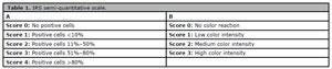
Table 1. IRS semi-quantitative scale.
Statistical analysis
This study applied different analysis techniques depending on the data distribution pattern. For example, One-way analysis of variance (ANOVA) was applied to normally distributed variables. Meanwhile, if the data were not normally distributed, the Kruskal-Wallis test was performed. Following that, the study conducted a post hoc Bonferroni correction for multiple comparisons. IL-1β expression followed normal distribution and was analyzed using ANOVA, whereas IL-8 expression and lesion size were analyzed using the Kruskal-Wallis test (Harada et al., 1999). The work has been reported in line with the ARRIVE guidelines 2.0.
RESULTS
This study consisted of three stages: stem cell preparation, induction of the endometriosis model, and evaluation of BMMSC effects on IL-1β, IL-8, and lesion area. Stem cells were confirmed as mesenchymal by immunophenotyping (CD73+, CD90+, CD105+, and CD45-), and their homing capacity was verified by PKH26 luminescence (Iwabe et al., 1998; Uccelli et al., 2008; Andres et al., 2015; Becker et al., 2017; Viganò et al., 2018). To validate the model, mice from all groups (K+-P3) were euthanized on day 14, showing established peritoneal lesions.
Isolation and culture of bone marrow mesenchymal stem cells
Bone marrow aspirates from mouse tibiae were processed according to standard protocols in the Stem Cell Laboratory, Institute of Tropical Disease, Universitas Airlangga. Cultured cells exhibited fibroblast-like morphology, appearing elongated, flattened, and spindle-shaped with large nuclei. Subculturing was performed every 5 days until the fourth passage. By passage 4, cultures displayed the characteristic swirling growth pattern of mesenchymal stem cells (Figure 1).
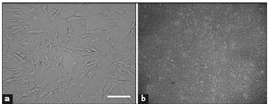
Figure 1. Mus musculus bone marrow mesenchymal stem cell culture. (a) Mesenchymal stem cell morphology. Cells appear to be small cell bodies (fibroblast-shaped), which are long and flattened with a large nucleus; (b) swirling pattern in passage four [inverted microscope, 40×.
Characterization of bone marrow mesenchymal stem cells
Stem cells developed in vitro can be characterized using both genotypic and phenotypic approaches. In this study, phenotypic identification was performed by immunocytochemistry using monoclonal antibodies conjugated with fluorescein isothiocyanate (FITC) (F3651; Sigma, St. Louis, MO). BMMSCs were confirmed by the expression of CD73, CD90, and CD105, and the absence of CD45 to exclude hematopoietic contamination (Figure 2). Immunofluorescence analysis demonstrated strong CD73 and CD90 expression, while no green luminescence was detected for CD45, consistent with the ISCT minimal criteria (Vernon & Wilson, 1985; Iwabe et al., 1998; Grümmer, 2006).
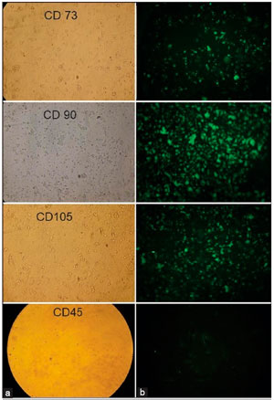
Figure 2. Immunohistocytochemical examination of CD 73, CD90, CD105, and CD 45. (a) Observation of bone marrow mesenchymal stem cells without fluorescence; (b) fluorescent observation of bone marrow mesenchymal stem cells (fluorescent microscope, 100×).
PKH26 fluorescence was observed in the membranes of transplanted BMMSCs within ovarian tissue (Figure 3), confirming their ability to home to the target site (Viganò et al., 2018; Piriyev et al., 2025).
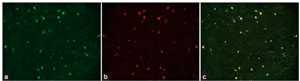
Figure 3. PKH26 luminescence in ovarian preparations of mice with endometriosis labeled PKH26. (a) Green filter, (b) red filter, (c) red-green filter (fluorescent microscope, 4.2×).
Figure 4 indicates that immunohistochemical staining patterns of IL-1β were observed in K+, P1, P2, and P3 groups (A, B, C, and D, respectively). Strong positive reactions were evident in the K+ group, whereas weak positive reactions were noted in the P1 group (100×). Most positively stained cells were macrophages (red arrows) (E) and mesothelial cells (black arrows) (F), both showing cytoplasmic expression of IL-1β (400×) (Iwabe et al., 1998; Mahnke et al., 2000; Piriyev et al., 2025).
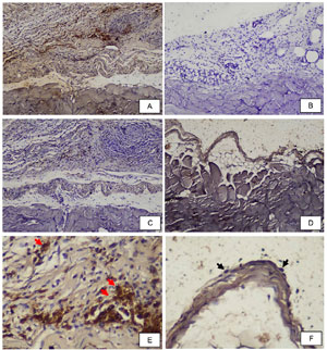
Figure 4. IL-1β protein expression in peritoneal endometriosis.
Figure 5 shows that the control group (K+) exhibited the highest IL-1β and IL-8 expression, whereas BMMSC-treated animals (P1) showed the lowest levels. Dienogest alone (P3) or in combination with BMMSCs (P2) produced some reduction but remained markedly less effective than BMMSC monotherapy (von Wolff et al., 1999; Ueda et al., 2002; Sanchez et al., 2016; 2017; Muzii et al., 2023).
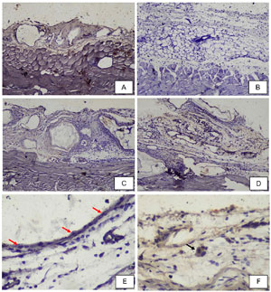
Figure 5. IL-8 protein expression in peritoneal endometriosis.
Immunohistochemical Results
Table 2 shows that the control group (K+) demonstrated the highest IL-1β and IL-8 expression, whereas BMMSC-treated animals (P1) exhibited the lowest. Dienogest alone (P3) or in combination with BMMSCs (P2) provided some reduction but were markedly less effective than BMMSC monotherapy (von Wolff et al., 1999; Ueda et al., 2002; Sanchez et al., 2016; 2017; Muzii et al., 2023).
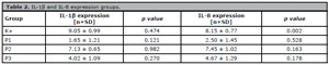
Table 2. IL-1β and IL-8 expression groups.
Values are mean ± SD. Statistical analysis by ANOVA (IL-1β) and Kruskal-Wallis test (IL-8).
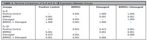
Table 3. Pairwise Comparison of IL-8 and IL-1β Expression Between Groups.
Post Hoc Bonferroni test
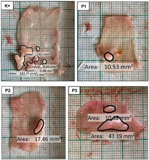
Figure 6. Macroscopic appearance of implant lesions.
ImageJ v1.54 (NIH, USA) analysis confirmed that BMMSC therapy was the only intervention associated with a significant reduction. The mean lesion size was 6.91 mm2, with 50% of BMMSC-treated mice completely free of lesions, compared to large, hypervascular lesions in the control group (mean 52.89 mm2, maximum 227.33 mm2). Neither Dienogest nor combination therapy demonstrated significant effects (Table 4).

Table 4. Lesion area (mm2) across groups.GroupMean ± SD [mm2]Range [mm2]T1 → K+ (Control)52.89±64.724.49 - 227.33T2 → P1 (BMMSC)6.91±7.250 - 19.51T3 → P2 (BMMSC + Dienogest)18.18±19.670 - 64.16T4 → P3 (Dienogest)23.28±16.910 - 53.57Values are expressed as mean ± standard deviation (SD) and range. Lesion size was analyzed using the Kruskal-Wallis test with Bonferroni post hoc.
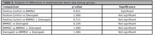
Table 5. Analysis of differences in endometriotic lesion size among groups.
Post-hoc Bonferroni test following Kruskal-Wallis (H=10.397, p=0.01)].
DISCUSSION
The BMMSCs characterized in this study exhibited the expected immunophenotypic profile of mesenchymal stem cells, expressing CD73, CD90, and CD105 while lacking CD45, in accordance with the International Society for Cellular Therapy (ISCT) criteria (Iwabe et al., 1998). This confirmed that the transplanted cells were genuine BMMSCs with immunomodulatory and regenerative potential. Moreover, PKH26 fluorescence verified their ability to migrate and engraft into ovarian tissue, demonstrating successful homing (Piriyev et al., 2025).
Endometriosis is strongly associated with chronic pelvic inflammation, leading to elevated peritoneal cytokine concentrations that disrupt folliculogenesis (Mahnke et al., 2000; Muzii et al., 2023). In the present study, immunohistochemistry revealed high expression of IL-1β and IL-8 in glandular epithelial and stromal cells of the control group, whereas BMMSC treatment markedly suppressed both cytokines. These findings are consistent with earlier evidence implicating IL-1β and IL-8 in lesion proliferation, angiogenesis, fibrotic progression, and reduced oocyte competence (Mahnke et al., 2000; Szukiewicz, 2022; Piriyev et al., 2025).
BMMSCs are recognized for their capacity to downregulate inflammatory mediators through paracrine mechanisms, including secretion of IL-10, prostaglandin E₂, and TGF-β, as well as inhibition of NF-κB signaling (Xu et al., 2025). Such mechanisms likely account for the robust cytokine suppression observed in the BMMSC group.
Role of inflammatory cytokines in endometriosis
Endometriosis is characterized by a chronic inflammatory peritoneal environment. Elevated levels of IL-1β, IL-8, and TNF-α are consistently detected in the peritoneal and follicular fluid of affected women, where they contribute to angiogenesis, fibrotic remodeling, granulosa cell apoptosis, and impaired steroidogenesis (Iwabe et al., 1998; Mahnke et al., 2000; Witz, 2000; Ueda et al., 2002; Khan et al., 2014; Da Broi et al., 2016; Giacomini et al., 2017; Sanchez et al., 2017; Wu et al., 2017). These cytokines have also been implicated in poor oocyte competence and reduced IVF success rates (Barnhart et al., 2002; Omland et al., 2005; Gupta et al., 2008; Da Broi et al., 2016; Giacomini et al., 2017; Sanchez et al., 2017; Wu et al., 2017; Viganò et al., 2018). Our findings confirm that IL-1β and IL-8 expression were highest in untreated controls, reinforcing their central role in disease persistence.
Limitations of hormonal therapy
Hormonal therapies, including Dienogest, primarily act by suppressing ovulation and inducing endometrial atrophy (Andres et al., 2015; Bedaiwy et al., 2017; Wu et al., 2022; Muzii et al., 2023). Although they provide symptomatic relief, they do not directly modulate the inflammatory microenvironment, and recurrence is common once treatment is discontinued (Andres et al., 2015; Becker et al., 2017; Bedaiwy et al., 2017; Szukiewicz, 2022; Wu et al., 2022; Muzii et al., 2023; Piriyev et al., 2025). In the present study, Dienogest did not significantly reduce IL-1β, IL-8, or lesion size compared with controls, consistent with clinical evidence of incomplete efficacy (Wu et al., 2022; Muzii et al., 2023; Piriyev et al., 2025).
Mechanisms of BMMSC therapy
BMMSCs exert their therapeutic effects primarily through paracrine signaling, secreting prostaglandin E₂, IL-10, and TGF-β, and inhibiting NF-κB activation (Dominici et al., 2006; Uccelli et al., 2008; Chen et al., 2018; Ghaneialvar et al., 2018; Schmelzer et al., 2019; Bian et al., 2022; Műzes & Sipos, 2022; Kulesza et al., 2023). These mechanisms suppress inflammatory cascades and modulate immune cell activity. In addition, BMMSC-derived extracellular vesicles and secretomes protect granulosa cells from apoptosis, reduce oxidative stress, and promote follicular repair (Chen et al., 2018; Rajabzadeh et al., 2019; Bozorgmehr et al., 2020; Na & Kim, 2020; Liao et al., 2021; Cui & Jing, 2024; Kavaldzhieva et al., 2025). In the present study, BMMSC monotherapy achieved the most pronounced suppression of IL-1β and IL-8 and significantly reduced lesion size, consistent with their established immunomodulatory and regenerative roles.
Lack of synergy with Dienogest
The absence of synergistic effects between BMMSCs and Dienogest may be attributed to potential interactions between hormonal suppression and MSC regenerative pathways. Hormonal therapy could interfere with MSC homing or paracrine activity, thereby diminishing their therapeutic efficacy. Similar observations have been reported in studies where immunomodulatory therapies showed reduced effects when combined with hormonal suppression20-22.
Clinical implications
BMMSC therapy offers several potential advantages over conventional management. By directly targeting inflammation and tissue remodeling, BMMSCs may provide disease-modifying and fertility-preserving benefits, which are particularly valuable for women seeking pregnancy (Barnhart et al., 2002; Omland et al., 2005; Da Broi et al., 2016; Sanchez et al., 2016; 2017; Giacomini et al., 2017; Wu et al., 2017; Rajabzadeh et al., 2019; Bozorgmehr et al., 2020; Liao et al., 2021; Cui & Jing, 2024; Kavaldzhieva et al., 2025).
Study limitations
Although this study offers important findings, several limitations must be acknowledged. First, the observation period was relatively short (14 days post-treatment). Second, angiogenic and fibrotic markers such as VEGF and TGF-β were not evaluated, despite their central roles in endometriosis progression lin (Xu et al., 2025). Finally, murine models cannot fully reproduce the complexity of human disease (Vernon & Wilson, 1985; Grümmer, 2006). Future studies should address these limitations through longer observation periods, broader biomarker assessment, and eventual translation into clinical trials.Mechanistically, the therapeutic advantage of BMMSCs is attributed primarily to paracrine effects rather than cellular differentiation. Their secretome has been shown to suppress inflammation, inhibit angiogenesis, alleviate oxidative stress, and prevent granulosa cell apoptosis. By reshaping the peritoneal microenvironment, BMMSCs disrupt the self-sustaining cycle of cytokine release and lesion survival. The lack of synergy between BMMSCs and Dienogest may be explained by hormonal suppression interfering with BMMSC regenerative and immunomodulatory pathways (Dominici et al., 2006; Uccelli et al., 2008; Chen et al., 2018; Na & Kim, 2020).
Comparison with previous studies and novelty
Previous studies have reported beneficial effects of mesenchymal stem cells in endometriosis models (Chen et al., 2018; Rajabzadeh et al., 2019; Bozorgmehr et al., 2020; Na & Kim, 2020; Liao et al., 2021; Cui & Jing, 2024; Kavaldzhieva et al., 2025). However, these investigations generally focused on MSC transplantation alone without direct comparison to standard pharmacological therapy. To our knowledge, this is the first study to perform a head-to-head evaluation of BMMSCs, Dienogest, and their combination in a murine endometriosis model. The novelty of our findings lies in demonstrating that BMMSC monotherapy not only suppressed pro-inflammatory cytokines (IL-1β and IL-8) but also significantly reduced lesion size, whereas Dienogest alone did not. This provides unique preclinical evidence of the superior disease-modifying potential of BMMSCs compared with standard care.From a clinical perspective, BMMSC therapy may represent a disease-modifying and fertility-preserving approach for endometriosis. The strong suppression of IL-1β and IL-8, together with lesion regression, provides compelling preclinical evidence. Nonetheless, several limitations must be acknowledged. The observation period was relatively short, angiogenic and fibrotic markers such as VEGF and TGF-β were not assessed (Xu et al., 2025), and murine models cannot fully recapitulate the complexity of human disease (Vernon & Wilson, 1985; Grümmer, 2006). Another limitation is that BMMSC characterization was performed by immunofluorescence (CD73+, CD90+, CD105+, CD45+) but was not validated by flow cytometry due to facility limitations. Future studies should include longer-term evaluation, broader biomarker panels, and flow cytometric profiling to comply fully with ISCT recommendations (Dominici et al., 2006; Chen et al., 2018; Na & Kim, 2020).From a translational perspective, BMMSC therapy holds promise as a fertility-preserving and disease-modifying approach for endometriosis. The strong suppression of IL-1β and IL-8, together with lesion regression, suggests potential applications in women seeking pregnancy while minimizing recurrence. Future studies should extend the observation period, evaluate angiogenic and fibrotic markers such as VEGF and TGF-β (Xu et al., 2025), and advance toward clinical trials to determine long-term efficacy and safety. To our knowledge, this is the first study to directly compare BMMSCs with standard hormonal therapy in endometriosis models, reinforcing their potential as a regenerative therapeutic strategy.
CONCLUSION
BMMSC monotherapy demonstrated superior efficacy compared with Dienogest or combination therapy in suppressing IL-1β and IL-8 expression and in reducing lesion size in murine endometriosis models. These findings underscore BMMSCs as a promising regenerative, disease-modifying, and fertility-preserving therapeutic strategy for the management of endometriosis (Dominici et al., 2006; Uccelli et al., 2008; Chen et al., 2018; Ghaneialvar et al., 2018; Rajabzadeh et al., 2019; Schmelzer et al., 2019; Bozorgmehr et al., 2020; Liao et al., 2021; Bian et al., 2022; Műzes & Sipos, 2022; Kulesza et al., 2023; Cui & Jing, 2024; Kavaldzhieva et al., 2025).
ACKNOWLEDGMENTS
The authors thank the Stem Cell Laboratory, Institute of Tropical Disease, Universitas Airlangga, and the Faculty of Medicine, Universitas Sriwijaya, Universitas Muhammadiyah Palembang for technical and laboratory support.
REFERENCES
Andres Mde P, Lopes LA, Baracat EC, Podgaec S. Dienogest in the treatment of endometriosis: systematic review. Arch Gynecol Obstet. 2015;292:523-9. PMID: 25749349 DOI: 10.1007/s00404-015-3681-6 Medline
Barnhart K, Dunsmoor-Su R, Coutifaris C. Effect of endometriosis on in vitro fertilization. Fertil Steril. 2002;77:1148-55. PMID: 12057720 DOI: 10.1016/S0015-0282(02)03112-6 Medline
Becker CM, Gattrell WT, Gude K, Singh SS. Reevaluating response and failure of medical treatment of endometriosis: a systematic review. Fertil Steril. 2017;108:125-36. PMID: 28668150 DOI: 10.1016/j.fertnstert.2017.05.004 Medline
Bedaiwy MA, Alfaraj S, Yong P, Casper R. New developments in the medical treatment of endometriosis. Fertil Steril. 2017;107:555-65. PMID: 28139238 DOI: 10.1016/j.fertnstert.2016.12.025 Medline
Bian D, Wu Y, Song G, Azizi R, Zamani A. The application of mesenchymal stromal cells (MSCs) and their derivative exosome in skin wound healing: a comprehensive review. Stem Cell Res Ther. 2022;13:24. PMID: 35073970 DOI: 10.1186/s13287-021-02697-9 Medline
Bozorgmehr M, Gurung S, Darzi S, Nikoo S, Kazemnejad S, Zarnani AH, Gargett CE. Endometrial and Menstrual Blood Mesenchymal Stem/Stromal Cells: Biological Properties and Clinical Application. Front Cell Dev Biol. 2020;8:497. PMID: 32742977 DOI: 10.3389/fcell.2020.00497 Medline
Chen X, Wang Q, Li X, Wang Q, Xie J, Fu X. Heat shock pretreatment of mesenchymal stem cells for inhibiting the apoptosis of ovarian granulosa cells enhanced the repair effect on chemotherapy-induced premature ovarian failure. Stem Cell Res Ther. 2018;9:240. PMID: 30257708 DOI: 10.1186/s13287-018-0964-4 Medline
Cui X, Jing X. Stem cell-based therapeutic potential in female ovarian aging and infertility. J Ovarian Res. 2024;17:171. PMID: 39182123 DOI: 10.1186/s13048-024-01492-3 Medline
Da Broi MG, de Albuquerque FO, de Andrade AZ, Cardoso RL, Jordão Junior AA, Navarro PA. Increased concentration of 8-hydroxy-2’-deoxyguanosine in follicular fluid of infertile women with endometriosis. Cell Tissue Res. 2016;366:231-42. PMID: 27250533 DOI: 10.1007/s00441-016-2428-4 Medline
Dominici M, Le Blanc K, Mueller I, Slaper-Cortenbach I, Marini F, Krause D, Deans R, Keating A, Prockop Dj, Horwitz E. Minimal criteria for defining multipotent mesenchymal stromal cells. The International Society for Cellular Therapy position statement. Cytotherapy. 2006;8:315-7. PMID: 16923606 DOI: 10.1080/14653240600855905 Medline
Ghaneialvar H, Soltani L, Rahmani HR, Lotfi AS, Soleimani M. Characterization and Classification of Mesenchymal Stem Cells in Several Species Using Surface Markers for Cell Therapy Purposes. Indian J Clin Biochem. 2018;33:46-52. PMID: 29371769 DOI: 10.1007/s12291-017-0641-x Medline
Giacomini E, Sanchez AM, Sarais V, Beitawi SA, Candiani M, Viganò P. Characteristics of follicular fluid in ovaries with endometriomas. Eur J Obstet Gynecol Reprod Biol. 2017;209:34-8. PMID: 26895700 DOI: 10.1016/j.ejogrb.2016.01.032 Medline
Grümmer R. Animal models in endometriosis research. Hum Reprod Update. 2006;12:641-9. PMID: 16775193 DOI: 10.1093/humupd/dml026 Medline
Gupta S, Goldberg JM, Aziz N, Goldberg E, Krajcir N, Agarwal A. Pathogenic mechanisms in endometriosis-associated infertility. Fertil Steril. 2008;90:247-57. PMID: 18672121 DOI: 10.1016/j.fertnstert.2008.02.093 Medline
Harada T, Enatsu A, Mitsunari M, Nagano Y, Ito M, Tsudo T, Taniguchi F, Iwabe T, Tanikawa M, Terakawa N. Role of cytokines in progression of endometriosis. Gynecol Obstet Invest. 1999;47:34-9. PMID: 10087426 DOI: 10.1159/000052857 Medline
Iwabe T, Harada T, Tsudo T, Tanikawa M, Onohara Y, Terakawa N. Pathogenetic significance of increased levels of interleukin-8 in the peritoneal fluid of patients with endometriosis. Fertil Steril. 1998;69:924-30. PMID: 9591504 DOI: 10.1016/S0015-0282(98)00049-1 Medline
Kavaldzhieva K, Mladenov N, Markova M, Belemezova K. Mesenchymal Stem Cell Secretome: Potential Applications in Human Infertility Caused by Hormonal Imbalance, External Damage, or Immune Factors. Biomedicines. 2025;13:586. PMID: 40149563 DOI: 10.3390/biomedicines13030586 Medline
Khan KN, Fujishita A, Kitajima M, Hiraki K, Nakashima M, Masuzaki H. Intra-uterine microbial colonization and occurrence of endometritis in women with endometriosis†. Hum Reprod. 2014;29:2446-56. PMID: 25205755 DOI: 10.1093/humrep/deu222 Medline
Kulesza A, Paczek L, Burdzinska A. The Role of COX-2 and PGE2 in the Regulation of Immunomodulation and Other Functions of Mesenchymal Stromal Cells. Biomedicines. 2023;11:445. PMID: 36830980 DOI: 10.3390/biomedicines11020445 Medline
Liao Z, Liu C, Wang L, Sui C, Zhang H. Therapeutic Role of Mesenchymal Stem Cell-Derived Extracellular Vesicles in Female Reproductive Diseases. Front Endocrinol. 2021;12:665645. PMID: 34248842 DOI: 10.3389/fendo.2021.665645 Medline
Mahnke JL, Dawood MY, Huang JC. Vascular endothelial growth factor and interleukin-6 in peritoneal fluid of women with endometriosis. Fertil Steril. 2000;73:166-70. PMID: 10632434 DOI: 10.1016/S0015-0282(99)00466-5 Medline
Műzes G, Sipos F. Mesenchymal Stem Cell-Derived Secretome: A Potential Therapeutic Option for Autoimmune and Immune-Mediated Inflammatory Diseases. Cells. 2022;11:2300. PMID: 35892597 DOI: 10.3390/cells11152300 Medline
Muzii L, Di Tucci C, Galati G, Carbone F, Palaia I, Bogani G, Perniola G, Tomao F, Kontopantelis E, Di Donato V. The Efficacy of Dienogest in Reducing Disease and Pain Recurrence After Endometriosis Surgery: a Systematic Review and Meta-Analysis. Reprod Sci. 2023;30:3135-43. PMID: 37217824 DOI: 10.1007/s43032-023-01266-0 Medline
Na J, Kim GJ. Recent trends in stem cell therapy for premature ovarian insufficiency and its therapeutic potential: a review. J Ovarian Res. 2020;13:74. PMID: 32576209 DOI: 10.1186/s13048-020-00671-2 Medline
Omland AK, Abyholm T, Fedorcsák P, Ertzeid G, Oldereid NB, Bjercke S, Tanbo T. Pregnancy outcome after IVF and ICSI in unexplained, endometriosis-associated and tubal factor infertility. Hum Reprod. 2005;20:722-7. PMID: 15591078 DOI: 10.1093/humrep/deh664 Medline
Piriyev E, Schiermeier S, Römer T. Hormonal Treatment of Endometriosis: A Narrative Review. Pharmaceuticals (Basel). 2025;18:588. PMID: 40284023 DOI: 10.3390/ph18040588 Medline
Rajabzadeh N, Fathi E, Farahzadi R. Stem cell-based regenerative medicine. Stem Cell Investig. 2019;6:19. PMID: 31463312 DOI: 10.21037/sci.2019.06.04 Medline
Sanchez AM, Somigliana E, Vercellini P, Pagliardini L, Candiani M, Vigano P. Endometriosis as a detrimental condition for granulosa cell steroidogenesis and development: From molecular alterations to clinical impact. J Steroid Biochem Mol Biol. 2016;155:35-46. PMID: 26407755 DOI: 10.1016/j.jsbmb.2015.07.023 Medline
Sanchez AM, Vanni VS, Bartiromo L, Papaleo E, Zilberberg E, Candiani M, Orvieto R, Viganò P. Is the oocyte quality affected by endometriosis? A review of the literature. J Ovarian Res. 2017;10:43. PMID: 28701212 DOI: 10.1186/s13048-017-0341-4 Medline
Santamaria X, Massasa EE, Taylor HS. Migration of cells from experimental endometriosis to the uterine endometrium. Endocrinology. 2012;153:5566-74. PMID: 22968642 DOI: 10.1210/en.2012-1202 Medline
Schmelzer E, McKeel DT, Gerlach JC. Characterization of Human Mesenchymal Stem Cells from Different Tissues and Their Membrane Encasement for Prospective Transplantation Therapies. Biomed Res Int. 2019;2019:6376271. PMID: 30941369 DOI: 10.1155/2019/6376271 Medline
Szukiewicz D. Epigenetic regulation and T-cell responses in endometriosis - something other than autoimmunity. Front Immunol. 2022;13:943839. PMID: 35935991 DOI: 10.3389/fimmu.2022.943839 Medline
Uccelli A, Moretta L, Pistoia V. Mesenchymal stem cells in health and disease. Nat Rev Immunol. 2008;8:726-36. PMID: 19172693 DOI: 10.1038/nri2395 Medline
Ueda M, Yamashita Y, Takehara M, Terai Y, Kumagai K, Ueki K, Kanda K, Yamaguchi H, Akise D, Hung YC, Ueki M. Survivin gene expression in endometriosis. J Clin Endocrinol Metab. 2002;87:3452-9. PMID: 12107265 DOI: 10.1210/jcem.87.7.8682 Medline
Vernon MW, Wilson EA. Studies on the surgical induction of endometriosis in the rat. Fertil Steril. 1985;44:684-94. PMID: 4054348 DOI: 10.1016/S0015-0282(16)48988-0 Medline
Viganò P, Ottolina J, Sarais V, Rebonato G, Somigliana E, Candiani M. Coagulation Status in Women With Endometriosis. Reprod Sci. 2018;25:559-65. PMID: 28681683 DOI: 10.1177/1933719117718273 Medline
von Wolff M, Classen-Linke I, Heid D, Krusche CA, Beier-Hellwig K, Karl C, Beier HM. Tumour necrosis factor-alpha (TNF-alpha) in human endometrium and uterine secretion: an evaluation by immunohistochemistry, ELISA and semiquantitative RT-PCR. Mol Hum Reprod. 1999;5:146-52. PMID: 10065870 DOI: 10.1093/molehr/5.2.146 Medline
Witz CA. Interleukin-6: another piece of the endometriosis-cytokine puzzle. Fertil Steril. 2000;73:212-4. PMID: 10685517 DOI: 10.1016/S0015-0282(99)00556-7 Medline
Wu G, Bersinger NA, Mueller MD, von Wolff M. Intrafollicular inflammatory cytokines but not steroid hormone concentrations are increased in naturally matured follicles of women with proven endometriosis. J Assist Reprod Genet. 2017;34:357-64. PMID: 28074436 DOI: 10.1007/s10815-016-0865-3 Medline
Wu Y, Liu Y, Jia H, Luo C, Chen H. Treatment of endometriosis with dienogest in combination with traditional Chinese medicine: A systematic review and meta-analysis. Front Surg. 2022;9:992490. PMID: 36386543 DOI: 10.3389/fsurg.2022.992490 Medline
Xu X, Li J, Lin H, Lin Z, Ji G. The role of TGF-β superfamily in endometriosis: a systematic review. Front Immunol. 2025;16:1638604. PMID: 40873582 DOI: 10.3389/fimmu.2025.1638604 Medline