JBRA Assisted Reproduction 2026;30(3):641-648
REVIEW
doi: 10.5935/1518-0557.20260026
Endometriosis as a cause of reduced IVF success rates: guilty beyond reasonable doubt?
1Clínica Hope Reprodução Humana, São Paulo, São Paulo, Brazil
2Science For Everymind, São Paulo, São Paulo, Brazil
3Fertility - FertGroup Medicina Reprodutiva, São Paulo, São Paulo, Brazil
CONFLICT OF INTERESTS
The authors have no competing interests to declare.
ABSTRACT
Endometriosis is a chronic inflammatory disease that affects around 10% of women of reproductive age and is frequently associated with infertility. Despite its high prevalence in assisted reproductive technology (ART) cycles, the exact mechanisms by which endometriosis may impair in vitro fertilization (IVF) outcomes remain under debate. This integrative narrative review critically examines the evidence on how endometriosis may affect oocyte quality and competence, ovarian reserve, and endometrial receptivity. While several studies have suggested molecular and cellular alterations in the follicular microenvironment and endometrial environment, population-based registry analyses have reported conflicting results regarding their clinical impact. Instead, the most reproducible negative impact appears to be a reduction in the number of oocytes retrieved, particularly in cases involving endometriomas or after ovarian surgery. Similarly, while endometrial alterations have been observed in women with endometriosis, large registry-based studies of donor oocyte cycles suggest only a marginal impairment of endometrial receptivity, if any. Taken together, reasonable doubt remains regarding the extent to which endometriosis influences fertility outcomes in IVF cycles.
Keywords: endometriosis, in vitro fertilization, oocytes, ovarian reserve, endometrial receptivity, assisted reproductive technology
INTRODUCTION
Endometriosis is a chronic, benign, estrogen-dependent inflammatory disease characterized by the presence of endometrial-like tissue outside the uterine cavity. Its pathogenesis is most commonly attributed to the retrograde menstruation of viable endometrial cells into the pelvic cavity. Endometriosis affects 5 to 10% of reproductive-age women worldwide and, to date, there is no medical or surgical cure (Ghiasi et al., 2020; As-Sanie et al., 2025; La Marca et al., 2025).
Endometriosis is classified into four subtypes, which may coexist and have implications for diagnosis and treatment: (1) superficial peritoneal endometriosis, involving lesions located on the peritoneal surface; (2) deep endometriosis, characterized by lesions infiltrating pelvic structures such as the uterosacral ligaments, bowel, or urinary tract; (3) endometriomas, cystic ovarian formations lined by endometrial tissue; and (4) extrapelvic endometriosis, involving sites beyond the pelvis, including the diaphragm, abdominal wall, and central nervous system (As-Sanie et al., 2025). Moreover, the American Society for Reproductive Medicine (ASRM) classifies endometriosis into four stages (Stages I-IV) based on the extent, location, depth of the lesions, and the presence of adhesions (Canis et al., 1997).
Dysmenorrhea, nonmenstrual pelvic pain, and dyspareunia are the most common symptoms of endometriosis and have frequently been associated with infertility (As-Sanie et al., 2025). Approximately 30-50% of women with endometriosis are estimated to experience infertility (Meuleman et al., 2009; Evans & Decherney, 2017). Moreover, according to a recent systematic review, the prevalence of endometriosis among women diagnosed with unexplained infertility is approximately 44% (Van Gestel et al., 2024). As a result, many couples pursue assisted reproductive technologies (ART) for infertility treatment, with endometriosis representing approximately 10% of the indications for in vitro fertilization (IVF) (Somigliana et al., 2017).
This review critically examines the evidence on the relationship between endometriosis and infertility, aiming to challenge prevailing assumptions and encourage a more nuanced understanding of its impact on oocyte quality and competence, ovarian reserve, and endometrial receptivity. This article was conceived as an interpretative narrative review aimed at conceptually integrating molecular, cellular, and clinical evidence related to endometriosis-associated infertility. The literature search was conducted in PubMed for studies published up to January 2025, using combinations of the following keywords: endometriosis, endometrioma, deep endometriosis, oocyte quality, oocyte competence, embryo quality, ovarian reserve, endometrial receptivity and IVF outcomes. References were selected based on their scientific relevance and contribution to conceptual and mechanistic discussion.
FROM ASSUMPTION TO INTERROGATION: TRACING THE DEBATE ON ENDOMETRIOSIS AND INFERTILITY
Several factors have been proposed as contributors to endometriosis-related infertility, including: (i) dyspareunia, (ii) mechanical factors such as adhesions or anatomical distortion, (iii) diminished ovarian reserve due to endometrioma, (iv) reduced oocyte quality, (v) sperm dysfunction, (vi) impaired tubo-ovarian interaction or embryo transport difficulties, (vii) disrupted ovulation, and (viii) endometrial receptivity dysfunction, among others. Many of these potential causes of infertility in endometriosis are associated with the systemic inflammatory environment induced by the disease (Bonavina & Taylor, 2022).
Reactive oxygen species, immune system dysregulation, and disruption of cellular architecture due to impaired differentiation, proliferation, and apoptosis are critical mechanisms that could negatively impact the reproductive environment (Simopoulou et al., 2021).
The direct causal relationship between tubal dysfunction secondary to adhesions in endometriosis and infertility has been extensively documented and considered irrefutable within the scientific community. Likewise, dyspareunia attributed to endometriosis is widely acknowledged as a critical factor compromising sexual function and quality of life. However, outside the context of tubal dysfunction and pain, the association between endometriosis and infertility has been called into question, as some authors have highlighted the lack of supporting evidence (Ata & Somigliana, 2024).
Generating high-quality scientific evidence on the true impact of endometriosis on fertility remains a major challenge. Several methodological limitations complicate the interpretation of existing studies. First, there is considerable heterogeneity in disease stage, lesion phenotype, and prior surgical history, which makes patient populations difficult to compare. Second, isolating the specific contribution of endometriosis itself is often problematic, as adenomyosis frequently coexists and may independently influence reproductive outcomes. Moreover, differences in the type, extent, and timing of surgical interventions can significantly affect ovarian reserve and endometrial function, further confounding results. Finally, when considering evidence derived from ART cycles, additional variability arises from the diversity of treatment strategies applied at different stage of the cycle (before ovarian stimulation or prior to embryo transfer). Together, these factors make it difficult to accurately determine the extent to which endometriosis itself contributes to infertility.
OOCYTE QUALITY VS. COMPETENCE: WHAT DOES ENDOMETRIOSIS REALLY AFFECT?
In the scientific literature, the concepts of quality and competence are often confused or used interchangeably. However, although related, these terms have distinct definitions and encompass independent characteristics that are essential to consider (Figure 1). Oocyte competence refers to the oocyte’s functional ability to successfully undergo and complete critical reproductive processes, including maturation, fertilization, early embryonic development, and progression to a viable blastocyst (Rienzi et al., 2012; Palomba et al., 2017). Thus, a competent oocyte is defined by its ability to successfully complete these critical developmental milestones. In contrast, oocyte quality is generally associated with the morphological and cytoplasmic features of the oocyte, observable structural and functional characteristics of a cell, including structural integrity and organelle distribution (Rienzi et al., 2012; Palomba et al., 2017). Distinguishing between these concepts is crucial, as certain pathological conditions, including endometriosis, may impact oocyte quality (e.g., altering morphology or specific organelle patterns) without necessarily compromising oocyte competence, such as its ability to reach the blastocyst stage. This conceptual distinction is frequently neglected in the literature, where the terms “quality” and “competence” are often used interchangeably, leading to misinterpretation of findings and clinical implications.
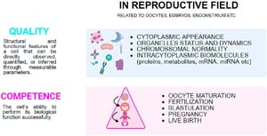
Figure 1. Conceptual distinction between cellular quality and competence in reproduction. Quality reflects intrinsic cellular features, including nuclear and cytoplasmic morphology, organelle integrity, chromosomal stability, and key molecular components. Competence denotes the functional capacity to achieve key developmental endpoints
Endometriosis is associated with alterations in the follicular microenvironment that may impair cumulus cell function and compromise oocyte quality at both the cellular and molecular levels. The importance of the follicular environment for ensuring oocyte quality and developmental competence is well established. Cumulus cells, which are directly exposed to this environment, support oocyte growth throughout folliculogenesis via transzonal projections, extracellular vesicles, and gap junctions. This intimate communication enables the regulation of oocyte meiotic reactivation, the transfer of key metabolic substrates such as pyruvate, and the delivery of molecules essential for maintaining intracellular homeostasis (Del Collado et al., 2018). Consequently, it is plausible that disruptions to the follicular environment may adversely affect oocyte quality and competence.
Endometriosis has been associated with decreased levels of estrogen and testosterone and increased levels of progesterone in the follicular fluid. These alterations are thought to result from local inflammation and impaired steroidogenesis in theca and granulosa cells, leading to reduced estradiol and testosterone production. Conversely, the elevated progesterone levels are likely a consequence of premature luteinization and altered steroid metabolism within the inflammatory microenvironment (Fan et al., 2023). Additionally, studies have demonstrated enhanced activation of apoptotic pathways and a higher number of apoptotic bodies in follicular cells of patients with endometriosis (Demirel et al., 2001; Goud et al., 2014; Sanchez et al., 2016). A larger prospective study involving patients with stage III and IV endometriosis identified distinct profiles of pro-inflammatory and anti-inflammatory cytokine signaling within the follicular fluid (Singh et al., 2016). Endometriosis has also been linked to increased endoplasmic reticulum stress and oxidative stress in granulosa and cumulus cells (Scutiero et al., 2017; Kunitomi et al., 2020; Lin et al., 2020).
At the oocyte level, transcriptomic profiling of cumulus cells from nine patients with stage I-II endometriosis identified 26 differentially expressed genes implicated in the regulation of inflammatory and immune pathways when compared to controls (Da Luz et al., 2022). Additionally, transcriptomic analysis of individual metaphase II (MII) oocytes from seven patients with endometriosis, contrasted with oocytes from healthy donors, revealed widespread gene expression alterations independent of ovarian laterality, affecting pathways involved in steroid metabolism, oxidative stress response, and cell cycle regulation (Ferrero et al., 2019). Endometriosis has also been associated with mitochondrial dysfunction and morphological changes (Xu et al., 2015), as well as disruptions in cortical granule dynamics and meiotic spindle integrity within oocytes during an in vitro maturation (IVM) experiment (Goud et al., 2014).
Beyond cellular and molecular alterations, endometriosis has further been related to compromised oocyte quality, including an increased proportion of oocytes presenting morphological abnormalities (Shebl et al., 2017; Kasapoglu et al., 2018; Robin et al., 2021). Notably, the influence on oocyte quality could be affected by endometriosis stage, as indicated by Shebl et al. (2017), who reported a much severe phenotype regarding oocyte morphology in stage IV. Moreover, a retrospective cohort study of more than 6,000 oocytes concluded that besides a decrease in the number of recovered oocytes and increase in some morphological abnormality in endometriosis oocytes, mainly by a presence of endometrioma, the oocyte quality scores Average Oocyte Quality Index (AOQI) and metaphase II oocyte morphological scoring system (MOMS) did not differ between endometriosis and control patients (Robin et al., 2021).
However, the critical question remains whether these alterations in oocyte quality translate into impaired developmental competence. A recent retrospective study including 248 patients with endometriosis, matched by age, stimulation protocol, treatment date, and anti-Müllerian hormone (AMH) levels to 248 controls, found no significant differences in the number of oocytes retrieved, 2PN embryos, blastocysts, or high-quality blastocysts (Invernici et al., 2022). Furthermore, the euploidy rate appears unaffected by endometriosis, as demonstrated by an analysis of 1,873 biopsied embryos from 261 patients, which showed no differences between endometriosis and male factor infertility groups. Clinical pregnancy rates following euploid embryo transfers were also comparable (Bishop et al., 2021). Conversely, a larger retrospective matched cohort study encompassing 3,071 IVF cycles reported that endometriosis negatively influences pregnancy outcomes, being associated with lower cumulative birth rates and diminished oocyte and embryo quantity and quality. This study additionally revealed increased macrophage infiltration and an imbalance between proand anti-inflammatory cytokines in follicular fluid from patients with endometriosis, indicating an altered follicular microenvironment as a potential mechanism (Zhou et al., 2022). Nevertheless, the meta-analyses available to date have not identified significant differences in total or good quality blastocyst numbers, nor in clinical pregnancy or live birth rates between women with and without endometriosis or endometrioma undergoing ART, suggesting no adverse impact on oocyte competence (Qu et al., 2022; Gayete-Lafuente et al., 2024).
Although there is evidence supporting the impact of endometriosis on oocyte quality, its effect on oocyte competence remains less well defined, with most available data suggesting no significant impairment. It is also important to acknowledge that the lack of consistent evidence for reduced oocyte competence in cohort studies and meta-analyses may partly reflect the heterogeneity of the study populations, including differences in disease phenotype, stage, prior surgical history, and clinical management. Nevertheless, it cannot be excluded that oocyte competence may indeed be affected in specific phenotypes or in advanced stages of the disease, such as stage III and IV endometriosis.
BESIDES OOCYTE QUALITY AND COMPETENCE: THE IMPACT OF ENDOMETRIOSIS ON OVARIAN RESERVE
In addition to oocyte quality and competence, endometriosis may also influence the number of oocytes retrieved. In theory, endometriosis may impair ovarian reserve through chronic systemic inflammation and direct disruption of ovarian function, contributing to reduced follicular recruitment and ovulatory alterations (Cobo et al., 2021).
A meta-analysis, including 26 studies, confirmed that women with endometriosis produce fewer oocytes; however, the analysis did not control for the bias of prior surgical intervention, which limits the causal interpretation of the isolated role of endometriosis (Qu et al., 2022). On the other hand, a recent meta-analysis demonstrates that ovarian endometriosis, endometriomas, reduce both the total oocyte and the number of MII, showing no significant differences in fertilization or blastulation rates (Gayete-Lafuente et al., 2024).
As reviewed by La Marca et al. (2025), the reduction in ovarian reserve observed in women with endometriomas may result from elevated concentrations of proinflammatory cytokines, proteolytic enzymes, and oxidative stress agents within the endometrioma fluid. These harmful substances can infiltrate the adjacent healthy ovarian cortex, causing damage or loss of primordial follicles and leading to a subsequent decline in AMH levels. Some authors have proposed that, in addition to inflammation and oxidative stress, an increased activation of the PI3K-AKT pathway associated with endometriosis may lead to the premature activation of dormant primordial follicles, thereby accelerating follicular depletion and contributing to a reduced ovarian reserve. According to this hypothesis, the local inflammatory response and subsequent fibrosis at endometriotic sites, alongside Wnt signaling dysregulation, could impair ovarian vascularization, nutrient supply to developing follicles and promote cell death and follicular atresia (Demirel et al., 2001; Takeuchi et al., 2019; Fan et al., 2023).
Furthermore, the presence of large endometriomas may impose mechanical stress on ovarian tissue, further compromising the ovarian reserve. Moreover, a pragmatic study by Cobo et al. (2021) showed that the most direct and measurable detrimental impact of endometriomas on ovarian reserve appears to be associated with cystectomy, particularly when performed in young women, showing lower cumulative live birth rates than nonoperated women in age-matched groups.
To date, available evidence suggests that the impact of endometriosis lies more in the quantity of oocytes retrieved rather than in their quality or competence. As a consequence of a reduced number of oocytes, fewer blastocysts become available for transfer, ultimately leading to a lower cumulative live birth rate in patients with endometriosis as shown in Zhou et al. (2022).
Given this quantitative decline, fertility preservation has been increasingly discussed as a potential strategy in the management of endometriosis. The ESHRE guideline (2022) recommends that, in cases of extensive ovarian endometriosis, clinicians should discuss fertility preservation with affected women, as its actual benefit in this population remains uncertain (Becker et al., 2022). In contrast, the most recent ASRM Committee Opinion on endometriosis and infertility does not specifically address fertility preservation, as its focus is mainly on the management of existing infertility rather than on preventive strategies (Practice Committee of the American Society for Reproductive Medicine, 2012). Despite the absence of formal recommendations from reproductive societies or clinical guidelines, fertility preservation has been proposed for patients over 35 years, those with bilateral or unilateral endometriomas (prior to surgery), diminished ovarian reserve, a history or risk of multiple surgeries, large endometriomas (>4 cm), severe endometriosis (stages III and IV), low endometriosis fertility index (EFI) scores, and a low probability of natural conception following surgery (La Marca et al., 2025).
ENDOMETRIAL ALTERATIONS IN ENDOMETRIOSIS: THEIR CLINICAL RELEVANCE
One of the major challenges in reproductive physiology, and consequently in IVF, is to understand the key factors that determine successful embryo implantation and to elucidate the complex cross-talk between the embryo and the endometrium, often referred to as the ‘black box’ of implantation. Implantation failure, particularly involving euploid embryos, remains one of the most enigmatic and complex issues in assisted reproductive technologies (Cimadomo et al., 2023). Therefore, any condition that might impair endometrial receptivity is a consistent focus of investigation, as is the case with endometriosis.
In endometriosis, alterations in molecular pathways, such as those related to epigenetic regulation, immune and inflammatory responses, hormonal signaling, and epithelial-mesenchymal transition, have been suggested to impair embryo endometrium cross-talk, potentially contributing to implantation failure (Guo et al., 2023). Moreover, endometriosis is commonly associated with an increased inflammation-related in-situ production of estradiol and progesterone resistance (de Ziegler et al., 2010).
Single-cell transcriptomic analyses have revealed that even the eutopic endometrium of women with endometriosis displays distinct cellular and molecular alterations compared to healthy controls. These changes involve transcriptional reprogramming and structural remodeling of epithelial and stromal cells, as well as dysregulation of immune related pathways (Fonseca et al., 2023). Likewise, a proinflammatory immune profile in the eutopic endometrium of women with endometriosis is affected, with increased CD8+ T cells linked to a higher risk of infertility (Wu et al., 2021). Several studies have reported alterations alterations in the endometrium, ranging from dysregulation of immune pathways to epigenetic factors (Hromadnikova et al., 2022; Shi et al., 2025). Nonetheless, while several studies have reported changes in the endometrial quality, it remains to be clarified whether such alterations compromise endometrial competence, the ability to support implantation and achieve a live birth.
One of the most widely discussed studies on this topic is the meta-analysis published in JAMA Network Open by Paffoni et al. (2024) which evaluated live birth rates in women with endometriosis who received donor oocytes, as it provides robust data on endometrial receptivity by excluding the potential confounding factor of impaired embryo quality in these patients. This systematic review and meta-analysis included data from four individual studies (two prospective and two retrospective) as well as from two major IVF registries: the Human Fertilization and Embryology Authority (HFEA, UK) and the Society for Assisted Reproductive Technology (SART, USA). In total, 7,212 donor oocyte cycles derived from the studies were examined, in addition to 162,082 cycles reported by the registries (137,182 from SART and 24,900 from HFEA). The adjusted live birth outcome showed a small, but statistically significant, difference in patients with endometriosis (OR, 0.89; 95% CI, 0.81-0.97), observed exclusively in the registry data, not in the meta-analysis of prospective or retrospective studies. The authors conclude that there appears to be a minimal reduction in live birth rates among recipients with a history of endometriosis undergoing ART cycles with donor oocytes. They also suggest that a marginal impairment of uterine receptivity may be present in women affected by endometriosis. However, whether this impairment is related to comorbidities or confounding factors remains unclear and should be further investigated in future studies.
These findings are consistent with those reported reported by Bishop et al. (2021), which showed no statistically significant differences in clinical outcomes following single euploid embryo transfer among patients with endometriosis, male factor infertility, and PGT-M cycles, as well as with a recent meta-analysis on clinical outcomes in endometriosis patients undergoing IVF (Qu et al., 2022). On the other hand, an interesting meta-analysis identified an approximately 12% reduction in live birth rates following ART specifically among patients with stage III-IV endometriosis, highlighting that the phenotypic expression and clinical impact of the disease may vary according to its stage and subtype (Horton et al., 2019).
Regarding cumulative live birth rates, when analyses control for confounding factors such as the number of oocytes retrieved and the number and quality of embryos obtained, it becomes more appropriate to assess the potential impact of endometriosis on endometrial receptivity. A recent matched cohort study, which included 100 women with endometriosis and 100 controls matched for age, as well as the number and quality of blastocysts, reported comparable cumulative live birth rates between the groups (Casalechi et al., 2023). These findings support the hypothesis that endometriosis does not significantly compromise endometrial receptivity in affected individuals.
This apparent discrepancy between molecular alterations and preserved clinical outcomes may reflect a certain degree of endometrial resilience, in which compensatory mechanisms maintain implantation capacity despite local disturbances. Likewise, it is plausible that compensatory molecular pathways or redundant mechanisms involved in implantation and early placentation mitigate the functional impact of the transcriptomic and proteomic disturbances observed in endometriosis.
Despite the lack of robust evidence supporting a negative impact of endometriosis on clinical pregnancy or live birth rates, a recent meta-analysis highlights that severe forms of the disease (stage III-IV and deep endometriosis) represent significant risk factors for placenta previa and preterm birth (Busnelli et al., 2024), suggesting that the impact of endometriosis may manifest later in pregnancy.
LESSONS FROM THERAPEUTIC INTERVENTIONS DURING ART
The appropriate timing, indication, and type of treatment for endometriosis in infertile patients remain topics of ongoing debate. Some authors have proposed structured management approaches to guide clinical decision-making in this population (Di Spiezio Sardo et al., 2025). In summary, for women under 35 years of age experiencing pelvic pain, conservative surgical treatment may be considered, and if spontaneous pregnancy does not occur within one year postoperatively, IVF (with or without prior medical therapy) may be indicated. For women over 35 years of age with pain, or for asymptomatic women, proceeding directly to IVF with possible pre-treatment using medical therapy is generally recommended. Furthermore, in cases of repeated IVF failure, surgical intervention may be considered (Di Spiezio Sardo et al., 2025).
For selected patients, hormonal or surgical treatment may precede IVF or embryo transfer. GnRH agonists have demonstrated anti-inflammatory, anti-angiogenic, and pro-apoptotic effects beyond their hypoestrogenic mechanism (Khan et al., 2010), while progestins may improve the endometrial environment by reducing inflammatory cytokines and oxidative stress (Tamura et al., 2019). However, evidence remains inconsistent. The most recent Cochrane meta-analysis, including eight RCTs, found low-certainty evidence and concluded that prolonged GnRH agonist pretreatment does not clearly improve pregnancy or live birth rates (Georgiou et al., 2019). Similarly, a network meta-analysis of nine RCTs found no benefit of hormonal pretreatment before ovarian stimulation, with higher live birth rates observed in women who did not receive pretreatment (Riemma et al., 2025).
Consistently, the ESHRE guideline states that neither hormonal nor surgical interventions should be routinely used before IVF to improve live birth rates (Becker et al., 2022). Surgery may be considered only to alleviate pain or improve follicle accessibility in specific cases, such as endometrioma or deep lesions. Overall, the absence of consistent benefit from pre-IVF interventions reinforces the notion that endometriosis exerts a limited direct impact on ART outcomes.
CONCLUSION: WEIGHING THE EVIDENCE: VERDICT YET TO BE REACHED
Most of the current evidence linking endometriosis and (in)fertility originates from ART cycles, which may not fully represent physiological reproductive conditions. Furthermore, many studies reporting clinical pregnancy rates aggregate data from women attempting natural conception and those undergoing ART, across varying stages of endometriosis, with or without comorbidities such as adenomyosis, and under highly heterogeneous treatment protocols, all factors that complicate the interpretation of definitive conclusions.
Although it is well established that endometriosis alters the reproductive tract milieu and, consequently, the local microenvironment, the precise repercussions of these alterations on gamete and endometrial function remain incompletely understood (Figure 2).
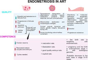
Figure 2. Schematic representation of the possible mechanisms by which endometriosis affects reproductive outcomes in assisted reproductive technology (ART) at multiple levels. The upper panel depicts alterations in ovarian, oocyte, and endometrial quality, including hormonal imbalances, oxidative stress, apoptosis, mitochondrial and spindle defects, and transcriptional dysregulation. The lower panel illustrates the downstream consequences on ovarian reserve, embryo development, and clinical outcomes, such as reduced numbers of mature oocytes and embryos and compromised embryo developmental and implantation rates.
Undeniably, endometriosis can lead to infertility when pelvic adhesions cause tubal obstruction or when severe pain interferes with sexual intercourse. In the context of ART. However, large-scale studies have not consistently demonstrated a detrimental effect of endometriosis on reproductive competence, although more severe phenotypes (stages III-IV) may be associated with greater adverse outcomes.
In summary, until new and robust evidence emerges to challenge current assumptions, reasonable doubt persists regarding the true extent to which endometriosis influences fertility outcomes in IVF cycles. It may now be time to broaden the investigative lens from a micro-level focus on cellular quality to a more integrated perspective that also encompasses cellular competence. Future research should employ matched-cohort designs that control for the type and stage of endometriosis, as well as for oocyte yield, to better isolate disease-specific effects. Ideally, these studies should integrate molecular and clinical analyses, linking oocyte quality assessments with measures of developmental and clinical competence, such as live birth and cumulative live birth rates. Moreover, studies using donor oocytes may help clarify whether endometrial receptivity is genuinely impaired in women with endometriosis. Collectively, such approaches may ultimately determine whether endometriosis compromises oocyte or embryonic competence and/or endometrial receptivity, thereby providing a more comprehensive understanding of its true impact on reproductive outcomes.
Authors’ Contribution
MC and MDC contributed to the conceptualization, investigation, and writing of the original draft. MDC also supervised the project and contributed to review and editing. VGA, GT, MAT, and AM contributed to investigation and manuscript review and editing. EB provided supervision and critical revision of the manuscript. All authors read and approved the final manuscript.
REFERENCES
As-Sanie S, Mackenzie SC, Morrison L, Schrepf A, Zondervan KT, Horne AW, Missmer SA. Endometriosis: A Review. JAMA. 2025;334:64-78. PMID: 40323608 DOI: 10.1001/jama.2025.2975 Medline
Ata B, Somigliana E. Endometriosis, staging, infertility and assisted reproductive technology: time for a rethink. Reprod Biomed Online. 2024;49:103943. PMID: 38733678 DOI: 10.1016/j.rbmo.2024.103943 Medline
Becker CM, Bokor A, Heikinheimo O, Horne A, Jansen F, Kiesel L, King K, Kvaskoff M, Nap A, Petersen K, Saridogan E, Tomassetti C, van Hanegem N, Vulliemoz N, Vermeulen N; ESHRE Endometriosis Guideline Group. ESHRE guideline: endometriosis. Hum Reprod Open. 2022;2022:hoac009. PMID: 35350465 DOI: 10.1093/hropen/hoac009 Medline
Bonavina G, Taylor HS. Endometriosis-associated infertility: From pathophysiology to tailored treatment. Front Endocrinol (Lausanne). 2022;13:1020827. PMID: 36387918 DOI: 10.3389/fendo.2022.1020827 Medline
Busnelli A, Di Simone N, Somigliana E, Greppi D, Cirillo F, Bulfoni A, Inversetti A, Levi-Setti PE. Untangling the independent effect of endometriosis, adenomyosis, and ART-related factors on maternal, placental, fetal, and neonatal adverse outcomes: results from a systematic review and meta-analysis. Hum Reprod Update. 2024;30:751-88. PMID: 39049473 DOI: 10.1093/humupd/dmae024 Medline
Casalechi M, Reschini M, Palermo MC, Stefano GD, Vercellini P, Benaglia L, Somigliana E, Viganò P. Is endometrial receptivity affected in women with endometriosis? Results from a matched pair case-control study of assisted reproductive technology treatments. Reprod Biomed Online. 2023;47:103414. PMID: 37879123 DOI: 10.1016/j.rbmo.2023.103414 Medline
Cimadomo D, Rienzi L, Conforti A, Forman E, Canosa S, Innocenti F, Poli M, Hynes J, Gemmell L, Vaiarelli A, Alviggi C, Ubaldi FM, Capalbo A. Opening the black box: why do euploid blastocysts fail to implant? A systematic review and meta-analysis. Hum Reprod Update. 2023;29:570-633. PMID: 37192834 DOI: 10.1093/humupd/dmad010 Medline
Cobo A, García-Velasco JA, Remohí J, Pellicer A. Oocyte vitrification for fertility preservation for both medical and nonmedical reasons. Fertil Steril. 2021;115:1091-101. PMID: 33933172 DOI: 10.1016/j.fertnstert.2021.02.006 Medline
Da Luz CM, Da Broi MG, Koopman LO, Plaça JR, da Silva-Jr WA, Ferriani RA, Meola J, Navarro PA. Transcriptomic analysis of cumulus cells shows altered pathways in patients with minimal and mild endometriosis. Sci Rep. 2022;12:5775. PMID: 35388025 DOI: 10.1038/s41598-022-09386-4 Medline
Demirel LC, Cengiz B, Ünlü C. Severe endometriosis and apoptotic granulosa cells. Fertil Steril. 2001;75:642. PMID: 11265664 DOI: 10.1016/S0015-0282(00)01771-4 Medline
Del Collado M, Andrade GM, Meirelles FV, da Silveira JC, Perecin F. Contributions from the ovarian follicular environment to oocyte function. Anim Reprod. 2018;15:261-70. PMID: 34178149 DOI: 10.21451/1984-3143-AR2018-0082 Medline
de Ziegler D, Borghese B, Chapron C. Endometriosis and infertility: pathophysiology and management. Lancet. 2010;376:730-8. PMID: 34178149 DOI: 10.21451/1984-3143-AR2018-0082 Medline
Di Spiezio Sardo A, Becker CM, Renner SP, Suvitie PA, Tarriel JE, Vannuccini S, Garcia Velasco JA, Verguts J, Mercorio A. Management of women with endometriosis in the 21st century. Curr Opin Obstet Gynecol. 2025;37:149-57. PMID: 40237624 DOI: 10.1097/GCO.0000000000001027 Medline
Evans MB, Decherney AH. Fertility and Endometriosis. Clin Obstet Gynecol. 2017;60:497-502. PMID: 28742581 DOI: 10.1097/GRF.0000000000000295 Medline
Ferrero H, Corachán A, Aguilar A, Quiñonero A, Carbajo-García MC, Alamá P, Tejera A, Taboas E, Muñoz E, Pellicer A, Domínguez F. Single-cell RNA sequencing of oocytes from ovarian endometriosis patients reveals a differential transcriptomic profile associated with lower quality. Hum Reprod. 2019;34:1302-12. PMID: 31211846 DOI: 10.1093/humrep/dez053 Medline
Gayete-Lafuente S, Vilà Famada A, Albayrak N, Espinós Gómez JJ, Checa Vizcaíno MÁ, Moreno-Sepulveda J. Indirect markers of oocyte quality in patients with ovarian endometriosis undergoing IVF/ICSI: a systematic review and meta-analysis. Reprod Biomed Online. 2024;49:104075. PMID: 38943812 DOI: 10.1016/j.rbmo.2024.104075 Medline
Georgiou EX, Melo P, Baker PE, Sallam HN, Arici A, Garcia-Velasco JA, Abou-Setta AM, Becker C, Granne IE. Long-term GnRH agonist therapy before in vitro fertilisation (IVF) for improving fertility outcomes in women with endometriosis. Cochrane Database Syst Rev. 2019;2019:CD013240. PMID: 31747470 DOI: 10.1002/14651858.CD013240.pub2 Medline
Goud PT, Goud AP, Joshi N, Puscheck E, Diamond MP, Abu-Soud HM. Dynamics of nitric oxide, altered follicular microenvironment, and oocyte quality in women with endometriosis. Fertil Steril. 2014;102:151-9.e5. PMID: 24825428 DOI: 10.1016/j.fertnstert.2014.03.053 Medline
Guo SW, Habiba M, Benagiano G. From Retrograde Menstruation to Endometrial Determinism and a Brave New World of “Root Treatment” of Endometriosis: Destiny or a Fanciful Utopia? Biomolecules. 2023;13:336. PMID: 36830705 DOI: 10.3390/biom13020336 Medline
Horton J, Sterrenburg M, Lane S, Maheshwari A, Li TC, Cheong Y. Reproductive, obstetric, and perinatal outcomes of women with adenomyosis and endometriosis: a systematic review and meta-analysis. Hum Reprod Update. 2019;25:592-632. PMID: 31318420 DOI: 10.1093/humupd/dmz012 Medline
Hromadnikova I, Kotlabova K, Krofta L. First-trimester predictive models for adverse pregnancy outcomes-a base for implementation of strategies to prevent cardiovascular disease development. Front Cell Dev Biol. 2024 Sep 4;12:1461547. PMID: 39296937 DOI: 10.3389/fcell.2024.1461547 Medline
Invernici D, Reschini M, Benaglia L, Somigliana E, Galati G, La Vecchia I, Vigano’ P, Vercellini P. The impact of endometriosis on IVF efficacy: qualitative and quantitative assessment of ovarian response and embryo development. Reprod Biomed Online. 2022;45:275-81. PMID: 35764471 DOI: 10.1016/j.rbmo.2022.04.010 Medline
Kasapoglu I, Kuspinar G, Saribal S, Turk P, Avcı B, Uncu G. Detrimental effects of endometriosis on oocyte morphology in intracytoplasmic sperm injection cycles: a retrospective cohort study. Gynecol Endocrinol. 2018;34:206-11. PMID: 29057690 DOI: 10.1080/09513590.2017.1391203 Medline
Khan KN, Kitajima M, Hiraki K, Fujishita A, Sekine I, Ishimaru T, Masuzaki H. Changes in tissue inflammation, angiogenesis and apoptosis in endometriosis, adenomyosis and uterine myoma after GnRH agonist therapy. Hum Reprod. 2010;25:642-53. PMID: 20008888 DOI: 10.1093/humrep/dep437 Medline
Kunitomi C, Harada M, Takahashi N, Azhary JMK, Kusamoto A, Nose E, Oi N, Takeuchi A, Wada-Hiraike O, Hirata T, Hirota Y, Koga K, Fujii T, Osuga Y. Activation of endoplasmic reticulum stress mediates oxidative stress-induced apoptosis of granulosa cells in ovaries affected by endometrioma. Mol Hum Reprod. 2020;26:40-52. PMID: 31869409 DOI: 10.1093/molehr/gaz066 Medline
La Marca A, Semprini M, Mastellari E, Donno V, Capuzzo M, Alboni C, Giulini S. Fertility preservation in women with endometriosis. Hum Reprod Open. 2025;2025:hoaf012. PMID: 40123895 DOI: 10.1093/hropen/hoaf012 Medline
Meuleman C, Vandenabeele B, Fieuws S, Spiessens C, Timmerman D, D’Hooghe T. High prevalence of endometriosis in infertile women with normal ovulation and normospermic partners. Fertil Steril. 2009;92:68-74. PMID: 18684448 DOI: 10.1016/j.fertnstert.2008.04.056 Medline
Paffoni A, Casalechi M, De Ziegler D, Cicinelli E, Somigliana E, Viganò P, Vitagliano A. Live Birth After Oocyte Donation In Vitro Fertilization Cycles in Women With Endometriosis: A Systematic Review and Meta-Analysis. JAMA Netw Open. 2024;7:e2354249. PMID: 38294811 DOI: 10.1001/jamanetworkopen.2023.54249 Medline
Palomba S, Daolio J, La Sala GB. Oocyte Competence in Women with Polycystic Ovary Syndrome. Trends Endocrinol Metab. 2017;28:186-98. PMID: 27988256 DOI: 10.1016/j.tem.2016.11.008 Medline
Riemma G, Cobellis L, Laganà AS, Etrusco A, Della Corte L, Torella M, Vastarella MG, Carotenuto RM, De Franciscis P. Efficacy of hormone pre-treatment before ART to improve reproductive outcomes in infertile women with endometriosis: Network meta-analysis of randomized controlled trials. Int J Gynaecol Obstet. 2025;170:1001-13. PMID: 40221832 DOI: 10.1002/ijgo.70134 Medline
Rienzi L, Balaban B, Ebner T, Mandelbaum J. The oocyte. Hum Reprod. 2012;27:i2-21. PMID: 22811312 DOI: 10.1093/humrep/des200 Medline
Robin C, Uk A, Decanter C, Behal H, Collinet P, Rubod C, Barbotin AL, Robin G. Impact of endometriosis on oocyte morphology in IVF-ICSI: retrospective study of a cohort of more than 6000 mature oocytes. Reprod Biol Endocrinol. 2021;19:160. PMID: 34656130 DOI: 10.1186/s12958-021-00798-x Medline
Sanchez AM, Somigliana E, Vercellini P, Pagliardini L, Candiani M, Vigano P. Endometriosis as a detrimental condition for granulosa cell steroidogenesis and development: From molecular alterations to clinical impact. J Steroid Biochem Mol Biol. 2016;155:35-46. PMID: 26407755 DOI: 10.1016/j.jsbmb.2015.07.023 Medline
Scutiero G, Iannone P, Bernardi G, Bonaccorsi G, Spadaro S, Volta CA, Greco P, Nappi L. Oxidative Stress and Endometriosis: A Systematic Review of the Literature. Oxid Med Cell Longev. 2017;2017:7265238. PMID: 29057034 DOI: 10.1155/2017/7265238 Medline
Shebl O, Sifferlinger I, Habelsberger A, Oppelt P, Mayer RB, Petek E, Ebner T. Oocyte competence in in vitro fertilization and intracytoplasmic sperm injection patients suffering from endometriosis and its possible association with subsequent treatment outcome: a matched case-control study. Acta Obstet Gynecol Scand. 2017;96:736-44. PMID: 27317364 DOI: 10.1111/aogs.12941 Medline
Singh AK, Dutta M, Chattopadhyay R, Chakravarty B, Chaudhury K. Intrafollicular interleukin-8, interleukin-12, and adrenomedullin are the promising prognostic markers of oocyte and embryo quality in women with endometriosis. J Assist Reprod Genet. 2016;33:1363-72. PMID: 27491770 DOI: 10.1007/s10815-016-0782-5 Medline
Somigliana E, Vigano P, Benaglia L, Busnelli A, Berlanda N, Vercellini P. Management of Endometriosis in the Infertile Patient. Semin Reprod Med. 2017;35:31-7. PMID: 27926972 Medline
Takeuchi A, Koga K, Satake E, Makabe T, Taguchi A, Miyashita M, Takamura M, Harada M, Hirata T, Hirota Y, Yoshino O, Wada-Hiraike O, Fujii T, Osuga Y. Endometriosis Triggers Excessive Activation of Primordial Follicles via PI3K-PTEN-Akt-Foxo3 Pathway. J Clin Endocrinol Metab. 2019;104:5547-54. PMID: 31219551 DOI: 10.1210/jc.2019-00281 Medline
Tamura H, Yoshida H, Kikuchi H, Josaki M, Mihara Y, Shirafuta Y, Shinagawa M, Tamura I, Taketani T, Takasaki A, Sugino N. The clinical outcome of Dienogest treatment followed by in vitro fertilization and embryo transfer in infertile women with endometriosis. J Ovarian Res. 2019;12:123. PMID: 31831028 DOI: 10.1186/s13048-019-0597-y Medline
Van Gestel H, Bafort C, Meuleman C, Tomassetti C, Vanhie A. The prevalence of endometriosis in unexplained infertility: a systematic review. Reprod Biomed Online. 2024;49:103848. PMID: 38943813 DOI: 10.1016/j.rbmo.2024.103848 Medline
Xu B, Guo N, Zhang XM, Shi W, Tong XH, Iqbal F, Liu YS. Oocyte quality is decreased in women with minimal or mild endometriosis. Sci Rep. 2015;5:10779. PMID: 26022105 DOI: 10.1038/srep10779 Medline