JBRA Assist. Reprod. 2026 2026;00(0):00-00
ORIGINAL ARTICLE
doi: 10.5935/1518-0557.20260065
Embryo Morphology and Euploidy Rate: A Cross-Sectional Analysis of Day-3 and Blastocyst-Stage Quality
1Faculty of Medicine of ABC, Santo André, SP, Brazil
2University of São Paulo, Ribeirão Preto, SP, Brazil
3Federal University of Ceará, CE, Brazil
4Federal University of Minas Gerais, MG, Brazil
CONFLICTS OF INTEREST
None.ABSTRACT
Objective: To evaluate the association between embryo morphology at the cleavage stage (Day 3) and the blastocyst stage and the likelihood of euploidy.
Methods: This retrospective cross-sectional study included 519 embryos from 250 patients who underwent controlled ovarian stimulation with either oral progestins or gonadotropin-releasing hormone (GnRH) antagonists, followed by intracytoplasmic sperm injection (ICSI) and preimplantation genetic testing for aneuploidy (PGT-A), between 2018 and 2023. Day-3 embryos were classified according to cell number (<8 vs. ≥8) and fragmentation grade (A, B, or C, according to the 2011 Istanbul Consensus). Blastocysts were graded using the Gardner classification and categorized as top quality (≥3BB) or non-top quality. Associations between morphology and ploidy were analyzed using the chi-square test or Fisher’s exact test, as appropriate, and expressed as odds ratios (ORs) with 95% confidence intervals (CIs).
Results: Among the 519 embryos, 140 (26.9%) were euploid and 379 (73.1%) were aneuploid. Embryos with ≥8 cells on Day 3 exhibited significantly less fragmentation (p<0.001) and were more likely to develop into top-quality blastocysts (50.1% vs. 37.7%, p=0.002). Top-quality blastocysts had nearly twice the euploidy rate of non-top-quality blastocysts (36.9% vs. 18.7%, p<0.001). No significant association was found between Day-3 cell number and euploidy (p=0.550).
Conclusions: Embryos with ≥8 cells on Day 3 were more likely to develop into top-quality blastocysts and exhibited less fragmentation. Although blastocyst morphology was associated with euploidy, Day-3 parameters alone were not predictive. These findings highlight the importance of combining morphological and genetic assessments for embryo selection.
Keywords: embryo morphology, Day-3 embryo, blastocyst quality, euploidy, PGT-A, IVF, embryo selection
INTRODUCTION
In vitro fertilization and embryo transfer have resulted in the birth of more than 12 million children worldwide, reflecting ongoing advances in assisted reproductive technologies (European IVF Monitoring Consortium (EIM) for the European Society of Human Reproduction and Embryology (ESHRE) et al., 2023). Despite increasing success rates, implantation after embryo transfer occurs in approximately 25% to 50% of cases, depending on several factors involved in the complex interplay between maternal and embryonic conditions (Bulletti et al., 2025). In complex infertility treatments, multiple oocytes are retrieved to maximize embryo formation. A major challenge in assisted reproduction is selecting embryos with the greatest likelihood of implantation and intrauterine development (Tognotti, 2014).
Embryo quality is considered one of the most important determinants of successful implantation (Franasiak & Scott Jr., 2014) and can be evaluated using several parameters, some of which are more invasive and costly than others. Morphological assessment remains the primary and most widely available method for estimating embryo viability before transfer. On Day 3, during the cleavage stage, embryos typically have 6-8 cells, and evaluation focuses on the number and symmetry of blastomeres, the degree of fragmentation, and cytoplasmic and nuclear appearance. Multinucleation, granularity, vacuolization, and dark cytoplasm indicate reduced quality (Alikani et al., 1999; Stone et al., 2005). On Day 5, during the blastocyst stage, grading is performed using the Gardner grading system, a noninvasive morphological method that assesses blastocyst expansion (scores 1-6), the inner cell mass (ICM), and the trophectoderm (TE), with the ICM and TE graded as A, B, or C. Embryos classified as ≥3BB are considered top quality (Gardner et al., 2000; Van Royen et al., 1999; Zhan et al., 2020; Working Group on the update of the ESHRE/ALPHA Istanbul Consensus, 2025).
Other adjunctive tools for assessing embryo quality include morphokinetic monitoring of embryo development and PGT-A. Although PGT-A enables chromosomal screening and may reduce miscarriage risk by facilitating the selection of euploid embryos, it requires an invasive biopsy and costly laboratory procedures. Its routine use in the general IVF population remains a matter of debate and is not currently universally recommended. Moreover, PGT-A remains inaccessible to many patients because of its cost and technical requirements (Scott et al., 2013; Gleicher & Orvieto, 2017; Bilibio et al., 2022). Noninvasive PGT-A approaches using cell-free DNA from spent culture medium have also been investigated (Huang et al., 2019a).
Because morphology is associated with implantation potential, it is conceivable that it may also reflect ploidy status, a key determinant of implantation success (Kong et al., 2016). Previous studies, although variable in sample size and design, have demonstrated that embryo morphology and developmental stage, particularly TE and ICM grades, are associated with implantation and euploidy rates (Capalbo et al., 2014; Minasi et al., 2016). Early cleavage-stage parameters, such as Day-3 cell number and fragmentation, may also be related to blastocyst development and live birth outcomes (della Ragione et al., 2007; Lee et al., 2015; Qiu et al., 2024; Wu et al., 2020).
This study aimed to assess whether simple morphological features at the cleavage and blastocyst stages are associated with embryo euploidy in a cohort of 519 embryos. Identifying noninvasive predictors could support embryo selection and biopsy prioritization, especially in settings with limited access to PGT-A (Qiu et al., 2024; Racowsky et al., 2000).
MATERIALS AND METHODS
Study Design
This retrospective cross-sectional study was conducted at Instituto Ideia Fértil and Centro Universitário FMABC in Santo André, Brazil, between January 2018 and December 2023. Data were collected from the electronic medical records of patients undergoing assisted reproductive treatment with controlled ovarian stimulation (COS) using either oral progestin or GnRH antagonist protocols. All included cases had a formal indication for PGT-A. The study protocol was approved by the institutional ethics committee (CAAE: 60721522.9.0000.0082; approval number: 5.654.344), and all data were anonymized before analysis. Given the retrospective nature of the study, the ethics committee waived the requirement for informed consent.
Participants and Eligibility Criteria
Eligible participants were women undergoing in vitro fertilization (IVF) cycles with controlled ovarian stimulation using either an oral progestin or a GnRH antagonist protocol who met institutional criteria for PGT-A. Indications included advanced maternal age (>35 years), recurrent pregnancy loss (≥2 miscarriages), abnormal semen analysis (reduced sperm concentration or abnormal Kruger morphology), and repeated implantation failure (defined as ≥4 transfers of good-quality embryos over at least three IVF cycles in women younger than 40 years). Cycles performed for preimplantation genetic testing for structural rearrangements (PGT-SR) were excluded. Patients were also excluded if they had incomplete medical records regarding stimulation protocols or embryo grading, canceled cycles, or a history of exposure to chemotherapy or radiotherapy. Only embryos that reached the blastocyst stage and underwent biopsy for PGT-A were included in the final analysis.
Ovarian Stimulation Protocols
All patients underwent controlled ovarian stimulation using recombinant follicle-stimulating hormone (rFSH) at daily doses of 100, 150, or 200 IU, beginning on cycle day 2 or 3. The dose was individualized according to patient age, antral follicle count (AFC), and previous response to stimulation. Patients aged ≤35 years with no previous ovarian surgery and an AFC >15 received 100 IU/day (designated the “progestin 100” protocol). Doses of 150 or 200 IU/day (the “progestin 150” and “progestin 200” protocols) were used for patients with advanced maternal age, low ovarian reserve, a single ovary, or a history of ovarian surgery. Two methods of pituitary suppression were used:
- In the progestin protocol, patients received 20 mg/day of an oral progestin-either medroxyprogesterone acetate (Provera® 10 mg; Pfizer) or dydrogesterone (Duphaston® 10 mg; Abbott)-beginning on the first day of stimulation and continuing until ovulation triggering.Ovulation was triggered with 0.2 mg of a GnRH agonist (Gonapeptyl® Daily; Ferring) when at least three follicles measured 17-22 mm in diameter. Oocyte retrieval was performed 35 hours after triggering under transvaginal ultrasound guidance in an outpatient surgical setting.
- In the GnRH antagonist protocol, daily injections of cetrorelix acetate (Cetrotide® 0.25 mg) or ganirelix acetate (Orgalutran® 0.25 mg) were initiated when the leading follicle reached 14 mm and continued until ovulation triggering.
Embryo Assessment
Embryo quality was assessed by two experienced embryologists. On Day 3, embryos were evaluated according to the number of blastomeres and degree of fragmentation, following the Istanbul Consensus (Alpha Scientists in Reproductive Medicine and ESHRE Special Interest Group of Embryology, 2011). Fragmentation was categorized as grade A (<10%), B (10%-25%), or C (>25%). Blastocysts formed on Day 5 or 6 were graded according to the Gardner system (Gardner et al., 2000), which considers the degree of expansion and the quality of the inner cell mass (ICM) and trophectoderm (TE). Blastocysts graded ≥3BB were classified as top quality. Only embryos that reached the blastocyst stage and underwent biopsy for PGT-A were included in the analysis.
Trophectoderm Biopsy and PGT-A
Trophectoderm biopsy was performed on Day 5 or 6 in expanded blastocysts. Five to eight cells were removed from the trophectoderm using laser-assisted biopsy and stored at -20°C until whole-genome amplification and next-generation sequencing were performed. PGT-A was used to determine embryo ploidy status. After biopsy, the blastocysts were vitrified for transfer in subsequent frozen embryo transfer cycles.
Statistical Analysis
All statistical analyses were performed using R software (version 4.4.1; R Core Team, Vienna, Austria).The normality of continuous variables was assessed using the Shapiro-Wilk test. Because most variables were not normally distributed, continuous variables are presented as medians and interquartile ranges (IQRs). Categorical variables are expressed as absolute and relative frequencies. For variables with incomplete data, percentages were calculated based on available cases, and the denominators are specified in the corresponding table footnotes. Group comparisons were performed using the chi-square test or Fisher’s exact test, as appropriate.A total of 521 embryos were initially identified. Only embryos with complete Day-3 morphological grading and available PGT-A results were included in embryo-level analyses. Two embryos lacked complete PGT-A results and were excluded, leaving 519 embryos in the final analytic dataset.Cleavage-stage embryo morphology was assessed according to the ESHRE/Alpha Istanbul Consensus criteria (Alpha Scientists in Reproductive Medicine and ESHRE Special Interest Group of Embryology, 2011), including Day-3 cell number and fragmentation grade (A, B, or C). Blastocyst quality was classified according to the Gardner grading system (Gardner et al., 2000), with embryos categorized as top quality (≥3BB) or non-top quality.Associations between Day-3 morphological subgroups and outcomes were initially evaluated using logistic regression models. For the primary analysis of predictors of aneuploidy, a mixed-effects logistic regression model with a random intercept for patient ID was constructed to account for the clustering of multiple embryos within the same IVF cycle.The multivariable model included covariates selected a priori based on clinical relevance and previous literature: female age (continuous), Day-3 cell number (<8 cells [reference] vs. ≥8 cells), blastocyst quality (non-top quality [reference] vs. top quality), and stimulation protocol (GnRH antagonist [reference] vs. progestin based). Each categorical variable was coded using a single reference category to ensure mutually exclusive and interpretable comparisons. Odds ratios (ORs) and 95% confidence intervals (CIs) were reported. P values were derived from Wald tests within the respective regression models.Data visualization and descriptive analyses were performed using the ggplot2, dplyr, tidyr, and readr packages. Publication-ready figures with annotated P values were generated using ggplot2 and ggpubr (Wickham, 2016; Wickham et al., 2023; Kassambara, 2023). A post hoc power analysis indicated that, with 519 embryos and an aneuploidy prevalence of 73%, the study had greater than 80% power to detect ORs ≥1.8 for categorical predictors at a two-sided α level of 0.05.
RESULTS
Baseline Characteristics
A total of 250 patients undergoing IVF cycles with PGT-A were included in the study. The median female age was 39 years (IQR, 37-41), and the median body mass index (BMI) was 25.4 kg/m2 (IQR, 22.2-29.2). The median duration of infertility was 4 years (IQR, 2-6). Descriptive statistics for other quantitative variables, including blastocyst formation and embryo quality, are presented in Table 1. Categorical baseline characteristics, including female and male smoking status, type of infertility (primary or secondary), stimulation protocol (progestin or GnRH antagonist), and presence of endometriosis, are presented as absolute and relative frequencies in Table 2.
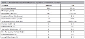
Table 1. Baseline characteristics of the study population (quantitative variables)
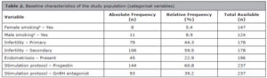
Table 2. Baseline characteristics of the study population (categorical variables)
a Female and male smoking status was defined according to documented tobacco use in the medical records, regardless of the duration of exposure.
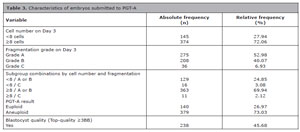
Table 3. Characteristics of embryos submitted to PGT-A
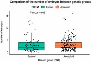
Figure 1. Embryo count per patient in each genetic group
Embryo Morphology and Genetic Outcomes
Figure 2 illustrates the distribution of cleavage-stage embryos according to Day-3 cell number and fragmentation grade. Embryos with ≥8 cells were significantly more likely to have lower fragmentation grades than those with <8 cells (χ2 test, p<0.0001). The odds of having ≥8 cells rather than <8 cells were approximately four times higher among embryos with better fragmentation grades (OR=3.94; 95% CI, 2.19-7.29). These findings indicate a strong association between a higher cell number and better morphological quality at the cleavage stage.
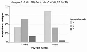
Figure 2. Distribution of embryos by fragmentation grade within each Day-3 cell-number category
In the logistic regression model evaluating the association between Day-3 subgroups and euploidy status, none of the subgroups differed significantly from the reference group (<8 cells with grade A or B morphology). ORs ranged from 0.44 to 0.79, and all p values were nonsignificant, indicating no association between Day-3 embryo morphology and the likelihood of euploidy (Table 4).

Table 4. Association between Day-3 embryo subgroups and euploidy status

Table 5. Association between Day-3 subgroups and top-quality blastocyst formation

Table 6. Mixed-effects logistic regression analysis of predictors of aneuploidy
DISCUSSION
The findings of this study support the use of standardized morphological grading systems to identify embryos with greater developmental potential (Gardner et al., 2000; Working Group on the update of the ESHRE/ALPHA Istanbul Consensus, 2025). All embryos were analyzed at a single fertility center under uniform laboratory conditions, using the same culture media and involving the same embryologists and clinicians in all procedures. This consistency strengthened embryo assessment and reduced variability. Furthermore, the study cohort included patients with diverse characteristics in terms of race, age, stimulation protocol, duration of infertility, and number of embryos per cycle.
The lack of an association between cleavage-stage morphology and euploidy is consistent with previous research showing that early developmental features are poor indicators of chromosomal competence (Munné et al., 2019; Desai et al., 2014). Early cleavage is influenced by cytoplasmic and metabolic factors that do not necessarily reflect meiotic or mitotic errors. Moreover, excluding embryos that failed to reach the blastocyst stage may have limited the ability to detect an association because slower-developing embryos, which often have greater fragmentation, are more likely to arrest and to be aneuploid.
No significant difference was found between the progestin-primed ovarian stimulation and GnRH antagonist protocols in terms of embryo yield or euploidy rate, consistent with previous studies demonstrating comparable efficacy between these regimens (Kuang et al., 2015; Huang et al., 2019b). This finding suggests that the type of stimulation does not compromise oocyte or embryo competence and supports the use of progestin-primed ovarian stimulation as a practical alternative that provides scheduling flexibility in IVF programs.
The main limitations of this study include its retrospective design, use of a convenience sample, and potential selection bias resulting from the exclusion of embryos that did not reach the blastocyst stage. Although standardized, morphological grading remains subjective and dependent on laboratory conditions. Nevertheless, the consistency of protocols and personnel at a single center strengthens the internal validity of the findings.
Clinically, these findings emphasize that embryo morphology, particularly at the blastocyst stage, remains a valuable marker of reproductive potential. In settings where PGT-A is unavailable or financially inaccessible, morphology can still guide embryo selection and patient counseling. Future prospective studies using time-lapse imaging and molecular biomarkers may help refine predictive models that combine morphological, kinetic, and genetic parameters.
CONCLUSION
In conclusion, Day-3 embryo morphology alone did not predict chromosomal status, whereas blastocyst quality was significantly associated with euploidy. Integrating morphological assessment with genetic testing may enhance embryo selection strategies and improve clinical outcomes in assisted reproduction.
REFERENCES
Alikani M, Cohen J, Tomkin G, Garrisi GJ, Mack C, Scott RT. Human embryo fragmentation in vitro and its implications for pregnancy and implantation. Fertil Steril. 1999;71:836-42. PMID: 10231042 DOI: 10.1016/S0015-0282(99)00092-8
Medline
Alpha Scientists in Reproductive Medicine and ESHRE Special Interest Group of Embryology. The Istanbul consensus workshop on embryo assessment: proceedings of an expert meeting. Hum Reprod. 2011;26:1270-83. PMID: 21502182 DOI: 10.1093/humrep/der037
Medline
Bilibio JP, Lorenzzoni PL, Oliveira BM, Nascimento FL, Meireles AJC, Nascimento FCD. Associations among morphological parameters, clinical factors and euploid blastocyst formation. JBRA Assist Reprod. 2022;26:199-207. PMID: 33899456 DOI: 10.5935/1518-0557.20210008
Medline
Bulletti FM, Sciorio R, Conforti A, De Luca R, Bulletti C, Palagiano A, Berrettini M, Scaravelli G, Pierson RA. Causes of embryo implantation failure: a systematic review and meta-analysis of procedures to increase embryo implantation potential. Front Endocrinol (Lausanne). 2025;15:1429193. PMID: 40028443 DOI: 10.3389/fendo.2024.1429193
Medline
Capalbo A, Rienzi L, Cimadomo D, Maggiulli R, Elliott T, Wright G, Nagy ZP, Ubaldi FM. Correlation between standard blastocyst morphology, euploidy and implantation: an observational study in two centers involving 956 screened blastocysts. Hum Reprod. 2014;29:1173-81. PMID: 24578475 DOI: 10.1093/humrep/deu033
Medline
della Ragione T, Verheyen G, Papanikolaou EG, Van Landuyt L, Devroey P, Van Steirteghem A. Developmental stage on day 5 and fragmentation rate on day 3 can influence the implantation potential of top-quality blastocysts in IVF cycles with single embryo transfer. Reprod Biol Endocrinol. 2007;5:2. PMID: 17257401 DOI: 10.1186/1477-7827-5-2
Medline
Desai N, Ploskonka S, Goodman LR, Austin C, Goldberg J, Falcone T. Analysis of embryo morphokinetics, multinucleation and cleavage anomalies using continuous time-lapse monitoring in blastocyst transfer cycles. Reprod Biol Endocrinol. 2014;12:54. PMID: 24951056 DOI: 10.1186/1477-7827-12-54
Medline
European IVF Monitoring Consortium (EIM) for the European Society of Human Reproduction and Embryology (ESHRE); Smeenk J, Wyns C, De Geyter C, Kupka M, Bergh C, Cuevas Saiz I, De Neubourg D, Rezabek K, Tandler-Schneider A, Rugescu I, Goossens V. ART in Europe, 2019: results generated from European registries by ESHRE†. Hum Reprod. 2023;38:2321-38. PMID: 37847771 DOI: 10.1093/humrep/dead197
Medline
Franasiak JM, Scott RT Jr. Embryonic aneuploidy: overcoming molecular genetics challenges improves outcomes and changes practice patterns. Trends Mol Med. 2014;20:499-508. PMID: 25113799 DOI: 10.1016/j.molmed.2014.06.006
Medline
Gardner DK, Lane M, Stevens J, Schlenker T, Schoolcraft WB. Blastocyst score affects implantation and pregnancy outcome: towards a single blastocyst transfer. Fertil Steril. 2000;73:1155-8. PMID: 10856474 DOI: 10.1016/S0015-0282(00)00518-5
Medline
Gleicher N, Orvieto R. Is the hypothesis of preimplantation genetic screening (PGS) still supportable? A review. J Ovarian Res. 2017;10:21. PMID: 28347334 DOI: 10.1186/s13048-017-0318-3
Medline
Huang L, Bogale B, Tang Y, Lu S, Xie XS, Racowsky C. Noninvasive preimplantation genetic testing for aneuploidy in spent medium may be more reliable than trophectoderm biopsy. Proc Natl Acad Sci U S A. 2019a;116:14105-12. PMID: 31235575 DOI: 10.1073/pnas.1907472116
Medline
Huang P, Tang M, Qin A. Progestin-primed ovarian stimulation is a feasible method for poor ovarian responders undergoing IVF/ICSI compared with a GnRH antagonist protocol: A retrospective study. J Gynecol Obstet Hum Reprod. 2019b;48:99-102. PMID: 30321608 DOI: 10.1016/j.jogoh.2018.10.008
Medline
Kong X, Yang S, Gong F, Lu C, Zhang S, Lu G, Lin G. The Relationship between Cell Number, Division Behavior and Developmental Potential of Cleavage Stage Human Embryos: A Time-Lapse Study. PLoS One. 2016;11:e0153697. PMID: 27077739 DOI: 10.1371/journal.pone.0153697
Medline
Kuang Y, Chen Q, Fu Y, Wang Y, Hong Q, Lyu Q, Ai A, Shoham Z. Medroxyprogesterone acetate is an effective oral alternative for preventing premature luteinizing hormone surges in women undergoing controlled ovarian hyperstimulation for in vitro fertilization. Fertil Steril. 2015;104:62-70.e3. PMID: 25956370 DOI: 10.1016/j.fertnstert.2015.03.022
Medline
Lee E, Illingworth P, Wilton L, Chambers GM. The clinical effectiveness of preimplantation genetic diagnosis for aneuploidy in all 24 chromosomes (PGD-A): systematic review. Hum Reprod. 2015;30:473-83. PMID: 25432917 DOI: 10.1093/humrep/deu303
Medline
Minasi MG, Colasante A, Riccio T, Ruberti A, Casciani V, Scarselli F, Spinella F, Fiorentino F, Varricchio MT, Greco E. Correlation between aneuploidy, standard morphology evaluation and morphokinetic development in 1730 biopsied blastocysts: a consecutive case series study. Hum Reprod. 2016;31:2245-54. PMID: 27591227 DOI: 10.1093/humrep/dew183
Medline
Munné S, Kaplan B, Frattarelli JL, Child T, Nakhuda G, Shamma FN, Silverberg K, Kalista T, Handyside AH, Katz-Jaffe M, Wells D, Gordon T, Stock-Myer S, Willman S; STAR Study Group. Preimplantation genetic testing for aneuploidy versus morphology as selection criteria for single frozen-thawed embryo transfer in good-prognosis patients: a multicenter randomized clinical trial. Fertil Steril. 2019;112:1071-9.e7. PMID: 31551155 DOI: 10.1016/j.fertnstert.2019.07.1346
Medline
Qiu P, Ye R, Li P, Huang H, Ding L. Effect of Day 3 cell number on the live birth rate of vitrified-warmed Day 5 single blastocyst transfer in young women. BMC Pregnancy Childbirth. 2024;24:289. PMID: 38641596 DOI: 10.1186/s12884-024-06468-1
Medline
Racowsky C, Jackson KV, Cekleniak NA, Fox JH, Hornstein MD, Ginsburg ES. The number of eight-cell embryos is a key determinant for selecting day 3 or day 5 transfer. Fertil Steril. 2000;73:558-64. PMID: 10689013 DOI: 10.1016/S0015-0282(99)00565-8
Medline
Scott RT Jr, Upham KM, Forman EJ, Zhao T, Treff NR. Cleavage-stage biopsy significantly impairs human embryonic implantation potential while blastocyst biopsy does not: a randomized and paired clinical trial. Fertil Steril. 2013;100:624-30. PMID: 23773313 DOI: 10.1016/j.fertnstert.2013.04.039
Medline
Stone BA, Greene J, Vargyas JM, Ringler GE, Marrs RP. Embryo fragmentation as a determinant of blastocyst development in vitro and pregnancy outcomes following embryo transfer. Am J Obstet Gynecol. 2005;192:2014-9; discussion 2019-20. PMID: 15970880. DOI: 10.1016/j.ajog.2005.02.048
Medline
Tognotti E. Infertilidade: da prática clínica à laboratorial. Barueri: Manole; 2014. Portuguese.
Van Royen E, Mangelschots K, De Neubourg D, Valkenburg M, Van de Meerssche M, Ryckaert G, Eestermans W, Gerris J. Characterization of a top-quality embryo, a step towards single-embryo transfer. Hum Reprod. 1999;14:2345-9. PMID: 10469708 DOI: 10.1093/humrep/14.9.2345
Medline
Wickham H. ggplot2: Elegant Graphics for Data Analysis. New York: Springer-Verlag; 2016.
Working Group on the update of the ESHRE/ALPHA Istanbul Consensus; Coticchio G, Ahlström A, Arroyo G, Balaban B, Campbell A, De Los Santos MJ, Ebner T, Gardner DK, Kovačič B, Lundin K, Magli MC, Mcheik S, Morbeck DE, Rienzi L, Sfontouris I, Vermeulen N, Alikani M. The Istanbul Consensus update: a revised ESHRE/ALPHA consensus on oocyte and embryo static and dynamic morphological assessment† ‡. Reprod Biomed Online. 2025;50:104955. PMID: 40300986 DOI: 10.1016/j.rbmo.2025.104955
Medline
Wu J, Zhang J, Kuang Y, Chen Q, Wang Y. The effect of Day 3 cell number on pregnancy outcomes in vitrified-thawed single blastocyst transfer cycles. Hum Reprod. 2020;35:2478-87. PMID: 32944763 DOI: 10.1093/humrep/deaa209
Medline
Zhan Q, Sierra ET, Malmsten J, Ye Z, Rosenwaks Z, Zaninovic N. Blastocyst score, a blastocyst quality ranking tool, is a predictor of blastocyst ploidy and implantation potential. F S Rep. 2020;1:133-41. PMID: 34223229 DOI: 10.1016/j.xfre.2020.05.004
Medline