JBRA Assisted Reproduction 2026;30(3):460-468
ORIGINAL ARTICLE
doi: 10.5935/1518-0557.20260030
Blastocyst Morphology in Oocyte Donors stimulated During the Late Follicular Phase with either rFSH:rLH or HP-hMG in a PPOS protocol: a Pilot Randomized Controlled Trial
1Grupo PRANOR, Lima, Peru
2Clínica Concebir, Lima, Peru
3Merck Healthcare KGaA, Darmstadt, Germany
CONFLICT OF INTERESTS
The authors have no competing interests to declare.
ABSTRACT
Objective: The aim of this study was to investigate whether there were differences in the number of good-quality blastocysts after 6 days of treatment with r-hFSH followed by late follicular phase treatment with r-hFSH:r-hLH in a 2:1 fixed ratio combination or HP-hMG in a PPOS protocol in a population of young women.
Methods: Healthy oocyte donors were randomized to Group 1 or Group 2 (40 women per group). All donors were treated with 200 mg/day oral progesterone from Day 1 and 225 IU/day r-hFSH for the first six days of ovarian stimulation. From Day 7 onwards, Group 1 received 225 IU/day r-hFHS:r-hLH 2:1 fixed ratio combination and Group 2 received 225 IU/day HP-hMG. Insemination was performed using intracytoplasmic sperm injection. Embryo quality was graded using the SART classification. The primary outcome was the number of blastocysts with good morphology (AA or AB).
Results: The mean (SD) number of good-quality blastocysts was significantly higher in Group 1 compared with Group 2 (3.4 [2.0] vs. 2.3 [2.0]; difference 0.9 (0.6 to 1.7); Kruskal-Wallis test p=0.0359). There was no difference in total gonadotropin dose, length of stimulation or number of oocytes retrieved between the groups.
Conclusion: Treatment with r-hFSH:r-hLH in a 2:1 fixed ratio combination starting in the late follicular phase resulted in a significantly higher number of good-quality blastocysts compared with HP-hMG in oocyte donors receiving PPOS.
Keywords: oocyte donor, progesterone-primed ovarian stimulation, PPOS, r-hFSH:r-hLH, HP-hMG
INTRODUCTION
In recent years, declining fertility rates has been observed worldwide; for example, in the USA, a 4% decrease in the number of live births was recorded between 2019 and 2020 (Eisenberg et al., 2021). Globally, the reasons for this decline are multifactorial, although delayed childbirth due to personal, professional or financial reasons may be a key contributing factor in developed countries (Mertes & Pennings, 2011). However, women who delay childbirth for whatever reason may face problems conceiving, owing to the age-related decline in fertility, as advanced maternal age (AMA) can impact oocyte and embryo competence through several mechanisms, with 35 years defined as the lowest age threshold to define AMA (Cimadomo et al., 2018).
Assisted reproductive technology (ART) may be an option for women who delay childbirth to achieve their desire to build a family; however, these women may still have a high chance of recurrent failed cycles (Bashiri et al., 2018) or low prognosis for live birth (Schmidt et al., 2012). In some cases, oocyte donation from healthy, fertile women who voluntarily undergo ovarian stimulation (OS) is an option for women who delay childbirth (Lutjen et al., 1984; Hammoud et al., 2009). While the legality and legislative framework for oocyte donation varies considerably among countries (Calhaz-Jorge et al., 2020; López et al., 2021), current indications for women seeking oocyte donation include hypergonadotropic hypogonadism, diminished ovarian reserve, having (or being a known or suspected carrier of) a significant genetic defect or a family history of a condition, poor oocyte and/or embryo quality or multiple previous failed attempts to conceive via ART. In addition, where permitted, oocyte donation may be an option for men who do not have a female partner or those who have a trans-female partner and are planning to use a gestational carrier (Lutjen et al., 1984). AMA is also an indication for women seeking oocyte donation, usually due to delayed childbirth and/or in conjunction with the above-mentioned indications (Cimadomo et al., 2018).
Several exogenous gonadotropin preparations are indicated for OS, including highly purified human menopausal gonadotropin (HP-hMG), which is purified from the urine of postmenopausal women and comprises FSH and LH-like activity derived from human chorionic gonadotropin (hCG), and recombinant human FSH (r-FSH) and LH (r-LH), which are produced using recombinant DNA technology (Lunenfeld et al., 2019). The recombinant products are available as separate products (EMA, 2022a; EMA, 2022b) or as a 2:1 fixed-ratio combination product (EMA, 2023). In the general population undergoing ART procedures, OS with r-hFSH:r-hLH 2:1 has been reported to yield a higher number of oocytes retrieved than HP-hMG (Pacchiarotti et al., 2010; Fábregues et al., 2013; Revelli et al., 2015; Kirshenbaum et al., 2021). However, there is little published evidence on oocyte quantity or oocyte quality following any specific treatment protocol in the context of oocyte donors. A recent systematic review and meta-analysis identified five studies that evaluated the effect of the type of gonadotropin used for OS in oocyte donors (Martinez et al., 2021). Three of these studies compared HP-FSH versus hMG (Söderström-Anttila et al., 1996), r-hFSH versus r-hFSH + hMG (Tesarik et al., 2002) or r-hFSH versus HP-hMG versus r-hFSH + HP-hMG (Melo et al., 2010), respectively, in a long agonist protocol; and two studies compared r-hFSH versus r-hFSH + r-hLH (Acevedo et al., 2006) or corifollitropin alfa versus hMG (Cruz et al., 2017), respectively, in a gonadotropin releasing hormone (GnRH) antagonist protocol. Although no difference in the number of oocytes retrieved was reported between the studies using GnRH antagonists versus agonists, no further conclusions could be drawn owing to the high degree of heterogeneity among the studies.
As an alternative to using GnRH analogs to prevent the premature luteinizing hormone (LH) surge during OS, recent work has focussed on the use of stimulation cycles using progesterone (progestin-primed OS [PPOS]) to prevent early luteinisation (La Marca & Capuzzo, 2019; Glujovsky et al., 2023). PPOS protocols are advantageous in that they can be administered orally, are flexible and economic for patients, and control over the LH surge is achieved rapidly (Kuang et al. 2015; Qin et al., 2016, Massin, 2017). These factors highlight the potential of PPOS to be a cost-effective and patient-friendly oral alternative to injected GnRH analogs in younger women, whose motivation for oocyte donation is usually solely altruistic and, therefore, presents unique challenges for physicians when balancing the risk and benefits to ensure the wellbeing and convenience of the donor (Kool et al., 2018).
Recent studies have demonstrated few or no differences between PPOS and a GnRH antagonist protocols in oocyte quality (Yildiz et al., 2019) or competence (Vaiarelli et al., 2024), oocyte and MII oocyte number and number of cleavage and blastocyst embryos (Duc Thang et al., 2024), oocyte maturation rates and trigger-to-oocyte retrieval interval (Ata et al., 2024). However, there is no consensus on which populations of women may most benefit from PPOS (e.g., poor responders, women with endometriosis, women of advanced maternal age, young women for oocyte donation procedures) (Glujovsky et al., 2023). Furthermore, there are no robust data on any potential interaction between supraphysiological levels of progesterone in PPOS and the different LH-like activity in the commonly used preparations, and the effect this may have on oocyte quality.
The objective of the present study was to investigate the number of good-quality blastocysts obtained from population of young women treated in the late follicular phase with r-FSH:r-LH in a 2:1 fixed ratio combination or HP-hMG, following 6 days of treatment with r-FSH in a PPOS protocol in a population of young women.
MATERIALS AND METHODS
Study design
The aim of this pilot randomized controlled trial was to compare the blastocyst morphology of donor oocytes following OS with r-FSH:r-LH in a 2:1 fixed ratio combination (Pergoveris; Merck KGaA, Darmstadt, Germany) or HP-hMG (Menopur; Ferring, West Drayton, UK), both starting from stimulation Day 7 following 6 days of stimulation with r-FSH alfa (Gonal-f; Merck KGaA, Darmstadt, Germany). The study was conducted from August 2022 to June 2023 at a single centre in Lima, Peru. The study design is shown in Figure 1.
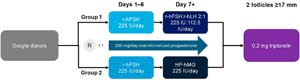
Figure 1. Study design.
Eligibility of gamete donors
Women undergoing OS for oocyte donation were screened under informed consent for inclusion and exclusion criteria through blood testing, and general medical evaluation.
Women were eligible as oocyte donors if they met all the following criteria: age 19-30 years at the time of providing informed consent; recorded levels of AMH ≥2 ng/mL, and BMI 21.0-29.9 kg/m2. All donors underwent physical, gynecological and psychological examinations. Additionally, they should be no family history of hereditary diseases or chromosomal disorders. All participants had a normal karyotype, and tested negative for sexually transmitted diseases and toxicological screening, and had an up-to-date negative cervical cancer screening. Recruitment of oocyte donors was done based on recommendations given by other donors and the reason for gamete donation was solely altruistic.
Sperm was obtained from the partners of the oocyte recipients (homologous) or from sperm donors (heterologous) in the case of poor semen quality (Boitrelle et al., 2021). Men aged 21-50 years were eligible as sperm donors if they met the criteria for donation at the treating clinic or another certified cryobank. Sperm quality was assessed according to semen volume, and sperm motility, quantity and morphology.
Randomization
Participants were randomized (1:1) using a computer-generated algorithm to either Group 1 (r-hFSH:r-hLH 2:1 fixed ratio combination) or Group 2 (HP-hMG) before receiving the first injection for OS. No blinding was performed.
Interventions
All donors were stimulated with 225 IU/day r-FSH (Gonal-f, Merck KGaA, Darmstadt, Germany) for the first six days. All donors received 200 mg/day oral micronized progesterone from Day 1 of stimulation (PPOS). From Day 7 onwards, oocyte donors in Group 1 were treated with 225 IU/day r-hFHS:r-hLH 2:1 fixed ratio combination (150 IU r-FSH: 75 IU r-hLH), and oocyte donors in Group 2 were treated with 225 IU/day HP-hMG (Figure 1).Follicular development was monitored by serial ultrasound scans. The first scan was conducted at the beginning of the cycle, with the second scan after 5 days of gonadotropin treatment, and then every 2 days until follicle size reached 16 mm; a final scan was conducted on the day of ovarian maturation trigger, when two follicles had reached a mean diameter ≥17 mm. Oocyte maturation was triggered using 0.2 mg triptorelin, with oocyte pick-up at 34-36 hours after triggering.
Insemination
Homologous sperm was obtained from the recipient’s partner in accordance with clinic protocols. Heterologous sperm was collected from the cryobank local to the treating centre or from another certified cryobank, and then transferred to the clinic as a frozen specimen in accordance with clinic standards. All inseminations were performed using intracytoplasmic sperm injection (ICSI).
Embryo culture and implantation
Zygotes were cultured individually until day 5/6 in drops of 25 μL of Global Total® media (Lifeglobal, Canada) under mineral oil at 37 °C in 7% CO2, 5% O2 and 88% N2 (adapted from Sepúlveda et al., 2009). Embryos were classified according to the Society for Assisted Reproductive Technology (SART) classification (Racowsky et al., 2010).
Embryo vitrification
Embryos were vitrified using a modified Cryotop method (Kuwayama et al., 2005). Embryos were equilibrated at room temperature for 12 minutes in the equilibrium solution (TCM-199 medium/20% synthetic serum substitute/7.5% (v/v) ethylene glycol/7.5% dimethyl sulfoxide). Embryos were then transferred to the vitrification solution (TCM-199 medium/20% synthetic serum substitute/15% ethylene glycol/15% dimethyl sulfoxide/0.5 M sucrose) and incubated for 1 minute before being placed in a cryoslip (one embryo per strip) and immediately submerged in liquid nitrogen.
Outcomes
Primary outcome
The primary outcome was the number of blastocysts with good morphology. All embryos were evaluated by two senior embryologists using the SART classification (Racowsky et al., 2010) and categorized as either good, fair or poor: a good grading was assigned to embryos with Grade A inner cell mass and trophectoderm Grade A or B (AA or AB blastocysts); a fair grading was assigned to embryos with Grade B inner cell mass and trophectoderm Grade A, B or C (BB, BC or BA blastocysts); a poor grading was assigned to embryos with Grade C inner cell mass (CC or CB blastocysts).
Secondary outcomes
Secondary outcomes were the total number of oocytes recovered and inseminated, the number of 2PN embryos, the total number of blastocysts, and the proportion of good-quality blastocysts, calculated as the proportion of embryos to be of good quality over all embryos assessed.
Data collection/monitoring
All donors were monitored for adverse events during OS, and were asked to self-report any adverse events occurring within two weeks after oocyte retrieval. The potential risks associated with the study were those common to all ART procedures: the risks of OS were ovarian hyperstimulation syndrome (OHSS) and ovarian torsion; the risks of egg retrieval were bleeding, risks associated with anaesthesia, infection, and trauma.
Monitoring involved regular transvaginal ultrasound scans and hormonal blood tests to monitor follicular development and ovarian response. Ultrasound scans were performed as previously described. Donors were scheduled for a follow-up clinical evaluation throughout the two weeks after oocyte retrieval to assess recovery and detect any delayed adverse events. Any adverse events reported by donors were to be documented in real time by the clinical team and coded using the Medical Dictionary for Regulatory Activities (MedDRA). The data collection process adhered to established clinical protocols and ethical guidelines, ensuring donor safety and compliance with research standards.
Statistical analysis
We estimated that 80 participants would be required to obtain 80% power to demonstrate a difference of at least one blastocyst in the number of good-morphology blastocysts, assuming a mean number of blastocysts in the HP-hMG group of 4.9, based on our previous observations and assuming a loss rate of 10%. Between-group differences in blastocyst morphology were assessed using the Kruskal-Wallis test. All other analyses were descriptive.
Ethical approval
This study protocol was approved by the local Ethics Committee. All participants provided informed consent after counselling. Consent was obtained by the investigator or a member of their team directly from the participants before that subject takes part in the study. Consent was documented either with a handwritten signature on paper or through an electronic signature following the Guidance on Use of Electronic Informed Consent, according to the patient’s preference. All participants received a copy of the Informed Consent Form.
RESULTS
Participants
Oocyte donors
Of the 80 donors recruited, seventy-nine were included in the analysis. One donor in Group 1 was excluded after their OS was cancelled. The baseline characteristics were similar between the women randomly assigned to either group (Table 1).
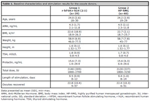
Table 1. Baseline characteristics and stimulation results for the oocyte donors.
Sperm donors
The mean paternal age was 36 (SD 10) years in Group 1 and 37 (SD 12) years in Group 2. Most semen samples were homologous (Group 1: 72.0%; Group 2: 27.7%) compared with heterologous (Group 1: 28.8%; Group 2: 27.8%) and the highest proportion of inseminations used fresh semen (Group 1: 48.4%; Group 2: 53.3%) rather than frozen semen (Group 1: 51.6%; Group 2: 46.7%).
Ovarian stimulation
The total FSH dose was 2140 (SD 205) IU in Group 1 and 2120 (SD 227) IU in Group 2. The length of stimulation was 9.5 (SD 0.9) days in Group 1 and 9.4 (SD 1.0) days in Group 2 (Table 1). The mean number of oocytes retrieved was 22.8 (SD 10.4) in Group 1 and 22.4 (SD 10.3) in Group 2 (Table 1).
Outcomes
Primary outcome
Overall, 97 embryo transfers were registered, 50 in Group A and 47 in Group B. The mean (SD) number of good-quality blastocysts was statistically significantly higher in the women in Group 1 compared with those in Group 2 (3.4 [2.0] vs. 2.3 [2.0]; difference 0.9 [95% CI 0.6 to 1.7]; Kruskal-Wallis test p=0.0359) (Table 2). There were no differences in the number of fair-quality (0.4 [95%CI -0.18 to 1.0]) or poor-quality (0.5 [95% CI -0.4 to 0.3]) blastocysts between the treatment groups.
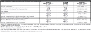
Table 2. Results of ovarian stimulation per stimulation cycle following ICSI
Secondary outcomes
Group 1 had a higher total number of blastocysts compared with Group 2 (difference 1.4 [95% CI 0.3 to 2.4]) (Table 2). There were no differences in the total number of oocytes inseminated (difference 0.3 [95% CI -0.4 to 1.0]), the total number of 2PN embryos (difference 0.2 [95% CI -0.7 to 1.1]), or the proportion of good-quality blastocysts (difference 8.2% [95% CI -20.0% to 25.6%]) between treatment groups (Table 2). Finally, ongoing pregnancy rates were similar among the group (62.5% for Group 1 and 58.9% for Group).
Safety
No adverse events were identified during the study. There were no reports of OHSS, ovarian torsion or other procedure-related complications.
DISCUSSION
The results of this randomized pilot study show that OS with r-FSH:r-LH in a 2:1 fixed ratio combination from Day 7 resulted in a significantly higher number of good-quality blastocysts compared with HP-hMG from Day 7 in oocyte donors using a PPOS protocol to prevent premature LH surge. These preliminary results using PPOS in healthy, young women show a potentially lower oocyte developmental competence when urinary gonadotrophins are used in combination with follicular phase progesterone in this population.
Premature LH surge can compromise oocyte yield in in-vitro fertilization (IVF)/ICSI and is one of the major reasons for cycle cancellation in OS (Messinis, 2006). However, the use of GnRH analogs (either GnRH agonists or GnRH antagonists (Macklon et al., 2006)) have recognised drawbacks. GnRH agonists increase the risk of OHSS through the requirement for hCG triggering, whereas GnRH antagonists can have variable effectiveness in some patient populations, including older patients and those with diminished ovarian reserve (Bosch et al., 2003; Reichman et al., 2014). Furthermore, both GnRH agonists and antagonists are injectable products, which increases the cost, complexity and patient burden of OS.
Studies using progesterone in the follicular phase have shown that it is a regulator of ovulation timing and a potentially convenient and flexible oral alternative to GnRH agonists for preventing early luteinization when used in a PPOS protocol in freeze-all cycles (Kuang et al., 2015). Most of the studies on PPOS published to date have compared the outcomes GnRH analog protocols and PPOS using locally available preparations of hMG for OS, although there are a few studies that directly compare the outcomes from different gonadotropin preparations. In a retrospective study covering two discrete time periods (2019 and 2020), during which two different OS regimens were used (2019: corifollitropin, recombinant FSH and GnRH antagonists, with ovulation triggered using GnRH agonist; 2020: PPOS protocol with human menopausal gonadotropin [hMG] and dual trigger [GnRH agonist and low dose hCG]), there was no difference in the ratio of the number of mature oocytes and serum AMH between the two periods (4.0 [2.3-7.1] vs. 4.0 [2.7-6.8]; p=0.80), although the total cost of drugs used for OS was lower in the second period €940 [€774-€1,096] vs. €520 [€434-€564]; p<0.001) (Filippi et al., 2023). In addition, a retrospective study in women who were stimulated with r-FSH (Gonal-f, Merck KGaA, Germany) or HP-hMG (Merional, IBSA Institut, Switzerland) in a PPOS protocol (Dydrogesterone [Duphaston® 10 mg, Abbott, Turkey]) reported that the total number of follicles, oocytes, MII and 2PN oocytes, fertilization rates and pregnancy rates were similar between the two groups, although higher doses of gonadotropin were used in the HP-hMG group compared with the r-FSH group (p=0.001) (Yetkinel et al., 2024). While these early results may demonstrate the promise of using PPOS cycles, they do not address any potential differences in the gonadotropin preparations that are commonly used in OS, specifically the FSH and LH activity in r-FSH:r-hLH 2:1, which are produced using complementary DNA technology and have a high level of purity and batch-to-batch consistency (Bassett & Driebergen, 2005; Lispi et al., 2006; Bassett et al., 2009; Lispi et al., 2023) or the LH-like activity in HP-hMG, which is mainly derived from hCG purified from pregnant women (Capolupo et al., 2024). Furthermore, there are no studies that investigate how these components may interact with the supraphysiological concentrations of progesterone used in PPOS.
Although LH and hCG share approximately 85% structural identity and bind to the same receptor (LHCGR) on the antral follicles, they cannot be considered equivalent, as they interact differently with the receptor and activate different cellular pathways (Casarini et al., 2018). The action of LH is preferentially exerted through phosphorylated extracellular-regulated kinase 1/2 (pERK1/2) and phosphorylated AKT (also known as protein kinase B), resulting in proliferative/antiapoptotic signals and partial agonism of progesterone production in vitro. In contrast, hCG displays notable cAMP/protein kinase A (PKA)-mediated steroidogenic and proapoptotic potential (Casarini et al., 2018). Therefore, on the one hand, r-hLH may have a positive effect on oocyte maturation, potentiated by r-FSH, as well as having an anti-apoptotic effect on cumulus cells and promoting the paracrine signalling required for cell expansion and oocyte signalling during folliculogenesis (Ruvolo et al., 2007; Huang et al., 2015). On the other hand, the hCG in HP-hMG may have a detrimental effect on oocyte development by activating the steroidogenic and proapoptotic pathways.
Progesterone contributes to normal mammalian ovarian function and has several critical functions during embryo development and implantation, including endometrial receptivity, embryonic survival during gestation, and transformation of the endometrial stromal cells to decidual cells (Salehnia & Zavareh, 2013). While hCG has greater steroidogenic potential than LH, reflecting its role during pregnancy to support increased progesterone production, the dual proliferative and androgen-stimulating roles exerted by LH in granulosa and theca cells, respectively, during folliculogenesis might not require induction of the steroidogenic pathway at the levels needed in pregnancy (Casarini et al., 2018). Furthermore, high levels of progesterone, such as the supraphysiological levels due to PPOS, may even have local doseand species-dependent effects on the developing oocyte (diZerega & Hodgen, 1982; Kim & Greenwald, 1987; Setty & Mills, 1987; Komatsu & Masubuchi, 2017). In a retrospective analysis of 1200 cycles of women stimulated with hMG in either a PPOS or GnRH antagonist protocol, at similar cumulative hMG doses, the number of follicles with diameter >10mm, the number of retrieved oocytes and the number of MII oocytes were lower in the PPOS protocol compared with the antagonist protocol, although the oocyte yields were rescued by higher doses of hMG (Long et al., 2021). These exploratory results we report here add weight to these preliminary data on the potential detrimental effect of the LH-like activity of hCG in conjunction with high levels of progesterone from PPOS, although this needs further investigation.
While we report a higher number of good-quality blastocysts, we did not observe a difference in the number of oocytes retrieved, which is contrary to the results of other studies that compare these two formulations. In a meta-analysis of six studies of OS with r-FSH:r-hLH 2:1 fixed ratio combination versus HP-hMG, a higher mean number of oocytes was retrieved in the rFSH:r-LH 2:1 fixed ratio combination cohort (1.43 [95% CI 0.18, 2.69]; p=0.025) (Dahan et al., 2026). However, the studies analysed reported on women undergoing routine IVF cycles using autologous oocytes, and there are few published data on the use of these treatments in donor cycles. In one study of donor cycles, treatment with r-FSH:r-hLH in a 2:1 ratio also resulted in a higher number of oocytes retrieved (16.5 [SD 4.1] vs. 11.8 [SD 2.6]; p=0.049) compared with treatment with urinary gonadotropins (HP-hMG plus uFSH) (Requena et al., 2014). This variance from the published literature warrants further investigation into the outcomes of different OS protocols specifically in donor cycles.
The strengths of this trial are it was a randomized pilot trial conducted at a single centre that recruited a representative population of women participating in oocyte donation. Some limitations also have to be acknowledged. Randomized treatment only commenced after stimulation Day 6, prior to which both cohorts were treated with r-FSH; therefore, the effect of the test compounds from Day 1 of stimulation could not be addressed, and no sperm DNA fragmentation was performed, also comparing the outcome to stimulation with rFSH alone.
CONCLUSIONS
In this randomized controlled pilot study we show that late follicular phase treatment with r-FSH:r-LH in a 2:1 fixed ratio combination resulted in a significantly higher number of good-quality blastocysts compared with late follicular phase HP-hMG following OS with r-FSH until Day 6 in oocyte donors. While these data need to be confirmed in larger clinical studies, optimizing oocyte quality in PPOS cycles may improve the chances of live birth resulting from a single cycle, thus reducing the burden on oocyte donors.
Funding statement: This analysis was funded by Merck (CrossRef Funder ID: 10.13039/100009945).
REFERENCES
Acevedo B, Gomez-Palomares JL, Ricciarelli E, Hernández ER. Triggering ovulation with gonadotropin-releasing hormone agonists does not compromise embryo implantation rates. Fertil Steril. 2006;86:1682-7. PMID: 17074344 DOI: 10.1016/j.fertnstert.2006.05.049 Medline
Ata B, Cakar A, Türkgeldi E, Yildiz S, Keles İ, Kalafat E. Should the trigger to oocyte retrieval interval be different in progestin-primed ovarian stimulation cycles? Reprod Biomed Online. 2024;48:103626. PMID: 38160486 DOI: 10.1016/j.rbmo.2023 Medline
Bashiri A, Halper KI, Orvieto R. Recurrent Implantation Failure-update overview on etiology, diagnosis, treatment and future directions. Reprod Biol Endocrinol. 2018;16:121. PMID: 30518389 DOI: 10.1186/s12958-018-0414-2 Medline
Bassett R, Lispi M, Ceccarelli D, Grimaldi L, Mancinelli M, Martelli F, Van Dorsselaer A. Analytical identification of additional impurities in urinary-derived gonadotrophins. Reprod Biomed Online. 2009;19:300-13. PMID: 19778474 DOI: 10.1016/s1472-6483(10)60163-0 Medline
Bassett RM, Driebergen R. Continued improvements in the quality and consistency of follitropin alfa, recombinant human FSH. Reprod Biomed Online. 2005;10:169-77. PMID: 15823219 DOI: 10.1016/s1472-6483(10)60937-6 Medline
Boitrelle F, Shah R, Saleh R, Henkel R, Kandil H, Chung E, Vogiatzi P, Zini A, Arafa M, Agarwal A. The Sixth Edition of the WHO Manual for Human Semen Analysis: A Critical Review and SWOT Analysis. Life (Basel). 2021;11:1368. PMID: 34947899 DOI: 10.3390/life11121368 Medline
Bosch E, Valencia I, Escudero E, Crespo J, Simón C, Remohí J, Pellicer A. Premature luteinization during gonadotropin-releasing hormone antagonist cycles and its relationship with in vitro fertilization outcome. Fertil Steril. 2003;80:1444-9. PMID: 14667881 DOI: 10.1016/j.fertnstert.2003.07.002 Medline
Calhaz-Jorge C, De Geyter CH, Kupka MS, Wyns C, Mocanu E, Motrenko T, Scaravelli G, Smeenk J, Vidakovic S, Goossens V. Survey on ART and IUI: legislation, regulation, funding and registries in European countries: The European IVF-monitoring Consortium (EIM) for the European Society of Human Reproduction and Embryology (ESHRE). Hum Reprod Open. 2020;2020:hoz044. PMID: 32042927 DOI: 10.1093/hropen/hoz044 Medline
Capolupo A, Petrocchi S, Melchiorre M, Jonas K, D’Hooghe T, Hanyaloglu A, Sunkara S, Palmese A, Ozgumus B, Amoresano A, Angiuoni G, Montenegro S, Simone P, Lispi M. Analytical Investigation of the Profile of Human Chorionic Gonadotropin in Highly Purified Human Menopausal Gonadotrophin Preparations. Int J Mol Sci. 2024;25:9405. PMID: 39273352 DOI: 10.3390/ijms25179405 Medline
Casarini L, Santi D, Brigante G, Simoni M. Two Hormones for One Receptor: Evolution, Biochemistry, Actions, and Pathophysiology of LH and hCG. Endocr Rev. 2018;39:549-92. PMID: 29905829 DOI: 10.1210/er.2018-00065 Medline
Cimadomo D, Fabozzi G, Vaiarelli A, Ubaldi N, Ubaldi FM, Rienzi L. Impact of Maternal Age on Oocyte and Embryo Competence. Front Endocrinol (Lausanne). 2018;9:327. PMID: 30008696 DOI: 10.3389/fendo.2018.00327 Medline
Cruz M, Alamá P, Muñoz M, Collado D, Blanes C, Solbes E, Requena A. Economic impact of ovarian stimulation with corifollitropin alfa versus conventional daily gonadotropins in oocyte donors: a randomized study. Reprod Biomed Online. 2017;34:605-10. PMID: 28366518 DOI: 10.1016/j.rbmo.2017.03.003 Medline
Dahan M, Schwarze JE, Gupta SS, Hayward B, Fischer R, Esteves SC, Chen MJ, Silverberg KM, Guzmán L, Santi D, Feferkorn I, D’Hooghe TM, Alviggi C. Comparative effectiveness of r-hFSH:r-hLH in a 2:1 ratio versus HP-hMG alone in ovarian stimulation for medically assisted reproduction treatment using IVF/ICSI: A systematic review and meta-analysis. Gynecol Obstet Invest. 2026:1-25. PMID: 41729769 DOI: 10.1159/000550420 Medline
diZerega GS, Hodgen GD. The interovarian progesterone gradient: a spatial and temporal regulator of folliculogenesis in the primate ovarian cycle. J Clin Endocrinol Metab. 1982;54:495-9. PMID: 6799537 DOI: 10.1210/jcem-54-3-495 Medline
Duc Thang L, Bao Long H, Thi Thu Trang D, Ngoc Quy P, Thi Mai Phuong G, Thi Hanh B, Trong Thach T, Thi Lien Huong N, Hoang L, Hugues JN.Non-inferiority of progestin-primed ovarian stimulation versus GnRH antagonist protocol: A propensity score-weighted analysis. J Formos Med Assoc. 2024;123:523-9. PMID: 38092654 DOI: 10.1016/j.jfma.2023.12.008 Medline
Eisenberg ML, Thoma ME, Li S, McLain AC. Trends in time-to-pregnancy in the USA: 2002 to 2017. Hum Reprod. 2021;36:2331-8. PMID: 34021350 DOI: 10.1093/humrep/deab107 Medline
Fábregues F, Creus M, Casals G, Carmona F, Balasch J. Outcome from consecutive ICSI cycles in patients treated with recombinant human LH and those supplemented with urinary hCG-based LH activity during controlled ovarian stimulation in the long GnRH-agonist protocol. Gynecol Endocrinol. 2013;29:430-5. PMID: 23350573 DOI: 10.3109/09513590.2012.754873 Medline
Filippi F, Reschini M, Polledri E, Cecchele A, Guarneri C, Vigano P, Fustinoni S, Platteau P, Somigliana E. Progestin-primed ovarian stimulation for fertility preservation in women with cancer: A comparative study. PLoS One. 2023;18:e0280238. PMID: 36976781 DOI: 10.1371/journal.pone.0280238 Medline
Glujovsky D, Pesce R, Miguens M, Sueldo C, Ciapponi A. Progestogens for prevention of luteinising hormone (LH) surge in women undergoing controlled ovarian hyperstimulation as part of an assisted reproductive technology (ART) cycle. Cochrane Database Syst Rev. 2023;11:CD013827. PMID: 38032057 DOI: 10.1002/14651858.CD013827 Medline
Hammoud AO, Gibson M, Stanford J, White G, Carrell DT, Peterson M. In vitro fertilization availability and utilization in the United States: a study of demographic, social, and economic factors. Fertil Steril. 2009;91:1630-5. PMID: 18539275 DOI: 10.1016/j.fertnstert.2007.10.038 Medline
Huang Y, Zhao Y, Yu Y, Li R, Lin S, Zhang C, Liu P, Qiao J. Altered amphiregulin expression induced by diverse luteinizing hormone receptor reactivity in granulosa cells affects IVF outcomes. Reprod Biomed Online. 2015;30:593-601. PMID: 25911599 DOI: 10.1016/j.rbmo.2015.03.001 Medline
Kim I, Greenwald GS. Stimulatory and inhibitory effects of progesterone on follicular development in the hypophysectomized follicle-stimulating hormone/luteinizing hormone-treated hamster. Biol Reprod. 1987;36:270-6. PMID: 3107623 DOI: 10.1095/biolreprod36.2.270 Medline
Kirshenbaum M, Gil O, Haas J, Nahum R, Zilberberg E, Lebovitz O, Orvieto R. Recombinant follicular stimulating hormone plus recombinant luteinizing hormone versus human menopausal gonadotropinsdoes the source of LH bioactivity affect ovarian stimulation outcome? Reprod Biol Endocrinol. 2021;19:182. PMID: 34886872 DOI: 10.1186/s12958-021-00853-7 Medline
Komatsu K, Masubuchi S. The concentration-dependent effect of progesterone on follicle growth in the mouse ovary. J Reprod Dev. 2017;63:271-7. PMID: 28321005 DOI: 10.1262/jrd.2016-154 Medline
Kool EM, Bos AME, van der Graaf R, Fauser BCJM, Bredenoord AL. Ethics of oocyte banking for third-party assisted reproduction: a systematic review. Hum Reprod Update. 2018;24:615-35. PMID: 29762669 DOI: 10.1093/humupd/dmy016 Medline
Kuang Y, Chen Q, Fu Y, Wang Y, Hong Q, Lyu Q, Ai A, Shoham Z. Medroxyprogesterone acetate is an effective oral alternative for preventing premature luteinizing hormone surges in women undergoing controlled ovarian hyperstimulation for in vitro fertilization. Fertil Steril. 2015;104:62-70.e3. PMID: 25956370 DOI: 10.1016/j.fertnstert.2015.03.022 Medline
Kuwayama M, Vajta G, Kato O, Leibo SP. Highly efficient vitrification method for cryopreservation of human oocytes. Reprod Biomed Online. 2005;11:300-8. PMID: 16176668 DOI: 10.1016/s1472-6483(10)60837-1 Medline
La Marca A, Capuzzo M. Use of progestins to inhibit spontaneous ovulation during ovarian stimulation: the beginning of a new era? Reprod Biomed Online. 2019;39:321-31. PMID: 31138494 DOI: 10.1016/j.rbmo.2019.03.212 Medline
Lispi M, Bassett R, Crisci C, Mancinelli M, Martelli F, Ceccarelli D, De Bellis C, Mendola D. Comparative assessment of the consistency and quality of a highly purified FSH extracted from human urine (urofollitropin) and a recombinant human FSH (follitropin alpha). Reprod Biomed Online. 2006;13:179-93. PMID: 16895630 DOI: 10.1016/s1472-6483(10)60613-x Medline
Lispi M, Humaidan P, Bousfield GR, D’Hooghe T, Ulloa-Aguirre A. Follicle-Stimulating Hormone Biological Products: Does Potency Predict Clinical Efficacy? Int J Mol Sci. 2023;24:9020. PMID: 37240364 DOI: 10.3390/ijms24109020 Medline
Long H, Yu W, Yu S, Yin M, Wu L, Chen Q, Cai R, Suo L, Wang L, Lyu Q, Kuang Y. Progesterone affects clinic oocyte yields by coordinating with follicle stimulating hormone via PI3K/AKT and MAPK pathways. J Adv Res. 2021;33:189-99.PMID: 34603789 DOI: 10.1016/j.jare.2021.02.008 Medline
López A, Betancourt M, Casas E, Retana-Márquez S, Juárez-Rojas L, Casillas F. The need for regulation in the practice of human assisted reproduction in Mexico. An overview of the regulations in the rest of the world. Reprod Health. 2021;18:241. PMID: 34838077 DOI: 10.1186/s12978-021-01293-7 Medline
Lunenfeld B, Bilger W, Longobardi S, Alam V, D’Hooghe T, Sunkara SK. The Development of Gonadotropins for Clinical Use in the Treatment of Infertility. Front Endocrinol (Lausanne). 2019;10:429. PMID: 31333582 DOI: 10.3389/fendo.2019.00429 Medline
Lutjen P, Trounson A, Leeton J, Findlay J, Wood C, Renou P. The establishment and maintenance of pregnancy using in vitro fertilization and embryo donation in a patient with primary ovarian failure. Nature. 1984;307:174-5. PMID: 6690997 DOI: 10.1038/307174a0 Medline
Macklon NS, Stouffer RL, Giudice LC, Fauser BC. The science behind 25 years of ovarian stimulation for in vitro fertilization. Endocr Rev. 2006;27:170-207. PMID: 16434510 DOI: 10.1210/er.2005-0015 Medline
Martinez F, Racca A, Rodríguez I, Polyzos NP. Ovarian stimulation for oocyte donation: a systematic review and meta-analysis. Hum Reprod Update. 2021;27:673-96. PMID: 33742206 DOI: 10.1093/humupd/dmab008 Medline
Massin N. New stimulation regimens: endogenous and exogenous progesterone use to block the LH surge during ovarian stimulation for IVF. Hum Reprod Update. 2017;23:211-20. PMID: 28062551 DOI: 10.1093/humupd/dmw047 Medline
Melo M, Bellver J, Garrido N, Meseguer M, Pellicer A, Remohí J. A prospective, randomized, controlled trial comparing three different gonadotropin regimens in oocyte donors: ovarian response, in vitro fertilization outcome, and analysis of cost minimization. Fertil Steril. 2010;94:958-64. PMID: 19931075 DOI: 10.1016/j.fertnstert.2009.05.005 Medline
Messinis IE. Ovarian feedback, mechanism of action and possible clinical implications. Hum Reprod Update. 2006;12:557-71. PMID: 16672246 DOI: 10.1093/humupd/dml020 Medline
Pacchiarotti A, Sbracia M, Frega A, Selman H, Rinaldi L, Pacchiarotti A. Urinary hMG (Meropur) versus recombinant FSH plus recombinant LH (Pergoveris) in IVF: a multicenter, prospective, randomized controlled trial. Fertil Steril. 2010;94:2467-9. PMID: 20537626 DOI: 10.1016/j.fertnstert.2010.04.035 Medline
Qin N, Chen Q, Hong Q, Cai R, Gao H, Wang Y, Sun L, Zhang S, Guo H, Fu Y, Ai A, Tian H, Lyu Q, Daya S, Kuang Y. Flexibility in starting ovarian stimulation at different phases of the menstrual cycle for treatment of infertile women with the use of in vitro fertilization or intracytoplasmic sperm injection. Fertil Steril. 2016;106:334-41.e1. PMID: 27114329 DOI: 10.1016/j.fertnstert.2016.04.006 Medline
Racowsky C, Vernon M, Mayer J, Ball GD, Behr B, Pomeroy KO, Wininger D, Gibbons W, Conaghan J, Stern JE. Standardization of grading embryo morphology. J Assist Reprod Genet. 2010;27:437-9. DOI: PMID: 20532975 10.1007/s10815-010-9443-2 Medline
Reichman DE, Zakarin L, Chao K, Meyer L, Davis OK, Rosenwaks Z. Diminished ovarian reserve is the predominant risk factor for gonadotropin-releasing hormone antagonist failure resulting in breakthrough luteinizing hormone surges in in vitro fertilization cycles. Fertil Steril. 2014;102:99-102. PMID: 24882557 DOI: 10.1016/j.fertnstert.2014.04.010 Medline
Requena A, Cruz M, Ruiz FJ, García-Velasco JA. Endocrine profile following stimulation with recombinant follicle stimulating hormone and luteinizing hormone versus highly purified human menopausal gonadotropin. Reprod Biol Endocrinol. 2014;12:10. PMID: 24476504 DOI: 10.1186/1477-7827-12-10 Medline
Revelli A, Pettinau G, Basso G, Carosso A, Ferrero A, Dallan C, Canosa S, Gennarelli G, Guidetti D, Filippini C, Benedetto C. Controlled Ovarian Stimulation with recombinant-FSH plus recombinant-LH vs. human Menopausal Gonadotropin based on the number of retrieved oocytes: results from a routine clinical practice in a real-life population. Reprod Biol Endocrinol. 2015;13:77. PMID: 26209525 DOI: 10.1186/s12958-015-0080-6 Medline
Ruvolo G, Bosco L, Pane A, Morici G, Cittadini E, Roccheri MC. Lower apoptosis rate in human cumulus cells after administration of recombinant luteinizing hormone to women undergoing ovarian stimulation for in vitro fertilization procedures. Fertil Steril. 2007;87:542-6. PMID: 17126339 DOI: 10.1016/j.fertnstert.2006.06.059 Medline
Salehnia M, Zavareh S. The effects of progesterone on oocyte maturation and embryo development. Int J Fertil Steril. 2013;7:74-81. PMID: 24520467. Medline
Schmidt L, Sobotka T, Bentzen JG, Nyboe Andersen A; ESHRE Reproduction and Society Task Force. Demographic and medical consequences of the postponement of parenthood. Hum Reprod Update. 2012;18:29-43. PMID: 21989171 DOI: 10.1093/humupd/dmr040 Medline
Sepúlveda S, Garcia J, Arriaga E, Diaz J, Noriega-Portella L, Noriega-Hoces L. In vitro development and pregnancy outcomes for human embryos cultured in either a single medium or in a sequential media system. Fertil Steril. 2009;91:1765-70. PMID: 18439587 DOI: 10.1016/j.fertnstert.2008.02.169 Medline
Setty SL, Mills TM. The effects of progesterone on follicular growth in the rabbit ovary. Biol Reprod. 1987;36:1247-52. PMID: 3113504 DOI: 10.1095/biolreprod36.5.1247 Medline
Söderström-Anttila V, Foudila T, Hovatta O. A randomized comparative study of highly purified follicle stimulating hormone and human menopausal gonadotrophin for ovarian hyperstimulation in an oocyte donation programme. Hum Reprod. 1996;11:1864-70. PMID: 8921055 DOI: 10.1093/oxfordjournals.humrep.a019508 Medline
Tesarik J, Mendoza C. Effects of exogenous LH administration during ovarian stimulation of pituitary down-regulated young oocyte donors on oocyte yield and developmental competence. Hum Reprod. 2002;17:3129-37. PMID: 12456612 DOI: 10.1093/humrep/17.12.3129 Medline
Vaiarelli A, Cimadomo D, Ruffa A, Rania E, Pittana E, Gallo C, Fiorenza A, Alviggi E, Alfano S, Carmelo R, Trabucco E, Alviggi C, Rosaria Campitiello M, Rienzi L, Maria Ubaldi F, Venturella R. Oocyte competence is comparable between progestin primed ovarian stimulation with Norethisterone acetate (NETA-PPOS) and GnRH-antagonist protocols: A matched case-control study in PGT-A cycles. Eur J Obstet Gynecol Reprod Biol. 2024;294:4-10. PMID: 38171151 DOI: 10.1016/j.ejogrb.2023.12.035 Medline
Yetkinel S, Aytaç PÇ, Durdağ GD, Yağınç DA, Kılıçdağ EB, Şimşek E. Comparison of highly purified human menopausal gonadotropin and recombinant follicle stimulating hormone use in patients undergoing in vitro fertilization with progestin-primed ovarian stimulation protocol: a single center retrospective analysis. Arch Gynecol Obstet. 2024;310:2657-62. PMID: 39358454 DOI: 10.1007/s00404-024-07756-z Medline
Yildiz S, Turkgeldi E, Angun B, Eraslan A, Urman B, Ata B. Comparison of a novel flexible progestin primed ovarian stimulation protocol and the flexible gonadotropin-releasing hormone antagonist protocol for assisted reproductive technology. Fertil Steril. 2019;112:677-83. PMID: 31371053 DOI: 10.1016/j.fertnstert.2019.06.009 Medline