JBRA Assist. Reprod. 2026;00(0):00-00
ORIGINAL ARTICLE
doi: 10.5935/1518-0557.20260033
Endometrioma may differentially affect eicosanoids in the follicular fluid and serum of infertile women with endometriosis
1Division of Human Reproduction, Department of Gynecology and Obstetrics, Ribeirão Preto Medical School, University of São Paulo, Ribeirão Preto-São Paulo, Brazil
2Chemistry Department, Faculty of Philosophy, Sciences and Letters at Ribeirão Preto, University of São Paulo, Ribeirão Preto, São Paulo, Brazil
3Department of Clinical Analyses, Toxicology and Food Science, School of Pharmaceutical Sciences of Ribeirão Preto, University of São Paulo, Ribeirão Preto, São Paulo, Brazil
4Brazilian National Institute of Hormones and Women’s Health, CNPq, Porto Alegre, RS, Brazil
CONFLICT OF INTERESTS
The authors have no competing interests to declare.
ABSTRACT
Objective: The etiopathogenesis of endometriosis-related infertility remains under investigation, including in women with endometriomas. Oocyte quality is believed to play an important role in the impairment of natural fertility in these patients, with systemic inflammation and oxidative stress acting as mediators of oocyte damage. Eicosanoids are bioactive lipids produced in response to inflammatory or oxidative conditions and are involved in various ovarian processes; their dysregulation may impair oocyte competence. This study aimed to compare the serum and follicular fluid eicosanoid profiles of women with and without endometriosis, including women with and without endometrioma.
Methods: In this prospective cohort study, blood and follicular fluid samples were collected on the day of oocyte retrieval from 18 infertile women with endometriosis (8 with endometrioma and 10 without endometrioma) and 20 patients with tubal and/or male-factor infertility. The eicosanoid profiles of the samples were analyzed by liquid chromatography-tandem mass spectrometry (LC-MS/MS).
Results: No differences were found in serum eicosanoids between the endometriosis and control groups. After subdividing the endometriosis group into women with and without endometrioma, we observed lower serum levels of 5-hydroxyeicosatetraenoic acid (5-HETE) in those with endometrioma (38.47pg/mL vs. 120.45pg/mL; p=0.01). Increased follicular levels of eicosapentaenoic acid (EPA) were found in women with endometriosis (4038.52pg/mL; p=0.03) and in those with endometrioma (5811.50pg/mL; p=0.005) compared with controls (1159.83 pg/mL).
Conclusion: The findings suggest that pelvic disease promotes a systemic pro-inflammatory alteration, whereas endometrioma induces a local anti-inflammatory response. This may represent a mechanism to prevent oocyte damage, highlighting different mechanisms underlying endometriosis-related infertility according to lesion type. Further studies should confirm these findings using appropriate sample sizes and methodology.
Keywords: endometrioma, infertility, eicosanoids, serum, follicular fluid
INTRODUCTION
Endometriosis is a chronic, benign, estrogen-dependent gynecological inflammatory condition (Eskenazi & Warner, 1997) characterized by the presence and growth of ectopic endometrial implants (Gupta et al., 2008) and is frequently associated with infertility (ASRM, 2012). The monthly fecundity rate in normal couples of reproductive age is 15% to 20%, whereas in infertile women with endometriosis this rate ranges from 2% to 10% (Hughes et al., 1993). Endometriosis is present in 25% to 50% of infertile women (Bulletti et al., 2010), and 30% to 50% of women with endometriosis struggle to conceive (Bulletti et al., 2010; Macer & Taylor, 2012). Despite evidence of reduced fertility in affected individuals, the mechanisms involved in the etiopathogenesis of infertility associated with endometriosis have not yet been fully elucidated, even in women with ovarian ectopic lesions (endometriomas).
Previous studies in oocyte donation programs reinforce the role of oocyte quality in the etiopathogenesis of endometriosis-related infertility, since patients with the disease appear to have the same implantation and pregnancy rates as patients without endometriosis when both receive oocytes from healthy donors (Simón et al., 1994). In contrast, patients who received oocytes from women with endometriosis showed a significantly lower implantation rate (Pellicer et al., 1994). Evidence also suggests a reduction in the number of mature oocytes retrieved from patients with endometriosis, especially those with moderate or severe disease, compared with women with other causes of infertility (Barbosa et al., 2014; Shebl et al., 2017), as well as lower fertilization rates (Barnhart et al., 2002).
Several authors have associated endometriosis with inflammation (Du et al., 2013; Malvezzi et al., 2019; Rana et al., 1996) and oxidative stress (OS) (Da Broi et al., 2016; 2018; Giorgi et al., 2016; Gupta et al., 2006; 2008), including the recruitment and activation of macrophages, release of cytokines, and increased production of reactive oxygen species (ROS) (Agarwal et al., 2003; Ruder et al., 2008). Studies have shown changes in inflammatory and oxidative markers in the peritoneal fluid of women with the disease (Fan et al., 2018; Polak et al., 2013). Furthermore, oxidative stress does not appear to be restricted to the lesion sites, as there is evidence of its occurrence at both systemic (Andrade et al., 2010; Da Broi et al., 2016; Liu et al., 2013; Prieto et al., 2012) and follicular levels (Choi et al., 2015; Da Broi et al., 2014; 2018; Giorgi et al., 2016; Singh et al., 2013).
Lipids can act as mediators of the inflammatory response (Calder, 2015; Yang et al., 2022) and are important targets of OS (Agarwal et al., 2003); their impact on reproductive potential has been the subject of several studies (Jungheim et al., 2011; Mirabi et al., 2017; Shaaker et al., 2012). Eicosanoids are metabolites of polyunsaturated fatty acids (PUFAs) and are characterized as bioactive molecules involved in pathophysiological events affecting most human cells and tissues (Harizi et al., 2008), including the acquisition of oocyte competence in the follicular microenvironment (Wathes et al., 2007; Wonnacott et al., 2010). These lipid mediators can be produced through reactions initiated by reactive oxygen species or by enzyme-mediated oxygenation reactions, including cyclooxygenase (COX), lipoxygenase (LOX), and cytochrome P450 (CYP) (Massey & Nicolaou, 2011). There is evidence that some eicosanoids may affect ovarian function and are involved in meiotic resumption and oocyte maturation in mice (Li et al., 2020). Moreover, eicosanoid levels in follicular fluid and their expression in cumulus cells appear to be related to oocyte developmental capacity (Ciepiela et al., 2015; Massoud et al., 2024). They may regulate follicular rupture and oocyte release and are also involved in the regulation of steroidogenesis, tissue remodeling, and neovascularization of luteinizing follicles (Du et al., 2013).
The pelvic microenvironment, which is notably rich in eicosanoids, likely plays a central role in the pathophysiology of endometriosis, as well as in the clinical sequelae of pain and infertility (Burney & Giudice, 2012; Motohashi et al., 2005; Numao et al., 2011). Peritoneal macrophages from women with endometriosis express higher levels of cyclooxygenase-2 (COX-2) and release significantly greater amounts of prostaglandins than macrophages from healthy women (Wu et al., 2002). Moreover, higher levels of prostaglandin E2 (PGE2) have been found in the follicular fluid of women with endometriosis, together with a decreased number of mature oocytes and poorer outcomes in assisted reproductive treatments in these patients (Du et al., 2013). These data suggest that the oxidative and inflammatory characteristics of the disease may affect ovarian eicosanoids involved in follicular development, thereby impairing oocyte competence.
Considering the systemic and follicular impact of endometriosis and the importance of lipid mediators such as eicosanoids in ovarian function, we questioned whether alterations in the eicosanoid profile could disrupt the follicular microenvironment and impair follicular recruitment, oocyte maturation, ovulation, fertilization, and/or subsequent embryo development (Leroy et al., 2005). To date, no study has evaluated the eicosanoid profile of patients with endometriosis. Thus, the objective of the present study was to compare the eicosanoid profile in the serum and follicular fluid of infertile women with endometriosis, with and without endometrioma, with that of women with tubal and/or male-factor infertility undergoing controlled ovarian stimulation (COS) for in vitro fertilization (IVF). We also aimed to correlate serum and follicular fluid eicosanoid levels.
MATERIALS AND METHODS
Study design
This single-center observational prospective cohort study was approved by the Brazilian National Commission of Ethics in Research (Ethics Approval Number: 3.349.839) and was conducted in accordance with the Code of Ethics of the World Medical Association. All participants provided written informed consent.
From September 2019 to September 2021, patients undergoing assisted reproduction treatment at the University Hospital were evaluated according to the eligibility criteria, and those considered eligible were interviewed. Patients who agreed to participate had serum and follicular fluid samples collected during oocyte retrieval.
Patients - Eligibility criteria
Infertility was defined according to the World Health Organization (WHO) as the failure to achieve pregnancy after 1 year of regular sexual intercourse without the use of contraceptive methods (WHO, 2025). Patients were considered eligible if they were younger than 38 years, had a body mass index (BMI) between 18.5kg/m2 and 30kg/m2, and had no other associated pathological conditions. In the endometriosis group, patients with infertility exclusively associated with endometriosis were selected, encompassing all stages of the disease (stages I to IV), diagnosed and classified by videolaparoscopy according to the ASRM criteria (1997), or diagnosed by transvaginal ultrasound based on the presence of endometriomas. In the control group, patients with tubal-factor infertility (without hydrosalpinx, salpingitis, or pelvic inflammatory disease) and/or male-factor infertility were included, with spermatozoa obtained from ejaculate. Male-factor infertility was defined as a total motile sperm count (TMSC) of less than 10 million.
Patients who, after recruitment, underwent ovarian stimulation protocols different from those proposed for the study, used medication incorrectly, had their cycle cancelled, or did not undergo oocyte retrieval were excluded.
Controlled ovarian stimulation protocol
Controlled ovarian stimulation was initiated 5 days after discontinuation of the combined oral contraceptive used for cycle scheduling. All women were monitored exclusively by transvaginal ultrasound and underwent a flexible GnRH antagonist protocol (Cetrotid®, Merck Serono, Rio de Janeiro, Brazil, or Orgalutran®, Merck & Co., New Jersey, USA), initiated when a follicle with a mean diameter of ≥14mm was identified and maintained until the day of administration of human chorionic gonadotropin (hCG) (Ovidrel®, Serono, Brazil). Controlled ovarian stimulation was performed using menotropin (Menopur®, Ferring, Aalst, Belgium) at a dose of 150 to 300 IU/day, alfa-coritropin (Elonva®, Schering-Plough, Brazil) at a dose of 100 µg for patients weighing up to 60 kg and 150µg for patients weighing more than 60kg, or recombinant FSH (FSHr; Gonal-F®, Serono, Brazil; Puregon®, Organon, Brazil) at a dose of 150 to 300IU/day. Six days after administration of one of the gonadotropins, the first transvaginal ultrasound (TVUS) was performed to monitor the cycle. When at least two follicles reached a mean diameter of 17mm, 250µg of recombinant hCG was administered on that day or the following day. Oocyte retrieval was performed 34 to 36 hours after administration of recombinant hCG.
Sample collection and storage
Blood samples were collected in BD Vacutainer® SST® II Advance® tubes (with clot activator and silica particles on the wall) at the time of venous puncture for induction of general anesthesia and oocyte retrieval. The blood samples were left at room temperature for 30 minutes to allow coagulation and were then centrifuged at 700 × g for 10 minutes at 4 °C to separate the serum. The supernatant was removed, and the samples were centrifuged again under the same conditions. Subsequently, the serum was transferred to individual 1.5-mL microtubes (Axygen, Corning Inc., NY, USA), and a stream of nitrogen gas was applied to remove oxygen from inside the microtube and minimize sample oxidation. The samples were stored at -80°C for subsequent lipid extraction and analysis.
Follicular fluid was obtained after oocyte retrieval, following inspection and removal of the oocytes from the follicular aspirate. It was centrifuged at 1000 × g for 10 minutes at 4°C to separate the liquid phase from the cellular components. It was then transferred to individual 1.5-mL microtubes (Axygen, Corning Inc., NY, USA), and a stream of nitrogen gas was applied to remove oxygen from inside the microtube and minimize sample oxidation. The samples were stored at -80 °C for subsequent lipid extraction and analysis.
Lipid extraction and LC-MS/MS
Eicosanoids were extracted from serum and follicular fluid samples using the solid-phase extraction (SPE) method (Sorgi et al., 2018). Sample purification was then performed using C18 SPE columns (Hypersep C18-500mg, 3 mL, Thermo Scientific, Bellefonte, PA, USA). The purified extracts were stored at -80°C to prevent metabolite degradation. Prior to analysis, the solvent was evaporated using a vacuum concentrator (Concentrator Plus, Eppendorf, Germany) at room temperature, and the extracts were resuspended in a MeOH+H2O solution (7:3, v/v) and stored at -80°C for LC-MS/MS analysis.
Serum and follicular fluid samples were analyzed using liquid chromatography-tandem mass spectrometry (LC-MS/MS). An ultra-high-performance liquid chromatography (UHPLC) system was interfaced with a TripleTOF5600+ mass spectrometer (Sciex, Foster City, CA, USA) equipped with a Turbo-V IonSpray source. Electrospray ionization (ESI) in negative ion mode was employed for MRM HR data acquisition. The mass range for product ion experiments was m/z 50 to 700, with a dwell time of 10 ms and a mass resolution of 35,000 achieved at m/z 400. Data acquisition was performed using Analyst Software (Sciex, Foster City, CA, USA).
The analyzed eicosanoids included: LTB4, 6-trans-LTB4, 20-OH-LTB4, LTC4, LTD4, 11-trans-LTD4, LTE4, LXA4, RvD1, RvD2, TXB2, PGB2, PGE2, PGD2, 15-keto-PGE2, 20-OH-PGE2, PGD2, PGJ2, 15-deoxy-PGJ2, 6-keto-PGF1α, PGF2α, 19-OH-PGB2, PGG2, maresin, 5-HETE, AA, 5-oxo-ETE, 20-HETE, 5,6-DIHETE, 12-HETE, 8-HETE, 11-HETE, 12-oxo-ETE, 12-oxo-LTB4, 15-oxo-ETE, 11,12-DIHETRE, 14,15-DIHETRE, EPA, DHA, 15-HETE, 5,6-DIHETRE.
The identification of lipid species obtained in the LC-MS analysis was performed using PeakView 2.1 software (Sciex, Foster City, CA, USA). MultiQuant software was used for quantitative analysis.
Study size
Considering the restrictive eligibility criteria and the difficulties imposed by the COVID-19 pandemic, all eligible women who agreed to participate in the study during the 25-month collection period were included.
Statistical analysis
Statistical analysis was performed using SAS® 9.3 software. Exploratory data analysis was conducted using measures of central tendency, measures of dispersion, and box plots. For the comparison of clinical characteristics between the endometriosis and control groups, variables with a normal distribution (age, basal FSH, BMI, and duration of infertility) were analyzed using Student’s t-test. Student’s t-test was also used to compare eicosanoid concentrations between the endometriosis and control groups. When comparing the endometriosis without endometrioma group, the endometriosis with endometrioma group, and the control group, ANOVA followed by Tukey’s post hoc test was used. Data are reported as mean ± standard deviation.
Serum and follicular fluid eicosanoid levels were correlated using Spearman’s correlation coefficient.
A significance level of 5% was adopted.
RESULTS
Flow chart
During the period from September 2019 to October 2021, a total of 999 medical records of women admitted for couple infertility were analyzed at the Assisted Reproduction Service of the Department of Gynecology and Obstetrics, Hospital das Clínicas, Ribeirão Preto Medical School, University of São Paulo. Among these, 145 belonged to patients who met the eligibility criteria. Of the 145 eligible women, 40 were discharged from the service because of spontaneous pregnancy, reaching the maximum number of three attempts stipulated by the public service, or discontinuing treatment, and 105 initiated the ovarian stimulation cycle and were invited to participate in the research. However, 11 women declined to participate, and 94 signed the informed consent form. Among them, 50 were excluded because of cycle cancellation due to poor ovarian response or other factors, such as treatment suspension during the COVID-19 pandemic. Thus, samples were collected from 44 patients. Of these, 21 were allocated to the endometriosis group and 23 to the control group. After collection, 6 samples were excluded for various reasons (1 insufficient follicular fluid sample, 2 hemolyzed blood samples, 2 samples collected after anesthetic administration, and 1 use of anti-dyslipidemic medication), including 3 from the endometriosis group and 3 from the control group. Therefore, the final sample comprised 20 patients in the control group and 18 in the endometriosis group. The endometriosis group was further subdivided into endometriosis without endometrioma and endometriosis with endometrioma, comprising 10 and 8 patients, respectively. The study flowchart is presented in Figure 1.
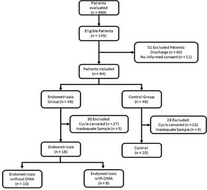
Figure 1. Flow chart of the study.
Clinical characteristics of patients
The comparison of clinical variables such as age, BMI, basal FSH, duration of infertility, and previous ovarian surgery between the endometriosis and control groups did not show statistically significant differences, indicating homogeneity between the groups. The results are presented in Table 1. When the endometriosis group was divided into subgroups with and without endometrioma, the results remained similar, with no statistically significant differences observed for any variable (Table 1).
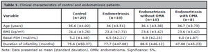
Table 1. Clinical characteristics of control and endometriosis patients.
Eicosanoid profile
In the analysis of the eicosanoid profile, a total of 41 analytes were evaluated in both serum and follicular fluid, as described in the study methodology. Of the 41 eicosanoids analyzed, five were detected in serum samples: 5-hydroxyeicosatetraenoic acid (5-HETE), 11-hydroxyeicosatetraenoic acid (11-HETE), 15-oxoeicosatetraenoic acid (15-oxo-ETE), eicosapentaenoic acid (EPA), and 15-hydroxyeicosatetraenoic acid (15-HETE). The same five eicosanoids were detected in follicular fluid samples, with the addition of prostaglandin E2 (PGE-2).
When comparing women with endometriosis with controls, no statistically significant differences were observed in the serum concentration of any eicosanoid (Table 2). To investigate whether the presence of endometrioma was related to potential changes in serum eicosanoid levels, the endometriosis group was divided into subgroups with and without endometrioma. This analysis revealed higher levels of 5-HETE in the endometriosis without endometrioma group (120.45pg/mL) than in the endometriosis with endometrioma group (38.47pg/mL; p=0.01; Table 2, Figure 2A). The other eicosanoids did not show significant differences in concentration between the groups (Table 2).
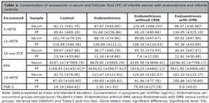
Table 2. Concentration of eicosanoids in serum and follicular fluid (FF) of infertile women with endometriosis and infertile controls.
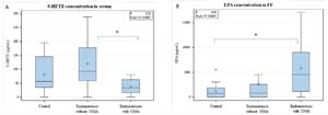
Figure 2. (A) Serum concentrations of 5-Hydroxyeicosatetraenoic acid (5-HETE) and (B) follicular concentrations of Eicosapentaenoic acid (EPA) among infertile women with endometriosis with endometrioma (OMA; n=8), without endometrioma (n=10), and controls (n=20; tubal and/or male factor of infertility).
Regarding follicular fluid eicosanoid concentrations, the endometriosis group showed higher EPA levels (4038.52pg/mL) than the control group (1159.83pg/mL; p=0.03; Table 2). The other eicosanoids did not show significant differences in concentration between the two groups (Table 2). When the endometriosis group was divided into subgroups with and without endometrioma, a statistically significant difference was observed for the same eicosanoid, EPA, with higher levels in the endometriosis with endometrioma group (5811.50pg/mL) than in the control group (1159.83pg/mL; p=0.005; Table 2, Figure 2B). The other eicosanoids did not show significant differences in concentration between the groups (Table 2).
Correlation between serum and follicular fluid eicosanoids
We observed a moderate positive correlation between 5-HETE in serum and 5-HETE in follicular fluid in control patients, as well as a weak positive correlation between serum 11-HETE and 15-oxo-ETE and follicular fluid 5-HETE in the control group. In patients with endometriosis, a weak positive correlation was also observed between serum 11-HETE and 15-HETE and follicular fluid 15-oxo-ETE. The correlation coefficient values are shown in Table 3.
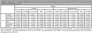
Table 3. Spearman’s correlation coefficient values of the eicosanoids levels between serum and follicular fluid (FF) in the control group and the endometriosis group.
DISCUSSION
Endometriosis is highly prevalent among women of reproductive age and is frequently associated with infertility (Burney & Giudice, 2012). However, the mechanisms involved in the etiopathogenesis of endometriosis-related infertility are not yet fully understood. Oocyte quality has been proposed as a key factor in the reduction of natural fertility in these patients (Sanchez et al., 2017), including in those with endometriomas (Casalechi et al., 2024; Somigliana et al., 2023), and inflammation and oxidative stress may mediate this oocyte damage (Barcelos et al., 2009; Da Broi & Navarro, 2016; Ferreira et al., 2019; Giorgi et al., 2016; Malvezzi et al., 2018). We hypothesized that the inflammatory process and oxidative stress associated with endometriosis may affect the systemic and follicular production of eicosanoids, bioactive lipid molecules that regulate several pathophysiological processes, including folliculogenesis, oocyte maturation, and ovulation (Leroy et al., 2005).
According to our findings, no significant difference was observed in serum eicosanoid concentrations between the control and endometriosis groups. However, when the endometriosis group was subdivided according to the presence or absence of endometrioma, a significant increase in 5-HETE was observed in the endometriosis without endometrioma group compared with the endometriosis with endometrioma group. 5-HETE is a potent chemotactic agent for neutrophils and eosinophils and may be involved in attracting these cells to ectopic endometrial tissue, thereby contributing to local and systemic inflammation (Bittleman & Casale, 1995). Additionally, studies have shown that 5-HETE can affect angiogenesis, cell proliferation, and apoptosis in various cell types, including ovarian cells (Miller et al., 2000; Zeng et al., 2002). Considering that ovarian follicles can be influenced by serum alterations, especially during the late stages of oocyte maturation (Leroy et al., 2004; Prieto et al., 2012; Edwards, 1974; Valckx et al., 2012), the pro-inflammatory changes found in this study may indicate a possible follicular impact in patients with pelvic lesions without endometrioma. Because pelvic endometriosis is characterized by lesions scattered throughout the abdominal cavity (Agarwal & Subramanian, 2010), whereas endometrioma is a localized ovarian lesion with an encapsulated structure (Hughesdon, 1957; Paffoni et al., 2019), this finding suggests greater systemic inflammatory repercussions in pelvic endometriosis than in endometrioma, based on the elevated 5-HETE levels. This interpretation should be confirmed in further studies with larger sample sizes. Pelvic endometriosis lesions can lead to the release of inflammatory cells and molecules into the bloodstream, resulting in chronic inflammation and oxidative stress, which can affect ovarian function and the quality of oocytes and embryos (Barcelos et al., 2009; Da Broi & Navarro, 2016; Ferreira et al., 2019; Malvezzi et al., 2019). Therefore, systemic inflammation and increased 5-HETE production may be involved in the etiopathogenesis of infertility associated with pelvic endometriosis. Further studies on the follicular repercussions of this serum eicosanoid would be valuable for a better understanding of this finding.
The analysis of eicosanoid levels in follicular fluid demonstrated an increase in EPA concentration in the endometriosis group compared with the control group. These findings became even more interesting when the endometriosis group was subdivided according to the presence or absence of endometrioma, revealing an elevated EPA concentration in the follicular fluid of women with endometrioma compared with controls. Once again, these data reflect distinct disease behavior according to lesion location. Endometriomas are pseudocysts formed by the accumulation of menstrual debris and focal bleeding (Hughesdon, 1957) and may be present in cases of moderate to severe disease, representing 30% to 40% of endometriosis lesions (Vercellini et al., 2003). Although they appear to have a detrimental influence on fertility, it remains unclear whether these ovarian lesions impair ovarian function by creating a cytotoxic follicular microenvironment, thereby affecting oocyte development (Paffoni et al., 2019), and/or by mechanically stretching the ovarian cortex, thus decreasing the number of follicles in the ovarian cortex (Bonavina & Taylor, 2022; Kitajima et al., 2011). As endometriomas are encapsulated lesions (Hughesdon, 1957; Paffoni et al., 2019), the observed alteration in follicular fluid may be a consequence of the nearby ovarian ectopic implants. EPA is known for its anti-inflammatory properties, including inhibition of pro-inflammatory prostaglandin and leukotriene synthesis (Calder, 2010; Serhan, 2005). The increased EPA concentration in the follicular fluid of women with endometrioma may reflect an imbalance in this eicosanoid within the follicular environment, probably as a compensatory response intended to protect against the local chronic inflammation that occurs in this type of lesion (Yland et al., 2020), although this needs to be confirmed with appropriate methodology. Such inflammation can lead to oxidative stress, with increased production of reactive oxygen species, which can result in cellular and tissue damage (Nakagawa et al., 2016). In this context, the elevation of EPA in follicular fluid may represent an attempt to reduce oxidative stress and protect ovarian and follicular cells from the chronic inflammation associated with endometrioma. There are limited studies directly assessing the association between EPA and infertility in patients with endometriosis. A study conducted by our group demonstrated that the addition of EPA to the oocyte maturation culture medium prevented meiotic damage caused by follicular fluid from women with endometriosis at all stages (Giorgi et al., 2018). Furthermore, there is evidence that EPA supplementation reduces pelvic pain associated with endometriosis (Netsu et al., 2008) and does not negatively affect ovarian reserve in patients with endometrioma. Interestingly, EPA levels in mouse oocytes were related to gamete maturation stage, with levels decreasing during meiotic resumption (Li et al., 2020). Considering that we found higher EPA concentrations in the follicular fluid of patients with endometrioma, we questioned whether this could negatively affect meiotic resumption in these patients and thus impair fertility. Altogether, these findings suggest that the follicular microenvironment in women with endometriosis is modulated differently depending on the presence or absence of endometrioma, since local inflammation and oxidative stress promoted by ovarian lesions may affect the adjacent follicular microenvironment, with clinical implications for fertility. However, further studies are needed to better understand the relationship between EPA and infertility in patients with endometriosis and endometrioma.
When evaluating the correlation between eicosanoid levels in serum and follicular fluid, we observed a moderate positive correlation only between 5-HETE in serum and follicular fluid in the control group, suggesting that serum concentrations of this eicosanoid may reflect those in the follicular environment under physiological conditions. In the endometriosis group, we did not observe any strong or moderate correlation between serum and follicular eicosanoid concentrations, which may be due to follicular anti-inflammatory defense mechanisms attempting to preserve gamete quality, as suggested in this study by the higher follicular EPA concentrations in patients with endometriosis than in controls. This finding discourages the use of serum eicosanoids to directly infer the follicular microenvironment.
This study has some limitations. First, the sample size was small because of the restrictive eligibility criteria adopted and treatment suspension during the COVID-19 pandemic, which limits the generalizability of the findings. However, these restrictions were necessary to avoid confounding factors related to oxidative stress and impaired oocyte quality, thereby increasing the internal validity of the study. Second, all patients underwent controlled ovarian stimulation, and the data may not be extrapolated to natural cycles. Moreover, only 5 eicosanoids were identified in serum and 6 in follicular fluid. The non-detection of several eicosanoid molecules in the samples may be explained by (1) the short half-life of these molecules, which are rapidly metabolized by enzymes, reducing their stability and availability for detection; and (2) the sensitivity of the analytical technique, because although the methodology used has high specificity, some molecules may have been present at very low concentrations, below the detection limit of the equipment.
In summary, no significant differences were observed in serum eicosanoid concentrations between infertile control women and women with endometriosis. However, after subdividing the endometriosis group, higher serum concentrations of 5-HETE were found in infertile women with endometriosis without endometrioma than in those with endometrioma, suggesting that pelvic disease promotes a systemic increase in this pro-inflammatory eicosanoid, which may contribute to the pathogenesis of endometriosis-related infertility. On the other hand, higher follicular concentrations of EPA were observed in infertile women with endometriosis than in controls, particularly in those with endometrioma, which may exert a protective effect on the follicular microenvironment against the chronic inflammation associated with this type of lesion, thereby helping to prevent oocyte damage. Collectively, these findings contribute to advancing our understanding of the mechanisms underlying infertility related to endometriosis. Clarifying the role of eicosanoids in the pathophysiology of endometriosis and oocyte quality may facilitate the development of innovative therapeutic approaches aimed at improving both natural fertility and assisted reproduction outcomes in infertile women with endometriosis who have poor gamete and embryo quality.
This study was presented at Endofeminina 2023, the World Congress on Endometriosis 2023, and the Congresso Brasileiro de Reprodução Humana 2025.
ACKNOWLEDGEMENTS
We thank the staff of the Human Reproduction Division, University Hospital, Department of Gynecology and Obstetrics, Ribeirão Preto Medical School, University of São Paulo (FMRP-USP), and the staff of the Center of Excellence in Lipid Quantification and Identification (Ceqil), Faculdade de Ciências Farmacêuticas de Ribeirão Preto (FCFRP-USP).
Ethical Approval
This study was approved by the Brazilian National Commission of Ethics in Research (Ethics Approval Number: 3.349.839) and was conducted in accordance with the Code of Ethics of the World Medical Association.
Funding
This study received financial support from the Coordenação de Aperfeiçoamento de Pessoal de Nível Superior - Brasil (CAPES) - Finance Code 001 (CAPES; grant number 88887.369797/2019), from the Brazilian National Institute of Hormones and Women’s Health/Conselho Nacional de Desenvolvimento Científico e Tecnológico (CNPq) (CNPq/INCT grant number 118389/2024-6; CNPq grant number 303461/2019-5), and from the Foundation for the Support of Teaching, Research and Assistance of the Hospital das Clínicas of the Faculty of Medicine of Ribeirão Preto, University of São Paulo (FAEPA).
REFERENCES
Agarwal A, Saleh RA, Bedaiwy MA. Role of reactive oxygen species in the pathophysiology of human reproduction. Fertil Steril. 2003;79:829-43. PMID: 12749418 DOI: 10.1016/s0015-0282(02)04948-8 Medline
Andrade AZ, Rodrigues JK, Dib LA, Romão GS, Ferriani RA, Jordão Junior AA, Navarro PA. Serum markers of oxidative stress in infertile women with endometriosis. Rev Bras Ginecol Obstet. 2010;32:279-85. PMID: 20945013 DOI: 10.1590/s0100-72032010000600005 Medline
Barbosa MA, Teixeira DM, Navarro PA, Ferriani RA, Nastri CO, Martins WP. Impact of endometriosis and its staging on assisted reproduction outcome: systematic review and meta-analysis. Ultrasound Obstet Gynecol. 2014;44:261-78. PMID: 24639087 DOI: 10.1002/uog.13366 Medline
Barcelos ID, Vieira RC, Ferreira EM, Martins WP, Ferriani RA, Navarro PA. Comparative analysis of the spindle and chromosome configurations of in vitro-matured oocytes from patients with endometriosis and from control subjects: a pilot study. Fertil Steril. 2009;92:1749-52. PMID: 19523612 DOI: 10.1016/j.fertnstert.2009.05.006 Medline
Barnhart K, Dunsmoor-Su R, Coutifaris C. Effect of endometriosis on in vitro fertilization. Fertil Steril. 2002;77:1148-55. PMID: 12057720 DOI: 10.1016/s0015-0282(02)03112-6 Medline
Bittleman DB, Casale TB. 5-Hydroxyeicosatetraenoic acid (HETE)-induced neutrophil transcellular migration is dependent upon enantiomeric structure. Am J Respir Cell Mol Biol. 1995;12:260-7. PMID: 7873191 DOI: 10.1165/ajrcmb.12.3.7873191 Medline
Bonavina G, Taylor HS. Endometriosis-associated infertility: From pathophysiology to tailored treatment. Front Endocrinol (Lausanne). 2022;13:1020827. PMID: 36387918 DOI: 10.3389/fendo.2022.1020827 Medline
Bulletti C, Coccia ME, Battistoni S, Borini A. Endometriosis and infertility. J Assist Reprod Genet. 2010;27:441-7. PMID: 20574791 DOI: 10.1007/s10815-010-9436-1 Medline
Burney RO, Giudice LC. Pathogenesis and pathophysiology of endometriosis. Fertil Steril. 2012;98:511-9. PMID: 22819144 DOI: 10.1016/j.fertnstert.2012.06.029 Medline
Calder PC. Omega-3 fatty acids and inflammatory processes. Nutrients. 2010;2:355-74. PMID: 22254027 DOI: 10.3390/nu2030355 Medline
Calder PC. Functional Roles of Fatty Acids and Their Effects on Human Health. JPEN J Parenter Enteral Nutr. 2015;39:18S-32S. PMID: 26177664 DOI: 10.1177/0148607115595980 Medline
Casalechi M, Di Stefano G, Fornelli G, Somigliana E, Viganò P. Impact of endometriosis on the ovarian follicles. Best Pract Res Clin Obstet Gynaecol. 2024;92:102430. PMID: 38311379 DOI: 10.1016/j.bpobgyn.2023.102430 Medline
Choi YS, Cho S, Seo SK, Park JH, Kim SH, Lee BS. Alteration in the intrafollicular thiol-redox system in infertile women with endometriosis. Reproduction. 2015;149:155-62. PMID: 25376627 DOI: 10.1530/REP-14-0438 Medline
Ciepiela P, Bączkowski T, Drozd A, Kazienko A, Stachowska E, Kurzawa R. Arachidonic and linoleic acid derivatives impact oocyte ICSI fertilization--a prospective analysis of follicular fluid and a matched oocyte in a ‘one follicle--one retrieved oocyte--one resulting embryo’ investigational setting. PLoS One. 2015;10:e0119087. PMID: 25763593 DOI: 10.1371/journal.pone.0119087 Medline
Da Broi MG, de Albuquerque FO, de Andrade AZ, Cardoso RL, Jordão Junior AA, Navarro PA. Increased concentration of 8-hydroxy-2’-deoxyguanosine in follicular fluid of infertile women with endometriosis. Cell Tissue Res. 2016;366:231-42. PMID: 27250533 DOI: 10.1007/s00441-016-2428-4 Medline
Da Broi MG, Jordão AA Jr, Ferriani RA, Navarro PA. Oocyte oxidative DNA damage may be involved in minimal/mild endometriosis-related infertility. Mol Reprod Dev. 2018;85:28-136. PMID: 29247565 DOI: 10.1002/mrd.22943 Medline
Da Broi MG, Malvezzi H, Paz CC, Ferriani RA, Navarro PA. Follicular fluid from infertile women with mild endometriosis may compromise the meiotic spindles of bovine metaphase II oocytes. Hum Reprod. 2014;29:315-23. PMID: 24166595 DOI: 10.1093/humrep/det378 Medline
Da Broi MG, Navarro PA. Oxidative stress and oocyte quality: ethiopathogenic mechanisms of minimal/mild endometriosis-related infertility. Cell Tissue Res. 2016;364:1-7. PMID: 26685866 DOI: 10.1007/s00441-015-2339-9 Medline
Du YB, Gao MZ, Shi Y, Sun ZG, Wang J. Endocrine and inflammatory factors and endometriosis-associated infertility in assisted reproduction techniques. Arch Gynecol Obstet. 2013;287:123-30. PMID: 23053311 DOI: 10.1007/s00404-012-2567-0 Medline
Edwards RG. Follicular fluid. J Reprod Fertil. 1974;37:189-219. PMID: 4274030 DOI: 10.1530/jrf.0.0370189 Medline
Eskenazi B, Warner ML. Epidemiology of endometriosis. Obstet Gynecol Clin North Am. 1997;24:235-58. PMID: 9163765 DOI: 10.1016/s0889-8545(05)70302-8 Medline
Fan YY, Chen HY, Chen W, Liu YN, Fu Y, Wang LN. Expression of inflammatory cytokines in serum and peritoneal fluid from patients with different stages of endometriosis. Gynecol Endocrinol. 2018;34:507-12. PMID: 29308924 DOI: 10.1080/09513590.2017.1409717 Medline
Ferreira EM, Giorgi VSI, Rodrigues JK, de Andrade AZ, Junior AAJ, Navarro PA. Systemic oxidative stress as a possible mechanism underlying the pathogenesis of mild endometriosis-related infertility. Reprod Biomed Online. 2019;39:785-94. PMID: 31540845 DOI: 10.1016/j.rbmo.2019.06.011 Medline
Giorgi VS, Da Broi MG, Paz CC, Ferriani RA, Navarro PA. N-Acetyl-Cysteine and l-Carnitine Prevent Meiotic Oocyte Damage Induced by Follicular Fluid From Infertile Women With Mild Endometriosis. Reprod Sci. 2016;23:342-51. PMID: 26342050 DOI: 10.1177/1933719115602772 Medline
Gupta S, Goldberg JM, Aziz N, Goldberg E, Krajcir N, Agarwal A. Pathogenic mechanisms in endometriosis-associated infertility. Fertil Steril. 2008;90:247-57. PMID: 18672121 DOI: 10.1016/j.fertnstert.2008.02.093 Medline
Harizi H, Corcuff JB, Gualde N. Arachidonic-acid-derived eicosanoids: roles in biology and immunopathology. Trends Mol Med. 2008;14:461-9. PMID: 18774339 DOI: 10.1016/j.molmed.2008.08.005 Medline
Hughes EG, Fedorkow DM, Collins JA. A quantitative overview of controlled trials in endometriosis-associated infertility. Fertil Steril. 1993;59:963-70. PMID: 8486196. Medline
Hughesdon PE. The structure of endometrial cysts of the ovary. J Obstet Gynaecol Br Emp. 1957;64:481-7.PMID: 13463645 DOI: 10.1111/j.1471-0528.1957.tb06276.x Medline
Jungheim ES, Macones GA, Odem RR, Patterson BW, Lanzendorf SE, Ratts VS, Moley KH. Associations between free fatty acids, cumulus oocyte complex morphology and ovarian function during in vitro fertilization. Fertil Steril. 2011;95:1970-4. PMID: 21353671 DOI: 10.1016/j.fertnstert.2011.01.154 Medline
Kitajima M, Defrère S, Dolmans MM, Colette S, Squifflet J, Van Langendonckt A, Donnez J. Endometriomas as a possible cause of reduced ovarian reserve in women with endometriosis. Fertil Steril. 2011;96:685-91. PMID: 21802672 DOI: 10.1016/j.fertnstert.2011.06.064 Medline
Leroy JL, Vanholder T, Delanghe JR, Opsomer G, Van Soom A, Bols PE, de Kruif A. Metabolite and ionic composition of follicular fluid from different-sized follicles and their relationship to serum concentrations in dairy cows. Anim Reprod Sci. 2004;80:201-11. PMID: 15036497 DOI: 10.1016/S0378-4320(03)00173-8 Medline
Leroy JL, Vanholder T, Mateusen B, Christophe A, Opsomer G, de Kruif A, Genicot G, Van Soom A. Non-esterified fatty acids in follicular fluid of dairy cows and their effect on developmental capacity of bovine oocytes in vitro. Reproduction. 2005;130:485-95. PMID: 16183866 DOI: 10.1530/rep.1.00735 Medline
Li L, Zhu S, Shu W, Guo Y, Guan Y, Zeng J, Wang H, Han L, Zhang J, Liu X, Li C, Hou X, Gao M, Ge J, Ren C, Zhang H, Schedl T, Guo X, Chen M, Wang Q. Characterization of Metabolic Patterns in Mouse Oocytes during Meiotic Maturation. Mol Cell. 2020;80:525-540.e9. PMID: 33068521 DOI: 10.1016/j.molcel.2020.09.022 Medline
Liu F, He L, Liu Y, Shi Y, Du H. The expression and role of oxidative stress markers in the serum and follicular fluid of patients with endometriosis. Clin Exp Obstet Gynecol. 2013;40:372-6. PMID: 24283168. Medline
Macer ML, Taylor HS. Endometriosis and infertility: a review of the pathogenesis and treatment of endometriosis-associated infertility. Obstet Gynecol Clin North Am. 2012;39:535-49. PMID: 23182559 DOI: 10.1016/j.ogc.2012.10.002 Medline
Malvezzi H, Da Broi MG, Meola J, Rosa-E-Silva JC, Ferriani RA, Navarro PA. Peritoneal fluid of women with endometriosis reduces SOD1 in bovine oocytes in vitro maturation. Cell Tissue Res. 2018;372:621-8. PMID: 29464366 DOI: 10.1007/s00441-018-2805-2 Medline
Malvezzi H, Hernandes C, Piccinato CA, Podgaec S. Interleukin in endometriosis-associated infertility-pelvic pain: systematic review and meta-analysis. Reproduction. 2019;158:1-12. PMID: 30933927 DOI: 10.1530/REP-18-0618 Medline
Massey KA, Nicolaou A. Lipidomics of polyunsaturated-fatty-acid-derived oxygenated metabolites. Biochem Soc Trans. 2011;39:1240-6. PMID: 21936796 DOI: 10.1042/BST0391240. Medline
Massoud G, Spann M, Vaught KC, Das S, Dow M, Cochran R, Baker V, Segars J, Singh B. Biomarkers Assessing the Role of Cumulus Cells on IVF Outcomes: A Systematic Review. J Assist Reprod Genet. 2024;41:253-75. PMID: 37947940 DOI: 10.1007/s10815-023-02984-9 Medline
Miller TA, Ghosh J, Myers CE, Macdonald TL. 5-HETE congeners as modulators of cell proliferation. Bioorg Med Chem Lett. 2000;10:1913-6. PMID: 10987416 DOI: 10.1016/s0960-894x(00)00370-x Medline
Mirabi P, Chaichi MJ, Esmaeilzadeh S, Ali Jorsaraei SG, Bijani A, Ehsani M, Hashemi Karooee SF. The role of fatty acids on ICSI outcomes: a prospective cohort study. Lipids Health Dis. 2017;16:18. PMID: 28109274 DOI: 10.1186/s12944-016-0396-z Medline
Motohashi E, Kawauchi H, Endo H, Kondo H, Kitasato H, Kuramoto H, Majima M, Unno N, Hayashi I. Regulatory expression of lipoxin A4 receptor in physiologically estrus cycle and pathologically endometriosis. Biomed Pharmacother. 2005;59:330-8. PMID: 15996849. DOI: 10.1016/j.biopha.2005.05.005 Medline
Nakagawa K, Hisano M, Sugiyama R, Yamaguchi K. Measurement of oxidative stress in the follicular fluid of infertility patients with an endometrioma. Arch Gynecol Obstet. 2016;293:197-202. PMID: 26246415 DOI: 10.1007/s00404-015-3834-7 Medline
Netsu S, Konno R, Odagiri K, Soma M, Fujiwara H, Suzuki M. Oral eicosapentaenoic acid supplementation as possible therapy for endometriosis. Fertil Steril. 2008;90:1496-502. PMID: 18054352 DOI: 10.1016/j.fertnstert.2007.08.014 Medline
Numao A, Hosono K, Suzuki T, Hayashi I, Uematsu S, Akira S, Ogino Y, Kawauchi H, Unno N, Majima M. The inducible prostaglandin E synthase mPGES-1 regulates growth of endometrial tissues and angiogenesis in a mouse implantation model. Biomed Pharmacother. 2011;65:77-84. PMID: 21247731 DOI: 10.1016/j.biopha.2010.12.008 Medline
Paffoni A, Bolis V, Ferrari S, Benaglia L, Vercellini P, Somigliana E. The Gametotoxic Effects of the Endometrioma Content: Insights From a Parthenogenetic Human Model. Reprod Sci. 2019;26:573-9. PMID: 29779474 DOI: 10.1177/1933719118777637 Medline
Polak G, Wertel I, Barczyński B, Kwaśniewski W, Bednarek W, Kotarski J. Increased levels of oxidative stress markers in the peritoneal fluid of women with endometriosis. Eur J Obstet Gynecol Reprod Biol. 2013;168:187-90. PMID: 23351670 DOI: 10.1016/j.ejogrb.2012.12.043 Medline
Practice Committee of the American Society for Reproductive Medicine. Endometriosis and infertility: a committee opinion. Fertil Steril. 2012;98:591-8. PMID: 22704630 DOI: 10.1016/j.fertnstert.2012.05.031 Medline
Prieto L, Quesada JF, Cambero O, Pacheco A, Pellicer A, Codoceo R, Garcia-Velasco JA. Analysis of follicular fluid and serum markers of oxidative stress in women with infertility related to endometriosis. Fertil Steril. 2012;98:126-30. PMID: 22578534 DOI: 10.1016/j.fertnstert.2012.03.052 Medline
Rana N, Braun DP, House R, Gebel H, Rotman C, Dmowski WP. Basal and stimulated secretion of cytokines by peritoneal macrophages in women with endometriosis. Fertil Steril. 1996;65:925-30. PMID: 8612850. Medline
Revised American Society for Reproductive Medicine classification of endometriosis: 1996. Fertil Steril. 1997;67:817-21. PMID: 9130884 DOI: 10.1016/s0015-0282(97)81391-x Medline
Ruder EH, Hartman TJ, Blumberg J, Goldman MB. Oxidative stress and antioxidants: exposure and impact on female fertility. Hum Reprod Update. 2008;14:345-57. PMID: 18535004 DOI: 10.1093/humupd/dmn011 Medline
Sanchez AM, Vanni VS, Bartiromo L, Papaleo E, Zilberberg E, Candiani M, Orvieto R, Viganò P. Is the oocyte quality affected by endometriosis? A review of the literature. J Ovarian Res. 2017;10:43. PMID: 28701212 DOI: 10.1186/s13048-017-0341-4 Medline
Serhan CN. Novel omega -- 3-derived local mediators in anti-inflammation and resolution. Pharmacol Ther. 2005;105:7-21. PMID: 15626453 DOI: 10.1016/j.pharmthera.2004.09.002 Medline
Shaaker M, Rahimipour A, Nouri M, Khanaki K, Darabi M, Farzadi L, Shahnazi V, Mehdizadeh A. Fatty acid composition of human follicular fluid phospholipids and fertilization rate in assisted reproductive techniques. Iran Biomed J. 2012;16:162-8. PMID: 23023218 DOI: 10.6091/ibj.1081.2012 Medline
Shebl O, Sifferlinger I, Habelsberger A, Oppelt P, Mayer RB, Petek E, Ebner T. Oocyte competence in in vitro fertilization and intracytoplasmic sperm injection patients suffering from endometriosis and its possible association with subsequent treatment outcome: a matched case-control study. Acta Obstet Gynecol Scand. 2017;96:736-44. PMID: 27317364 DOI: 10.1111/aogs.12941 Medline
Simón C, Gutiérrez A, Vidal A, de los Santos MJ, Tarín JJ, Remohí J, Pellicer A. Outcome of patients with endometriosis in assisted reproduction: results from in-vitro fertilization and oocyte donation. Hum Reprod. 1994;9:725-9. PMID: 8046030 DOI: 10.1093/oxfordjournals.humrep.a138578 Medline
Singh AK, Chattopadhyay R, Chakravarty B, Chaudhury K. Markers of oxidative stress in follicular fluid of women with endometriosis and tubal infertility undergoing IVF. Reprod Toxicol. 2013;42:116-24. PMID: 23994512 DOI: 10.1016/j.reprotox.2013.08.005 Medline
Somigliana E, Li Piani L, Paffoni A, Salmeri N, Orsi M, Benaglia L, Vercellini P, Vigano’ P. Endometriosis and IVF treatment outcomes: unpacking the process. Reprod Biol Endocrinol. 2023;21:107. PMID: 37936154 DOI: 10.1186/s12958-023-01157-8 Medline
Sorgi CA, Peti APF, Petta T, Meirelles AFG, Fontanari C, Moraes LAB, Faccioli LH. Comprehensive high-resolution multiple-reaction monitoring mass spectrometry for targeted eicosanoid assays. Sci Data. 2018;5:180167. PMID: 30129930 DOI: 10.1038/sdata.2018.167 Medline
Valckx SD, De Pauw I, De Neubourg D, Inion I, Berth M, Fransen E, Bols PE, Leroy JL. BMI-related metabolic composition of the follicular fluid of women undergoing assisted reproductive treatment and the consequences for oocyte and embryo quality. Hum Reprod. 2012;27:3531-9. PMID: 23019302 DOI: 10.1093/humrep/des350 Medline
Vercellini P, Chapron C, De Giorgi O, Consonni D, Frontino G, Crosignani PG. Coagulation or excision of ovarian endometriomas? Am J Obstet Gynecol. 2003;188:606-10. PMID: 12634628 DOI: 10.1067/mob.2003.7 Medline
Wathes DC, Abayasekara DR, Aitken RJ. Polyunsaturated fatty acids in male and female reproduction. Biol Reprod. 2007;77:190-201. PMID: 17442851 DOI: 10.1095/biolreprod.107.060558 Medline
Wonnacott KE, Kwong WY, Hughes J, Salter AM, Lea RG, Garnsworthy PC, Sinclair KD. Dietary omega-3 and -6 polyunsaturated fatty acids affect the composition and development of sheep granulosa cells, oocytes and embryos. Reproduction. 2010;139:57-69. PMID: 19789173 DOI: 10.1530/REP-09-0219 Medline
Wu MH, Sun HS, Lin CC, Hsiao KY, Chuang PC, Pan HA, Tsai SJ. Distinct mechanisms regulate cyclooxygenase-1 and -2 in peritoneal macrophages of women with and without endometriosis. Mol Hum Reprod. 2002;8:1103-10. PMID: 12468643 DOI: 10.1093/molehr/8.12.1103 Medline
Yang T, Zhao J, Liu F, Li Y. Lipid metabolism and endometrial receptivity. Hum Reprod Update. 2022;28:858-89. PMID: 35639910 DOI: 10.1093/humupd/dmac026 Medline
Yland J, Carvalho LFP, Beste M, Bailey A, Thomas C, Abrão MS, Racowsky C, Griffith L, Missmer SA. Endometrioma, the follicular fluid inflammatory network and its association with oocyte and embryo characteristics. Reprod Biomed Online. 2020;40:399-408. PMID: 32057676 DOI: 10.1016/j.rbmo.2019.12.005 Medline
Zeng ZZ, Yellaturu CR, Neeli I, Rao GN. 5(S)-hydroxyeicosatetraenoic acid stimulates DNA synthesis in human microvascular endothelial cells via activation of Jak/STAT and phosphatidylinositol 3-kinase/Akt signaling, leading to induction of expression of basic fibroblast growth factor 2. J Biol Chem. 2002;277:41213-9. PMID: 12193593 DOI: 10.1074/jbc.M204508200 Medline