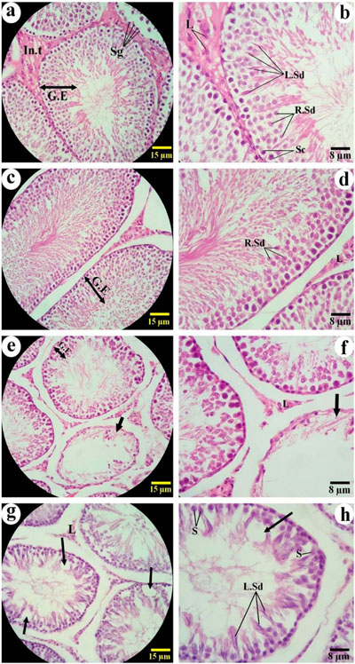
Figure 6. Histological evaluation of testicular tissue using H&E staining in experimental groups (Magnification ×100, ×400, n=6). Panels (a) and (b): Control group; (c) and (d): Chlorogenic acid (CGA)-treated group; (e) and (f): Busulfan-treated group; (g) and (h): Busulfan + CGA co-treated group. Large arrows indicate seminiferous tubules devoid of germ cells, while small arrows show focal areas lacking cellular components within the seminiferous epithelium. Cell types are labeled as follows: Spermatogonia (Sg), Spermatocytes (Sc), Round Spermatids (R.Sd), Elongated Spermatids (L.Sd), Sertoli cells (S), and Leydig cells (L). Hematoxylin and Eosin (H&E) staining was used to visualize histological structure.