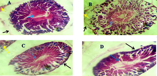
Figure 7. Effect of EECS Treatment on Testicular Histology in RDP-exposed Male Wistar Rats (H&E, Mag.: ×400).
Plate A (Control): The seminiferous tubules display normal architecture with well-organized germinal epithelium. The yellow arrow indicates the intact interstitial space with normal Leydig cells. The blue arrow shows mature spermatozoa within the lumen of the seminiferous tubule. The white arrow points to well-aligned spermatogenic cells, and the black arrow highlights an intact basement membrane.
Plate B (RDP-treated group): The blue arrow reveals a distorted and reduced spermatozoa population in the lumen. The white arrow indicates disorganization and sloughing of spermatogenic cells into the lumen. The black arrow shows thinning and detachment of the basement membrane. The yellow arrow reflects an edematous interstitial space and possibly a reduction in Leydig cell density, indicating inflammatory changes.
Plate C (EECS-only group): Seminiferous tubular architecture is similar to that of the control. The blue arrow shows the presence of mature spermatozoa within the tubular lumen. The white arrow indicates a well-organized germinal epithelium. The black arrow confirms an intact basement membrane, and the yellow arrow shows healthy interstitial tissue, suggesting that EECS alone has no adverse effects and may support testicular integrity.
Plate D (RDP + EECS group): The blue arrow denotes the reappearance of spermatozoa in the lumen, albeit in reduced quantity. The white arrow points to partially improved spermatogenic layers, while the black arrow shows improved basement membrane integrity. This suggests a partial ameliorative effect of EECS against RDP-induced testicular damage.