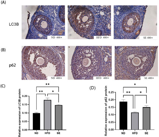
Figure 1. Immunohistochemical detection of LC3B and p62 expression in mouse ovarian tissues from ND, HFD, and SE groups(A) Representative LC3B staining in ovarian sections (ND, n=5; HFD, n=6; SE, n=7).(B) Representative p62 staining in ovarian sections (ND, n=7; HFD, n=10; SE, n=8).(C) Quantitative analysis of LC3B expression.(D) Quantitative analysis of p62 expression.Data are expressed as mean ± SEM. *p<0.05, **p<0.01.