JBRA Assist. Reprod. 2026 2026;00(0):00-00
ORIGINAL ARTICLE
doi: 10.5935/1518-0557.20260062
The impact of oral refined sugar consumption on male reproductive parameters: The role of cessation
1Department of Physiology, School of Basic Medical Sciences, Federal University of Technology, Akure, Nigeria
CONFLICT OF INTEREST
ABSTRACT
Objective: Refined sugar is a widely consumed dietary component that has been implicated in several health complications, including reproductive dysfunction. Despite growing concern regarding its potential endocrine-disrupt-ing effects, the reversibility of its impact on male reproduc-tive parameters remains underexplored. This study aimed to evaluate the effects of oral refined sugar consumption on male reproductive parameters in Wistar rats and to in-vestigate the role of sugar cessation in potential recovery.
Methods: Forty male Wistar rats were randomly divid-ed into eight groups (n=5): a control group, three treat-ment groups (low, medium, and high sugar doses), and corresponding cessation groups. Sugar dissolved in dis-tilled water was administered orally using an oropharyn-geal cannula for four weeks, followed by four weeks of withdrawal in the cessation groups. Reproductive parame-ters were then assessed.
Results: Refined sugar consumption resulted in statis-tically significant (p<0.05), dose-dependent reductions in relative testicular and epididymal weights, sperm count, motility, and morphology. Significant hormonal alterations were also observed, including reduced testosterone, LH, and FSH levels and increased estradiol levels (p<0.05). In addition, histological analysis revealed testicular degener-ation and vacuolization in all treatment groups. Notably, the cessation groups showed partial recovery in some pa-rameters, particularly the high-dose cessation group, with significant improvements (p<0.05) in sperm count and motility, a lower percentage of abnormal morphology, and increased testosterone, LH, and FSH levels.
Conclusion: Prolonged refined sugar intake impairs male reproductive health through structural and hormonal disruptions. However, cessation showed potential for par-tial recovery, underscoring the importance of dietary inter-vention in preserving male fertility.
Keywords: refined sugar, oral consumption, cessation, gonadal hormones, sperm parameters
INTRODUCTION
Sugar belongs to the carbohydrate family and is a ubiquitous component of modern diets, providing energy to the body. It exists in various forms and has different effects on human health (Johnson & Yon, 2010). Sugar, also known as sucrose, is a disaccharide composed of glucose and fructose molecules and is classified into two main categories: naturally occurring sugars and refined/ added sugars.
The average daily intake of sucrose varies widely across populations. According to the World Health Organization (WHO, 2015), free sugar intake should not exceed 10% of total energy intake. However, epidemiological data indicate that most individuals exceed this recommended threshold, with the average American consuming 27 pounds of sugar annually (Moynihan, 2016).
Refined sugar has been reported to have both beneficial and detrimental effects on the human body. Excessive sugar consumption has been implicated in several health disorders, including overweight/obesity, metabolic disorders, type 2 diabetes mellitus, cardiovascular disease, cancer, depression, and cognitive impairment (Moore et al., 1998). Recently, attention has shifted to the potential effects of refined sugar consumption on reproductive health, particularly in men.
A growing body of evidence suggests that high oral sugar consumption may have deleterious effects on sperm parameters and reproductive hormones, potentially leading to impaired fertility (Johnson et al., 2009). The rapid absorption of refined sugar into the bloodstream initiates a cascade of physiological responses, including insulin resistance, oxidative stress, and inflammation, which can negatively affect sperm quality, reproductive hormone balance, fertility, and overall male reproductive health (Yang et al., 2014). High refined sugar intake is associated with decreased sperm count, motility, and morphology, as well as altered reproductive hormone profiles, including reduced testosterone levels and disrupted gonadotropin secretion (Bener et al., 2009). These changes can impair fertility and increase the risk of reproductive disorders such as hypogonadism and erectile dysfunction. Beyond its physiological effects on sperm production and hormone levels, refined sugar has also been increasingly implicated in sexual dysfunction, particularly low libido. Testosterone, the primary male sex hormone, plays a central role not only in spermatogenesis but also in the regulation of sexual desire and performance (Adeyemi et al., 2024). The documented suppression of testosterone in high-sugar diets may therefore underlies both reduced fertility and decreased sexual drive (Chen et al., 2018). Clinical observations and animal studies support the hypothesis that dietary sugar can diminish sexual motivation and performance by altering neuroendocrine signaling and reducing the bioavailability of androgens essential for libido and sexual arousal.
The possible reversibility of the systemic effects of refined sugar, either with therapeutic agents or following simple withdrawal of sugar consumption without further intervention, has not been adequately demonstrated. The present study was therefore designed to explore the impact of different levels of refined sugar consumption on male reproductive parameters and the role of sugar cessation in these parameters.
MATERIAL AND METHODS
Study Design
Forty healthy male Wistar rats, 11 weeks of age and weighing 100-150 g, were purchased from a reputable animal vendor and housed in the animal facility of the Department of Physiology. The animals were acclimatized for two weeks under standard conditions. The rats were allowed free access to standard rat chow and water. Institutional ethical approval was obtained from the Centre for Research and Development (CERAD), Federal University of Technology, Akure, with reference number FUTA/ETH/25/234. Rats were randomly divided into eight groups of five rats each, as follows:
• Control (CNTL): received 0.002 mL/g body weight (BW) of distilled water daily for 4 weeks.Preparation and Dosage of Stock Sugar Solutions The LDS dosage was 20 mg/kg body weight (Ogli et al., 2023). Refined sugar (200 mg) was dissolved in 20 mL of distilled water to obtain a stock solution of 10 mg/mL. Each rat in the LDS group was administered a volume of stock solution calculated as follows: 0.002 mL × rat BW in grams. The MDS dosage was 30 mg/kg body weight (Ogli et al., 2023). Refined sugar (300 g) was dissolved in 20 mL of distilled water to obtain a stock solution of 15 mg/mL. Each rat was administered daily with a volume of stock solution calculated as follows: 0.002 mL × rat BW in grams.The HDS dosage was 60 mg/kg body weight (Ogli et al., 2023). Refined sugar (600 mg) was dissolved in 20 mL of distilled water to obtain a stock solution of 32 mg/mL. Each rat was administered daily with a volume of stock solution calculated as follows: 0.002 mL × rat BW in grams.Refined sugar was administered daily throughout the experiment by the oral route using an oropharyngeal cannula attached to a 2 mL syringe. All dilutions were prepared every 2 days and refrigerated to ensure freshness.
• Low-dose sugar (LDS) treatment group: treated with 20 mg/kg BW of refined sugar daily for 4 weeks (Ogli et al., 2023).
• Medium-dose sugar (MDS) treatment group: treated with 30 mg/kg BW of refined sugar daily for 4 weeks (Ogli et al., 2023).
• High-dose sugar (HDS) treatment group: treated with 60 mg/kg BW of refined sugar daily for 4 weeks (Ogli et al., 2023).
• Control cessation group (cCNTL): received 0.002 mL/g BW of distilled water for 4 weeks and was then withdrawn from treatment for another 4 weeks.
• Low-dose cessation group (cLDS): treated with 20 mg/kg BW of refined sugar for 4 weeks and then withdrawn from treatment for another 4 weeks.
• Medium-dose cessation group (cMDS): treated with 30 mg/kg BW of refined sugar for 4 weeks and then withdrawn from treatment for 4 weeks.
• High-dose cessation group (cHDS): treated with 60 mg/kg BW of refined sugar for 4 weeks and then withdrawn from treatment for 4 weeks.
Sample Collection
Animals were euthanized in two batches on two separate days. The first euthanasia was performed after four weeks of treatment for rats in the CNTL, LDS, MDS, and HDS groups. The second euthanasia was performed after a further four weeks of treatment cessation for rats in the cCNTL, cLDS, cMDS, and cHDS groups. Rats were sedated using diethyl ether inhalation, dissected, and euthanized by exsanguination.
Blood Sample Collection
Blood samples were collected by cardiac puncture into plain sample bottles and labeled appropriately. The blood samples were centrifuged at 4000 rpm for 15 minutes, and the serum was separated into fresh sample bottles for hormonal assays.
Measurement of Anthropometric Parameters
All rats were weighed weekly during the experimental period and immediately before euthanasia using a sensitive weighing balance, and the weight was recorded in grams (g). Following euthanasia by exsanguination, each animal was dissected, and the testes and epididymides were excised, freed of all fatty tissue, and weighed using a sensitive organ weighing balance. Relative organ weights were calculated as follows: organ weight/BW × 100.
Semen Analysis
The epididymis was carefully exposed, separated, and transferred onto a warm slide (27°C). The caudal part of the epididymis was used for all analyses. The samples were analyzed immediately as follows:.
-Determination of Epididymal Sperm Count:
The caudal epididymis was homogenized in a formalin-saline solution, and sperm cells were quantified using an improved Neubauer hemocytometer (LABART, Germany). A light microscope set at ×40 magnification was used to observe multiple fields for accurate enumeration. Total sperm concentration was computed using the following formula and expressed in millions per microliter (million/ μL): sperm count = (number of sperm × dilution factor) / volume of squares × 5.
-Determination of Sperm Motility:
Sperm motility was assessed using conventional microscopic techniques. After the sperm sample was gently expressed onto a prewarmed slide, two drops of 2.9% warm sodium citrate solution were added to preserve motility. A coverslip was carefully placed over the preparation, which was then examined under a microscope at ×40 magnification with reduced illumination. This method, based on protocols established by Zemjanis (1970) and refined by Oyeyemi & Ubiogoro (2005), allows accurate evaluation of motility patterns and spermatozoa integrity.
Determination of Sperm Morphology;
Sperm suspensions were used to prepare smears for the evaluation of sperm morphology and the rate of teratospermic abnormalities. A drop of sperm suspension was added to an equal volume of 1% eosin-Y/5% nigrosin, mixed, smeared onto prewarmed clean glass slides, and air-dried. Using a light microscope, 200 sperm cells were examined at ×100 magnification per animal to determine morphological abnormalities (Rezvanfar et al., 2008; Suresh et al., 2010). Sperm morphology was categorized based on the presence of one or more abnormal features (teratospermia), such as tail defects (short, irregular, coiled, or multiple tails), neck and midpiece defects (distended, irregular, bent midpiece, or abnormally thin midpiece), and head defects (round head, small or large head, double head, or detached head). Morphology was recorded as a percentage of the total number examined (Saalu, 2010).
Hormonal Assay
Serum levels of testosterone, luteinizing hormone (LH), follicle-stimulating hormone (FSH), and estradiol were determined by enzyme-linked immunosorbent assay (ELISA) according to the manufacturer’s instructions. The ELISA kits used in the study were manufactured by Monobind Inc., Lake Forest, CA 92630, USA.
Histopathological Analysis
The right testis from each animal was fixed in 10% neutral buffered formalin for 48 hours, dehydrated through ascending grades of alcohol (70%, 80%, 90%, and 100%), infiltrated and embedded in paraffin wax, sectioned at 5 μm thickness, and stained with hematoxylin and eosin (H&E). Slides were examined under a microscope at ×100 magnification and reported by a histologist blinded to the study design.
Statistical Analysis
Data were expressed as mean ± standard error of the mean (SEM). Data were entered into a Microsoft Office Excel 2022 spreadsheet and analyzed using GraphPad Prism version 10.0. One-way analysis of variance (ANOVA)was used to assess significant differences between groups, followed by Tukey’s multiple comparison test. p<0.05 was considered statistically significant.
RESULTS
Effect of Refined Sugar Consumption and Cessa-tion on Anthropometric Parameters of the Reproduc-tive Organs
Figures 1 and 2 show the effects of refined sugar consumption and cessation on testicular and epididymal weights and relative weights.
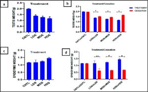
Figure 1. Effect of refined sugar consumption and cessation on weights of Testis and Epididymis.Values are expressed as Mean ± SEM; (*p<0.05, **p<0.001, #p<0.0001), CNTL = control, LDS = Low Dose Sugar, MDS = Medium Dose Sugar, HDS = High Dose Sugar, cCNTL, cLDS, cMDS, cHDS = Cessation groups
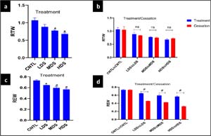
Figure 2. Effect of refined sugar consumption and cessation on relative testicular and epididymal weightsValues are expressed as Mean ± SEM; (*p<0.05, #p<0.0001), ns = not significant, RTW = Relative testicular weight, REW = Relative epididymal weight, CNTL = control, LDS = Low Dose Sugar, MDS = Medium Dose Sugar, HDS = High Dose Sugar, cCNTL = Cessation control group, cLDS, cMDS, cHDS = Cessation groups
The testicular weights of all three sugar-treated groups were significantly lower (p<0.05 for LDS and MDS; p<0.0001 for HDS) than those of the CNTL group (Figure 1a). As shown in Figure 1b, testicular weights in the cessation groups were significantly higher (p<0.05) than those in the corresponding treatment groups. As shown in Figure 1c, epididymal weight did not differ significantly (p>0.05) among the three sugar-treated groups compared with the CNTL group. The effect of refined sugar cessation on epididymal weight is presented in Figure 1d. Epididymal weight was significantly lower (p<0.05, p<0.0001, and p<0.0001) in cLDS, cMDS, and cHDS, respectively, compared with their corresponding sugar-treated groups.Relative testicular weights in the MDS and HDS groups were significantly lower than those in the CNTL group (p<0.05, Figure 2a), whereas relative testicular weights in the cessation groups did not differ significantly (p>0.05) from those in the corresponding treatment groups (Figure p>2b). As illustrated in Figure 2c, relative epididymal weight was significantly lower in all three sugar-treated groups (p<0.05 for LDS; p<0.0001 for MDS and HDS) than in the CNTL group. The effect of refined sugar cessation on relative epididymal weight is presented in Figure 2d. REW was significantly lower (p<0.05, p<0.0001, and p<0.0001) in cLDS, cMDS, and cHDS, respectively, compared with their corresponding sugar-treated groups.
Effect of Oral Refined Sugar Consumption on Sperm Parameters
The effect of oral refined sugar consumption on sperm parameters is presented in Table 1. Sperm count was significantly lower (p<0.0001) in all treatment groups than in the CNTL group. The percentage of sperm with normal motility was significantly reduced (p<0.0001), whereas the percentage of sperm with abnormal motility was significantly higher (p<0.0001) in both the MDS and HDS groups than in the CNTL group. Furthermore, the percentage of sperm with normal morphology was significantly lower (p<0.0001), whereas the percentage of sperm with abnormal morphology was significantly higher (p<0.0001) in all treatment groups than in the CNTL group.
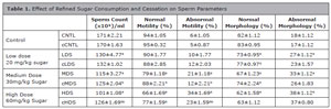
Table 1. Effect of Refined Sugar Consumption and Cessation on Sperm Parameters
asignificant difference versus CNTL at p<0.0001, n=5
bsignificant difference between the treatment group and cessation group at p<0.001,
bbsignificant difference between the treatment group and cessation group at p<0.0001
Effect of Refined Sugar Cessation on Sperm Pa-rameters
The effect of refined sugar cessation on sperm parameters is also presented in Table 1. Sperm count was significantly higher in the cMDS and cHDS groups (p<0.001 and p<0.0001, respectively) than in the MDS and HDS groups. Likewise, the percentage of sperm with normal motility improved significantly in the cMDS and cHDS groups (p<0.001) compared with the MDS and HDS groups, whereas the percentage of sperm with abnormal motility was significantly lower (p<0.01 and p<0.0001, respectively) in both groups compared with the MDS and HDS groups. The percentage of sperm with normal morphology was also significantly higher in cLDS (p<0.001) and cMDS (p<0.001) rats than in LDS and MDS rats, respectively.
Effect of Refined Sugar Consumption on Gonadal Hormones
The effect of refined sugar consumption on gonadal hormones in all experimental groups is presented in Figure 3. LH levels were significantly decreased (p<0.001) in all three sugar treatment groups compared with the CNTL group. Serum FSH levels were also significantly lower (p<0.05) in the MDS group than in the control group. However, FSH levels in the other groups did not differ significantly from those in the control group.
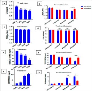
Figure 3. Effect of refined sugar consumption and cessation on Gonadal hormonesValues are expressed as Mean ± SEM; (*p<0.05, **p<0.001, #p<0.0001), ns = not significant, LH = luteinizing hormone, FSH = follicle stimulating hormone, CNTL = control, LDS = Low Dose Sugar, MDS = Medium Dose Sugar, HDS = High Dose Sugar, cCNTL, cLDS, cMDS, cHDS = Cessation group
Across all three sugar treatment groups, testosterone levels were significantly decreased (p<0.05), whereas estrogen levels were significantly increased (p<0.05) compared with the CNTL group.
Effect of Cessation of Refined Sugar on Gonadal Hormones
Gonadal hormone levels in the cessation groups are also presented in Figure 3. LH levels were significantly increased (p<0.05) in the cHDS group compared with the HDS group. Serum FSH levels showed a mild increase in all cessation groups compared with the corresponding sugar treatment groups; however, these increases were not statistically significant (p>0.05). Additionally, testosterone levels were higher in the cessation groups than in their corresponding sugar treatment groups; however, the difference was statistically significant only in cHDS rats compared with HDS rats (p<0.001). Conversely, estrogen levels declined significantly (p<0.001) in cMDS and cHDS compared with MDS and HDS, respectively.
Effect of Refined Sugar Consumption on Testicu-lar Histology
Testicular histopathological slides from the treatment groups are presented in Figure 4. As shown in Figure 4, the control (CNTL) rat showed normal testicular histoarchitecture, characterized by the normal arrangement of seminiferous tubules. In contrast, the slide from LDS rats revealed significant histoarchitectural distortion of the testis, with evidence of vacuolization and shrunken seminiferous tubules (Figure 4B). The slide from MDS rats also showed signs of vacuolization (Figure 4C), whereas the slide from HDS rats similarly showed vacuolization (Figure 4D).
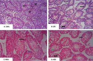
Figure 4. Effect of Refined Sugar Consumption on Testicular Histology. Histological photomicrograph of the testes showing slides from; (A) Control rats with normal histoarchitecture (B) Testes from LDS rat with vacuolization and shrunken seminiferous tubules (C) Testes from MDS rats and (D) Testes from HDS rats, both showing evidence of vacuolization (Magnification X100)
Effect of Refined Sugar Cessation on Testicular Histology
Figure 5 represents photomicrographs of histological slides of rat testes from the cessation groups.
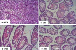
Figure 5. Effect of Refined Sugar Cessation on Testicular Histology. Histological photomicrograph of the testes showing slides from; (A) Control rats with normal histoarchitecture (B) Testes from LDS rat with vacuolization and shrunken seminiferous tubule (C) Testes from MDS rats and (D) Testes from HDS rats, both showing evidence of vacuolization (Magnification X100)
As shown in Figue 5 A, control (cCNTL) rats showed normal testicular histoarchitecture. Figures 5B, 5C, and 5D from cLDS, cMDS, and cHDS rat testes, respectively, showed evidence of vacuolization and shrunken seminiferous tubules.
DISCUSSION
This study provides significant insight into the detrimental effects of oral refined sugar consumption on male reproductive parameters in Wistar rats and explores the potential for recovery following cessation of sugar consumption. The findings demonstrate that refined sugar induces dose-dependent impairments in reproductive organ integrity, semen characteristics, hormonal balance, and testicular histoarchitecture, whereas cessation of sugar intake offers measurable but incomplete restorative effects.
This study revealed a dose-dependent decline in testicular weight (TW) and relative testicular weight (RTW), which is largely indicative of testicular atrophy, likely caused by degeneration of the seminiferous epithelium and impaired spermatogenesis (Rezvanfar et al., 2008). This result is consistent with previous studies associating high sugar intake with testicular structural damage through mechanisms involving oxidative stress and hormonal suppression (Rezvanfar et al., 2008).
The absence of significant differences in absolute epididymal weight (EW) among treatment groups, in contrast to the significant decline in relative epididymal weight (REW) in all sugar-treated groups, further supports REW as a more sensitive marker of subtle toxicological changes. The decline in REW may indicate epididymal dysfunction, impaired spermatogenesis, or impaired sperm maturation capacity (Saalu, 2010; Cooper, 2011). This is further supported in the present study by the observed dose-dependent decline in sperm count in all sugar treatment groups compared with the control group. Furthermore, the decline in both testicular and epididymal indices provides additional evidence of the damaging effect of refined sugar on the male reproductive system.
The partial restoration of testicular weight following cessation of oral sugar administration may suggest that removal of dietary sugar can initiate regenerative processes. However, relative epididymal weight remained significantly lower in cessation groups than in sugar-treated groups, implying persistent epididymal dysfunction. This finding cannot be satisfactorily explained within the scope of the present study, but it may provide a useful basis for future studies exploring the possibility of a slower recovery rate in epididymal tissue than in other testicular tissues. Furthermore, epididymal tissue is composed largely of smooth muscle fibers, which are known to heal by fibrosis following exposure to inflammation (El Ayadi et al., 2020). Therefore, it can be hypothesized that refined sugar consumption induces a significant degree of testicular inflammation, which, in the epididymis, a tubular smooth muscle tissue, resolves by fibrosis following cessation of sugar consumption.
Sperm count, motility, and morphology are critical indicators of male fertility. The current findings reveal a dose-dependent decline in these parameters, particularly in the medium- and high-dose sugar groups. This aligns with previous studies (León-Ramírez et al., 2021), which have linked high sugar consumption to poor semen quality through oxidative stress, mitochondrial dysfunction, and other mechanisms. The significant reduction in sperm motility and increase in abnormal morphology reflect both systemic and localized disruptions in spermatogenesis and sperm maturation.
Notably, the cessation groups showed considerable improvement in sperm quality, with significant recovery of sperm count and motility. This suggests that, although sugar-induced damage may impair spermatogenesis, some of these effects are reversible, particularly when dietary sugar is eliminated early enough (Chiu et al., 2014). However, incomplete recovery of morphology, especially in lower-dose cessation groups (cLDS), may indicate structural alterations that require extended time frames for the production of morphologically normal spermatozoa (Greco et al., 2005; Agarwal et al., 2023).
The endocrine profile in sugar-treated rats showed suppressed serum levels of testosterone, luteinizing hormone (LH), and follicle-stimulating hormone (FSH), with concurrent elevation of estradiol, particularly in rats treated with moderate to high doses of sugar. This pattern of hormonal imbalance supports prior hypotheses that sugar negatively affects the hypothalamic-pituitary-gonadal (HPG) axis, thereby predisposing to hypogonadism and increased systemic inflammation (Ma et al., 2022). Elevated estradiol levels in sugar-treated rats may indicate increased aromatization of testosterone, possibly caused by fat deposition or insulin resistance. Estradiol exerts negative feedback on gonadotropin release, which may have contributed to worsening the reproductive hormonal imbalance (Moenter et al., 2009).
Although cessation of sugar intake reversed some of these deleterious effects, notably increasing LH, FSH, and testosterone levels and reducing estradiol concentrations, these reversals did not appear sufficient to restore adequate gonadal function in the context of the present study. A longer duration of sugar cessation may have a more positive effect on gonadal hormone profiles and should be considered in subsequent studies. It is important to emphasize the observed increase in serum testosterone levels in high-dose sugar-treated rats after the period of sugar cessation, despite the persistence of testicular damage in the histological slides from the same group of rats. This phenomenon may be explained by compensatory endocrine activity of the few surviving Leydig cells, which may have produced testosterone beyond their usual capacity to compensate for the lost cells (Arokoyo et al., 2018).
The histological observations in this study further supported the biochemical and physiological findings, as evidenced by vacuolization and shrinking of the epididymis in the testes of sugar-treated rats. Although these histological observations were significantly limited by the relatively low quality of the photomicrographs, they are consistent with the work of León-Ramírez et al. (2023), who documented similar testicular architectural collapse following long-term exposure to high-sugar diets. These degenerative changes affect both the quantitative and qualitative properties of sperm, thereby contributing to subfertility or infertility.
The histological abnormalities observed after short-term oral administration of refined sugar were not sufficiently reversed after four weeks of sugar withdrawal. This observation suggests that, although metabolic and hormonal corrections may occur relatively quickly after sugar withdrawal, structural regeneration within the testes may depend on additional factors, including the duration of exposure to and withdrawal from refined sugar, as well as the endogenous repair capacity of the tissues. These aspects should be key focuses of subsequent studies on this topic.
This research provides compelling evidence of the reproductive risks associated with refined sugar consumption. Given the global rise in sugar-sweetened food intake, the findings have significant implications for male fertility, especially in the context of the increasing incidence of male factor infertility. The dose-dependent nature of our observations emphasizes the importance of moderation in sugar consumption, especially during critical reproductive windows. However, the fact that this study was carried out in animal subjects should not be overlooked, and the findings may not be exactly reproducible in humans. Direct human studies evaluating male reproductive parameters in mild, moderate, and high refined sugar consumers are necessary to validate the findings of the present study.
Furthermore, the partial reversibility of dysfunction after cessation offers hope for mitigation strategies. Lifestyle interventions, including reduced dietary sugar intake and possibly increased consumption of natural antioxidants, could play a key role in restoring male fertility parameters (Arokoyo et al., 2017). This aligns with WHO dietary recommendations and supports broader public health initiatives aimed at reducing refined sugar consumption.
CONCLUSION
Based on the findings of the current study, refined sugar consumption at low, medium, or high doses impairs male reproductive parameters to varying extents and may result in temporary or permanent damage to male reproductive health. In addition, cessation of sugar intake without further intervention resulted in partial recovery of some hormonal and sperm parameters, as well as some testicular anthropometric measures, suggesting that sugar-induced reproductive impairments may be partially reversible within a few weeks of dietary modification.
ACKNOWLEDGMENTS
The authors appreciate Mr. I.A. Oguntade of the Physiology Laboratory, School of Basic Medical Sciences, Federal University of Technology, Akure, for his technical contributions during the experimental phase of this study.
REFERENCES
Adeyemi D, Arokoyo D, Hamed M, Dare A, Oyedokun P, Akhigbe R. Cardiometabolic Disorder and Erectile Dysfunction. Cell Biochem Biophys. 2024;82:1751-62. PMID: 38907942 DOI: 10.1007/s12013-024-01361-2
Medline
Agarwal A, Cannarella R, Saleh R, Harraz AM, Kandil H, Salvio G, Boitrelle F, Kuroda S, Farkouh A, Rambhatla A, Zini A, Colpi G, Gül M, Kavoussi P, Hamoda TAA, Ko E, Calik G, Toprak T, Pinggera GM, Park HJ, et al. Impact of Antioxidant Therapy on Natural Pregnancy Outcomes and Semen Parameters in Infertile Men: A Systematic Review and Meta-Analysis of Randomized Controlled Trials. World J Mens Health. 2023;41:14-48. PMID: 36102104 DOI: 10.5534/wjmh.220067
Medline
Arokoyo DS, Oyeyipo IP, Du Plessis SS, Chegou NN, Aboua YG. Modulation of Inflammatory Cytokines and Islet Morphology as Therapeutic Mechanisms of Basella alba in Streptozotocin-Induced Diabetic Rats. Toxicol Res. 2018;34:325-32. PMID: 30370007 DOI: 10.5487/ TR.2018.34.4.325
Medline
Bener A, Al-Ansari AA, Zirie M, Al-Hamaq AO. Is male fertility associated with type 2 diabetes mellitus? Int Urol Nephrol. 2009;41:777-84. PMID: 19381857 DOI: 10.1007/s11255-009-9565-6
Medline
Chen L, Xie YM, Pei JH, Kuang J, Chen HM, Chen Z, Li ZW, Fu XY, Wang L, Lai SQ, Zhang ST, Chen ZJ, Lin JX. Sugar-sweetened beverage intake and serum testosterone levels in adult males 20-39 years old in the United States. Reprod Biol Endocrinol. 2018;16:61. PMID: 29935533 DOI: 10.1186/s12958-018-0378-2
Medline
Chiu YH, Afeiche MC, Gaskins AJ, Williams PL, Mendiola J, Jørgensen N, Swan SH, Chavarro JE. Sugar-sweetened beverage intake in relation to semen quality and reproductive hormone levels in young men. Hum Reprod. 2014;29:1575-84. PMID: 24812311 DOI: 10.1093/humrep/deu102
Medline
Cooper TG. The epididymis, cytoplasmic droplets and male fertility. Asian J Androl. 2011;13:130-8. PMID: 21076437 DOI: 10.1038/aja.2010.97
Medline
El Ayadi A, Jay JW, Prasai A. Current Approaches Targeting the Wound Healing Phases to Attenuate Fibrosis and Scarring. Int J Mol Sci. 2020;21:1105. PMID: 32046094 DOI: 10.3390/ijms21031105
Medline
Greco E, Iacobelli M, Rienzi L, Ubaldi F, Ferrero S, Tesarik J. Reduction of the incidence of sperm DNA fragmentation by oral antioxidant treatment. J Androl. 2005;26:349-53. PMID: 15867002 DOI: 10.2164/jandrol.04146
Medline
Johnson RK, Yon BA. Weighing in on added sugars and health. J Am Diet Assoc. 2010;110:1296-9. PMID: 20800119 DOI: 10.1016/j.jada.2010.06.013
Medline
Johnson RK, Appel LJ, Brands M, Howard BV, Lefevre M, Lustig RH, Sacks F, Steffen LM, Wylie-Rosett J; American Heart Association Nutrition Committee of the Council on Nutrition, Physical Activity, and Metabolism and the Council on Epidemiology and Prevention. Dietary sugars intake and cardiovascular health: a scientific statement from the American Heart Association. Circulation. 2009;120:1011-20. PMID: 19704096 DOI: 10.1161/ CIRCULATIONAHA.109.192627
Medline
León-Ramírez YM, Lara-García M, Pacheco P, Lara-García O, Martínez-Gómez M, Cuevas-Romero E, Rodríguez-Antolín J, Nicolás-Toledo L. Histomorphological testicular changes and decrease in the sperm count in pubertal rats induced by a high-sugar diet. Ann Anat. 2021;235:151678. PMID: 33515690 DOI: 10.1016/j.aanat.2021.151678
Medline
León-Ramírez YM, Sánchez EP, Pérez AC, Sánchez-Solís CN, Rodríguez-Antolín J, Nicolás-Toledo L. Dietary intervention in adult rats exposed to a high-sugar diet early in life permanently impairs sperm quality. Ann Endocrinol (Paris). 2023;84:779-89. PMID: 37343721 DOI: 10.1016/j. ando.2023.06.002
Medline
Ma X, Nan F, Liang H, Shu P, Fan X, Song X, Hou Y, Zhang D. Excessive intake of sugar: An accomplice of inflammation. Front Immunol. 2022;13:988481. PMID: 36119103 DOI: 10.3389/fimmu.2022.988481
Medline
Moenter SM, Chu Z, Christian CA. Neurobiological mechanisms underlying oestradiol negative and positive feedback regulation of gonadotrophin-releasing hormone neurones. J Neuroendocrinol. 2009;21:327-33. PMID: 19207821 DOI: 10.1111/j.1365-2826.2009.01826.x
Medline
Moore MC, Connolly CC, Cherrington AD. Autoregulation of hepatic glucose production. Eur J Endocrinol. 1998;138:240-8. PMID: 9539293 DOI: 10.1530/eje.0.1380240.
Medline
Moynihan P. Sugars and Dental Caries: Evidence for Setting a Recommended Threshold for Intake. Adv Nutr. 2016;7:149-56. PMID: 26773022 DOI: 10.3945/an.115.009365
Medline
Rezvanfar M, Sadrkhanlou R, Ahmadi A, Shojaei-Sadee H, Rezvanfar M, Mohammadirad A, Salehnia A, Abdollahi M. Protection of cyclophosphamide-induced toxicity in reproductive tract histology, sperm characteristics, and DNA damage by an herbal source; evidence for role of free-radical toxic stress. Hum Exp Toxicol. 2008;27:901-10. PMID: 19273545 DOI: 10.1177/0960327108102046
Medline
Saalu LC. The incriminating role of reactive oxygen species in idiopathic male infertility: an evidence based evaluation. Pak J Biol Sci. 2010;13:413-22. PMID: 20973394 DOI: 10.3923/pjbs.2010.413.422
Medline
Suresh S, Prithiviraj E, Prakash S. Effect of Mucuna pruriens on oxidative stress mediated damage in aged rat sperm. Int J Androl. 2010;33:22-32. PMID: 19207619 DOI: 10.1111/j.1365-2605.2008.00949.x
Medline
Yang Q, Zhang Z, Gregg EW, Flanders WD, Merritt R, Hu FB. Added sugar intake and cardiovascular diseases mortality among US adults. JAMA Intern Med. 2014;174:516-24. PMID: 24493081 DOI: 10.1001/jamainternmed.2013.13563
Medline