JBRA Assist. Reprod. 2026 2026;00(0):00-00
ORIGINAL ARTICLE
doi: 10.5935/1518-0557.20260083
Effect of platelet-rich plasma on patients with normal endometrium undergoing in vitro fertilization at a reproductive center in Mexico
1Chief of Endocrinology and Reproductive Medicine at Hospital Militar de Especialidades de la Mujer y Neona-tología, Mexico City
2Clinical Sciences Researcher at the Instituto Nacional de Cardiologia Ignacio Chavez, Mexico City
3Associated Researcher at the Faculty of Health Sciences, Universidad Anáhuac México, Huixquilucan
4Chief of the Reproductive Medicine Laboratory and Center at Hospital Militar de Especialidades de la Mujer y Neonatología, Mexico City
CONFLICTS OF INTEREST
No conflicts of interest were declared.
ABSTRACT
Objective: To compare the effect of platelet-rich plas-ma (PRP) on pregnancy rates versus the placebo group in patients with normal endometrium undergoing advanced assisted reproduction treatments.
Methods: Randomized, double-blind clinical trial that compared the use of PRP versus placebo (sterile saline solution) in patients undergoing endometrial preparation for frozen embryo transfer after in vitro fertilization. A total sample size of 52 patients was determined, and the pa-tients were randomized into two groups (n=26 per group). The groups were treated with either PRP or a placebo at the appropriate time. The impact on pregnancy rates and endometrial thickness was analyzed subsequently. Groups were compared using t-tests, U-tests, or chi-squared tests. A p-value<0.05 was considered statistically significant.
Results: The study analyzed an intervention group (PRP) and a control group (placebo). The overall pregnan-cy rate during the study period (n=52) was 38.4%, and the live birth rate was 33.3%. The pregnancy rate was 26.9% in the intervention group and 46.2% in the control group, with no significant difference between the groups (p=0.15). Similarly, no differences were found in endome-trial thickness and endometrial morphology between the groups analyzed (p>0.05).
Conclusion: There are no differences in favor of the PRP or control group in terms of achieving pregnancy, so the use of PRP in normal endometrium does not currently show advantages as an adjuvant to what is already used in women without other factors that alter endometrial re-ceptivity.
Keywords: In vitro fertilization, platelet-rich plasma, pregnancy rate, infertility
INTRODUCTION
Infertility is the inability to conceive after one year of continuous sexual intercourse; approximately 88.7% of women aged 30 and over have already given birth. However, since 2011-2013, the total fertility rate for economically active women has been 1.76 children per woman, which is almost one child fewer than for women not in the labor market (2.75 children per woman) (Sun et al., 2019).
Assisted reproductive technologies (ART) are infertility treatments that involve the manipulation of human oocytes, sperm, or embryos to establish a pregnancy (Remohi et al., 2017). These techniques can be classified as highand low-complexity; the latter include in vitro fertilization (IVF) and embryo transfer (ET). Endometrial proliferation and receptivity are fundamental cyclical processes for reproduction, closely regulated by ovarian function and its hormones (estrogen and progesterone). The endometrium prepares each month, proliferating in response to estrogen and becoming receptive in response to progesterone after ovulation. One factor influencing the success of reproductive treatment is endometrial thickness, which affects embryo receptivity. There is no consensus on the endometrial thickness threshold required for successful implantation; however, a thin endometrium, defined as less than 8 mm, has been identified as a risk factor for implantation failure. Therefore, an adequate endometrial thickness is 8 mm or greater (Makrigiannakis et al., 2021).
In women with a thin endometrium, the literature shows an increased endometrial thickness and in both chemical and clinical pregnancy rates following autologous Platelet-rich plasma (PRP) therapy. Although limited, the literature shows promising results, including increased endometrial thickness, elevated AMH, and decreased FSH levels, as well as improved chemical and clinical pregnancy rates. The lack of standardization in PRP preparation, along with the absence of large-scale randomized controlled trials, should be addressed in future studies (Sharara et al., 2021).
PRP is defined as a plasma fraction obtained from autologous blood with a platelet concentration higher than that of plasma under basal conditions. It not only has a high platelet count but also contains growth factors actively secreted by platelets and multiple proteins involved in cell adhesion, providing the necessary structural support for cell migration, proliferation, and tissue growth (Nazari et al., 2019).
The first clinical use of PRP in thin endometrium was reported by Chang et al. (2015) from China. This study involved five women undergoing IVF who had a thin endometrium following conventional hormone therapy. Endometrial thickness improved in all cases, and four women conceived successfully (Chang et al., 2015). Following this, several descriptive studies (case reports and series) and pilot studies were published, showing promising results. In 2017, a successful pregnancy was reported after the use of intrauterine PRP to improve endometrial receptivity in a 45-year-old woman with primary infertility and a history of two failed IVF cycles (Sharara et al., 2021). After multiple studies, it was concluded that PRP plays a positive role in promoting endometrial proliferation and improving embryo implantation and clinical pregnancy rates in women with thin endometrium during frozen embryo transfer cycles. Additionally, a significant improvement in endometrial vascularization has been reported (Eftekhar et al., 2018; Barros-Delgadillo & Castillo-Ruiz, 2017).
However, despite the above, there is limited research to determine whether its concomitant use with other assisted reproductive therapies helps improve pregnancy rates in the management of infertility. Many studies focus on pathologies in patients with implantation failure due to a thin endometrium (<7 mm), but there is no evidence supporting the use of PRP as an adjunct to improve clinical and chemical pregnancy rates in patients with adequate endometrial thickness (>7 mm). Therefore, the objective of this research was to evaluate the effect of platelet-rich plasma (PRP) on pregnancy rates in women with normal endometrium undergoing complex assisted reproductive treatments.
Although clinical trials have explored PRP use in patients with thin endometrium, there is no evidence supporting its application or effectiveness in women with normal endometrium undergoing complex assisted reproduction procedures. While the benefits of PRP for thin endometrium are well established, its effects on women with normal endometrium who struggle with infertility remain unclear. We hypothesized that PRP improves pregnancy rates in women who have not succeeded with IVF. We believe the many potential benefits of PRP could assist these women, who do not have a thin endometrium but still face fertility challenges.
MATERIAL AND METHODS
Study design and population
This prospective, randomized, double-blind clinical trial was conducted at the fertility clinic of the Military Hospital of Specialties for Women and Neonatology in Mexico City from March 2024 to June 2025. Patients with primary or secondary infertility were included and randomly assigned into two groups: the platelet-rich plasma (PRP) group and the sterile saline (placebo) group. The sampling was non-probabilistic. The sample size calculation was performed using the formula for studies with hypothesis testing of two proportions (focusing on the main outcome: achieving pregnancy after PRP use), as observed in a previous study (Nazari et al., 2019).At the start of the study, the main researcher randomized treatment for each patient using an online calculator (https://calcular.me/formar-equipos-al-azar), which generated a list allocating 26 patients to group A (PRP group) and 26 to group B (placebo group) (Figure 1).
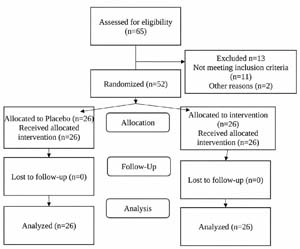
Figure 1. CONSORT Flow diagram
The attending physician at the infertility clinic identified patients who had started the endometrial preparation protocol and met the selection criteria, invited them to participate, obtained informed consent, scheduled the intervention appointment (preparation and placement of PRP or placebo), and notified the embryologist of the patient’s name and the appointment date. The embryologist received information on each participant and assigned her to the group according to the random list. On the day of the appointment, a nurse drew a blood sample and delivered subsamples to the clinical laboratory, which verified the platelet concentration, and to the embryologist, who prepared the PRP. A sample of the prepared PRP was sent to the clinical laboratory to verify the increase in platelet concentration. Once the quality of the PRP preparation was confirmed, the embryologist prepared the syringe with either PRP or a placebo, wrapped it in black tape to conceal its contents, and delivered it to the attending physician for intrauterine placement. Neither the patient nor the attending physician knew which treatment had been administered. Subsequently, the patient returned for the embryo transfer procedure. The researcher who performed the statistical analysis learned the participants’ group assignments at the end of the recruitment period, once the sample was complete.Inclusion criteria were: patients up to 40 years of age, with no history of recurrent pregnancy loss, with uterine cavity remodeling, and undergoing frozen-thawed embryo transfer cycles. Exclusion criteria were: patients with insufficient information in the clinical history, uterine anomalies, hematological disorders, implantation failure, fresh embryo transfer, thin endometrium, low ovarian reserve, and those who withdrew from the study. None of the patients met the criteria for hysteroscopy; in fact, major uterine surgeries such as myomectomy were exclusion criteria. All patients had normal transvaginal ultrasounds. Patients whose embryos were classified as poor quality according to the ASEBIR classification were excluded. Poor-quality embryos were excluded.This study was authorized by the Research and Ethics Committee of the Military Hospital for Women and Neonatology and strictly adheres to the current guidelines of the General Health Law, Chapter I, Article 17, on Ethical Aspects of Research Involving Human Subjects (Mexico). International standards for research involving human subjects were also followed, particularly regarding confidentiality, to ensure discretion at all times. Furthermore, the Declaration of Helsinki (Finland, 1964), as updated (Fortaleza, Brazil, 2013), and the preparation of informed consent forms were also observed.
Study protocol
Regarding the controlled ovarian stimulation technique, the antagonist protocol and stimulated endometrial preparation were used, with oral estradiol valerate at a fixed dose (6mg). An initial endovaginal ultrasound was then performed to confirm that the endometrium was ≥ 8 mm, and the initial endometrial size and morphology were recorded. The patient was then scheduled for a follow-up appointment 48 hours later to prepare PRP from autologous blood. A complete blood count was performed to determine the patient’s platelet concentration. The blood (15ml) was immediately centrifuged at 1200 rpm for 10 minutes in a vascular centrifuge, resulting in three layers: red blood cells at the bottom, plasma in the supernatant, and a buffy coat between them. The plasma layer and the buffy coat layer were collected in another tube and centrifuged again at 2500 rpm for 7 minutes. Platelet sediment was obtained and mixed with the supernatant to yield 1 ml of PRP. A second complete blood count was performed on the PRP, and the platelet concentration was assessed for any increase (Nazari et al., 2019; Eftekhar et al., 2018). The concentration of PRP obtained was, on average, three times the baseline, as corroborated by the blood count. PRP was administered 24 to 48 hours before the start of the single-dose progesterone. No stimuli were applied to activate platelets. However, leukocyte concentration was not determined.No ultrasound was performed after the PRP infusion. The PRP was used in the proliferative phase, before starting progesterone, that is, between 24 and 48 hours before, and with an endometrial thickness of at least 8 mm on ultrasound. Patients were scheduled for an outpatient procedure appointment. In the modified lithotomy position, a vaginal speculum was inserted after cleansing the vaginal cavity with a solution. The external cervical was visualized, and an intrauterine insemination catheter was inserted. With the catheter in place, the nurse provided the physician with a syringe containing either 1.0 mL of PRP or a placebo. The patient and the attending physician were unaware of the contents of the syringes administered to their respective groups. Double blinding was achieved by using syringes labeled with a sticker that completely concealed the contents. The only individuals who knew the contents of each syringe were the research support nurse and the embryologist responsible for preparing the PRP. Progesterone pearls 200mg (apply 400mg vaginally every 12 hours) were administered beginning 48 hours after this procedure and continued for six days. Progesterone is essential for endometrial receptivity, as it transforms the proliferative endometrium into a secretory one after ovulation, allowing embryo implantation. It prepares the uterus, promotes maturation, reduces contractions, and creates a suitable immune environment. Following this period, the embryo transfer was performed. On the same day as the embryo transfer, an endovaginal ultrasound was performed to measure endometrial thickness and morphology. Two weeks later, the beta-human chorionic gonadotropin (beta-hCG) level was measured in the blood to determine if pregnancy had been achieved. Ultrasound measurements were performed by a single operator.For endometrial preparation, estradiol valerate (6 mg) via an oral fixed-dose regimen; two ultrasounds: the first on day 2 or 3 of the cycle, the second a week later, when a normal endometrium was observed. From an ultrasound perspective, the endometrium is classified into three patterns: A, AB, and B. Pattern A is trilaminar, with three distinct hyperechoic (white) lines: one central and two lateral, with two hypoechoic (dark gray/black) areas between them. It is characteristic of the proliferative phase, when estrogen levels are high, and indicates healthy endometrial growth and maturation. In pattern AB, the endometrium becomes more homogeneous. Its echogenicity is similar to that of the surrounding myometrium. The triple line is less prominent or barely visible. This pattern is usually seen during the transition from the proliferative to the luteal phase (around ovulation). Finally, pattern BC is homogeneous and hyperechoic, with good myometrial differentiation. The central echogenic line is not visible, which is typical of the luteal phase (after ovulation) under the influence of progesterone. Global literature generally indicates that the presence of pattern A (trilaminar) is associated with higher implantation and pregnancy rates.For each case, the following variables were analyzed: age; baseline hormonal studies (anti-Müllerian hormone, follicle-stimulating hormone, and estradiol); antral follicle count; number of follicles on the day of ovulation capture; number of oocytes captured; number of mature oocytes (metaphase II); fertilized oocytes; fertilization technique; days of stimulation; number of embryos obtained and their quality; endometrial thickness and morphology; and whether there was a pregnancy, that is, whether it ended in pregnancy and how many of them ended in live newborns. Two embryos were transferred per patient. All patients who underwent transfer had progesterone levels below 1.2 ng/ ml on the day progesterone was started; progesterone was not measured on the day of transfer.The study’s data sources were medical records extracted from patient files and a structured, ad hoc personal epidemiological questionnaire. Platelet-rich plasma (PRP) has been used in many fields of medicine, but in reproductive medicine, research is limited on whether its use alongside other assisted reproductive therapies improves pregnancy rates in the management of infertility. Most studies focus on patients with implantation failure due to a thin endometrium (<8mm), but there is no evidence that PRP is used as an adjunct to improve clinical and chemical pregnancy rates in patients with adequate endometrial thickness (>7mm). Therefore, this research focused on evaluating the effect of PRP on pregnancy rates in women undergoing complex assisted reproductive treatments. The need to document variables of response to ovarian stimulation arose to ensure homogeneous and comparable groups and to gather evidence for or against PRP improving outcomes in these high-complexity treatments.
Statistical analysis
Qualitative variables were described as absolute counts and percentages, and compared between groups using the chi-square or Fisher’s exact test. For quantitative variables, the distribution was assessed using the Shapiro-Wilk test, and those with a normal distribution were described by their mean and standard deviation and compared between groups using the independent samples t-test. Those that did not have a normal distribution were described by median and interquartile range and compared between groups using the Mann-Whitney U test. SPSS version 20 statistical software was used to perform these analyses. A p-value <0.05 was considered statistically significant.
RESULTS
Table 1 describes the general characteristics of the participants and the main types of infertility. There were no statistically significant differences between the groups in any of these characteristics (age, body mass index, infertility factor, type of infertility, duration of infertility, and previous pregnancy). Regarding hormone levels in the population (Table 2), anti-Müllerian hormone, FSH, estradiol, prolactin, and TSH were all within normal ranges. No statistically significant differences were found between the groups.
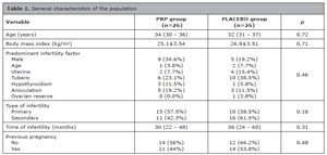
Table 1. General characteristics of the population
Statistical analysis: Mann-Whitney U test and chi-square test.
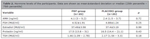
Table 2. Hormone levels of the participants. Data are shown as mean±standard deviation or median (25th percentile - 75th percentile)
AMH: Antimullerian hormone
FSH: Follicle-stimulating hormoneTSH: Thyroid-stimulating hormone
Statistical analysis: Mann-Whitney U test and chi-square test.
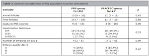
Table 3. General characteristics of the population (ovarian stimulation)
FIV: In Vitro Fertilization
MII: Metaphase IIICSI: Intracytoplasmic injection
Statistical analysis: Mann-Whitney U test and Chi-square test.
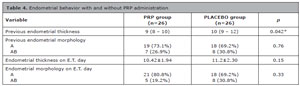
Table 4. Endometrial behavior with and without PRP administration
E.T.=Embryo transfer.
Statistical analysis: Mann-Whitney U test and Chi-square test.

Table 5. Pregnancy rate after embryo transfer
Statistical test used: chi-square.
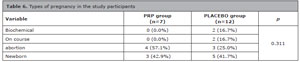
Table 6. Types of pregnancy in the study participants
*p<0.05=statistical significance; Statistical test used: chi-square.
DISCUSSION
The use of PRP has emerged in the medical field as a tool for improving health. Specifically, in the context of reproduction, the endometrium has been of interest. This is why we decided to conduct the present study, as embryo transfer and implantation are vital steps in achieving pregnancy. The endometrium is a complex but fundamental tissue for achieving pregnancy. To this day, we still lack knowledge of many details about it and its full role in implantation; that is, the mechanisms underlying conception are multifactorial. Studies worldwide are increasingly focused on understanding its receptivity (Hiraoka et al., 2023) and on how certain adjuvants can help achieve pregnancy (Sharara et al., 2021), which motivated the present study.
PRP used in patients with thin endometrium has, as previously mentioned, influenced pregnancy rates in this patient population and may also play a role in the immunological or endometrial response in patients undergoing reproductive treatments (Anitua et al., 2025). However, few studies demonstrate its effectiveness as an adjuvant in patients with a normal endometrium.
In fact, a very recent study also investigated the effect of PRP in patients with normal endometrium. However, it reported different results from ours, as the clinical pregnancy rate, embryo implantation rate, and live birth rate were higher in the platelet-rich plasma group than in the control group, even after logistic regression analysis (OR: 2.379, 95% CI 1.137-4.977, p=0.021; OR: 2.107, 95% CI 1.006-4.412, p=0.048) (Liu et al., 2025). Notably, in that study, PRP infusion was administered to some patients at 24, 48, or 72 hours.
In the present study, we observed an overall pregnancy rate of 38.4%, with a live birth rate of 46.2% in the placebo group. However, there was no statistically significant difference compared to the PRP group, which had a pregnancy rate of 26.9% (p=0.150). Therefore, there is no evidence to support the use of PRP in normal endometrium. These findings contrast with those of another study, in which the live birth rate was 44% in the PRP group (n=34) versus 20% in the control group (n=30) (Chang et al., 2019). That study also measured PDGF-AB and TGF-β levels in whole blood using an immunoenzymatic assay to assess appropriate PRP processing. In the present study, this situation was verified only by a control blood biometry, and leukocyte concentration was not evaluated.
Similarly, another study conducted with patients with recurrent implantation failure showed a higher percentage of live births, clinical pregnancies, and live births in patients treated with platelet-rich plasma (p<0.0001), with a higher rate of miscarriages in the control group (Nazari et al., 2022a). However, these findings contrast with those of the present study, which was carried out in patients without a history of implantation failure, who have a better reproductive prognosis.
Another study similarly focused on whether PRP use improved the pregnancy rate and found a higher live birth rate in the PRP group than in the control group (n=41/27), with rates of 29.7% vs. 18%, respectively (Xu et al., 2022). These figures were lower than those observed in our study, which found no differences between the groups. This could also be influenced by the fact that this group of patients already had a poor prognosis due to their medical history, compared to those in our study, who were generally undergoing at most their second embryo transfer, given our more stringent selection criteria.
With respect to the miscarriage rate in the PRP group (57.1%), it is known that many miscarriages in patients undergoing assisted reproductive technology are due to chromosomal abnormalities in the embryo. However, there is no evidence linking PRP use to the miscarriages in our study group. A study has shown that intrauterine infusion of PRP in patients with recurrent miscarriages undergoing IVF can increase the likelihood of a live birth (Nazari et al., 2022b). Regarding the types of pregnancies achieved in these patients, the control group had a higher proportion of live births (41.7% of pregnancies achieved) than the PRP group. Still, no difference was found between this variable and any other type of pregnancy, such as those ending in miscarriage or other types of loss. This contrasts with another study that reported miscarriages in 12% of patients undergoing platelet-rich plasma therapy (Frantz et al., 2020). In our study the intervention group had a high rate of miscarriage. However, our findings regarding the number of non-viable pregnancies are consistent with a pilot study where PRP treatment did not increase the implantation or live birth rate and therefore had no impact on the development of losses (Madhavan et al., 2018).
Similarly, despite the benefits of PRP described in other studies, our exploratory assessment did not show a significant change in endometrial thickness. This differs from a study in which, after the second application of PRP, endometrial thickness increased by more than 2 mm (Nazari et al., 2019), and also contrasts with another study that examined the effect of PRP on the endometrium in patients undergoing fresh and frozen-thawed embryo transfer with polycystic ovary syndrome and low ovarian reserve, which reported a significant increase in endometrial thickness in the PRP-treated group (Dogra et al., 2022). Regarding endometrial morphology, our study observed no differences between the groups, which aligns with another study that similarly found no increase in endometrial thickness, changes in morphology, or pregnancy rate in patients undergoing PRP infusion, particularly in those with recurrent implantation failure (Tehraninejad et al., 2021).
These conclusions from different studies differ widely due to a lack of standardization in the PRP technique. Moreover, most comparisons with previous literature, as discussed above, should be approached with caution, given that our study of women with normal endometrium may not generalize to most prior studies conducted in women with thin endometrium, recurrent implantation failure, or other reproductive contexts.
Limitations
Despite the study’s prospective and exploratory nature, the findings should not be considered definitive, given differences between our population and other groups with different infertility characteristics. Pregnancy rates between the groups (26.9% vs. 46.2%) show a clinically relevant difference but are not statistically significant. Given the available sample size, a type II error is possible. Further studies of this type, but with a larger number of participants, are needed to validate our results. This is true even with an adequate sample size calculation; ultimately, a multicenter study would be beneficial.The assessment of changes in endometrial thickness between the proliferative phase and the day of transfer was exploratory and is reported as observed, as is clinical practice. Since these changes are indeed subjective and operator-dependent, and were exploratory, they are not suitable for strong causal inference regarding the PRP effect.PGT-A analysis is not currently a mandatory indication for all patients, according to consensus in ESHRE, ASRM, and AMMR. Furthermore, PGT-A was not performed because it was unavailable at the hospital and outside the scope of the study. In the absence of embryo genetic data, miscarriages cannot be attributed to embryonic chromosomal abnormalities, and since the study is underpowered to evaluate miscarriage differences, the observed miscarriage pattern should be interpreted with caution. Another major limitation was the lack of determination of leukocyte concentration in the PRP sample. Although we did not stimulate the plasma sample to activate platelets, we cannot rule out variations in leukocyte concentrations among patients, which may lead to heterogeneity in the PRP’s biological properties. Also, progesterone was not quantified on the day of embryo transfer to confirm it was above 10 ng/mL, which would have ensured cycle adequacy and ruled out an effect on the implantation outcome.
CONCLUSIONS
In in vitro fertilization (IVF) treatments in patients with normal endometrium, PRP infusion showed pregnancy rates similar to those of placebo infusion. The study did not find statistically significant evidence of benefit from PRP. The use of PRP in women with normal endometrium did not demonstrate advantages as an adjunct to conventional management in women without other factors that alter endometrial receptivity; therefore, its routine use in this population is not recommended.
Study ID at clinical trials. Gov: HMEMN/0039/24
REFERENCES
Anitua E, de la Fuente M, Prado R, Alkhraisat MH. Platelet-rich plasma activation: are there differential effects in reproductive medicine? J Ovarian Res. 2025;18:84. PMID: 40275404 DOI: 10.1186/s13048-025-01667-6
Medline
Chang Y, Li J, Chen Y, Wei L, Yang X, Shi Y, Liang X. Autologous platelet-rich plasma promotes endometrial growth and improves pregnancy outcome during in vitro fertilization. Int J Clin Exp Med. 2015;8:1286-90. PMID: 25785127
Medline
Chang Y, Li J, Wei LN, Pang J, Chen J, Liang X. Autologous platelet-rich plasma infusion improves clinical pregnancy rate in frozen embryo transfer cycles for women with thin endometrium. Medicine (Baltimore). 2019;98:e14062. PMID: 30653117 DOI: 10.1097/MD.0000000000014062
Medline
Dogra Y, Singh N, Vanamail P. Autologous platelet-rich plasma optimizes endometrial thickness and pregnancy outcomes in women with refractory thin endometrium of varied aetiology during fresh and frozen-thawed embryo transfer cycles. JBRA Assist Reprod. 2022;26:13-21. PMID: 34296832 DOI: 10.5935/1518-0557.20210037
Medline
Eftekhar M, Neghab N, Naghshineh E, Khani P. Can autologous platelet rich plasma expand endometrial thickness and improve pregnancy rate during frozen-thawed embryo transfer cycle? A randomized clinical trial. Taiwan J Obstet Gynecol. 2018;57:810-3. PMID: 30545532 DOI: 10.1016/j.tjog.2018.10.007
Medline
Frantz N, Ferreira M, Kulmann MI, Frantz G, Bos-Mikich A, Oliveira R. Platelet-rich plasma as an effective alternative approach for improving endometrial receptivity - a clinical retrospective study. JBRA Assist Reprod. 2020;24:442-6. PMID: 32293825 DOI: 10.5935/1518-0557.20200026
Medline
Hiraoka T, Osuga Y, Hirota Y. Current perspectives on endometrial receptivity: a comprehensive overview of etiology and treatment. J Obstet Gynaecol Res. 2023;49:2397-409. PMID: 37527810 DOI: 10.1111/jog.15759
Medline
Liu XH, He LX, Li M, Tian ZP, Qin AQ, Yao J. Effects of autologous platelet-rich plasma intrauterine perfusion on clinical outcomes in recurrent implantation failure patients with non-thin endometrium undergoing frozen-thawed embryo transfer. Arch Gynecol Obstet. 2025;311:1423-33. PMID: 40105934 DOI: 10.1007/s00404-025-07983-y
Medline
Makrigiannakis A, Makrigiannakis F, Vrekoussis T. Approaches to improve endometrial receptivity in case of repeated implantation failures. Front Cell Dev Biol. 2021;9:613277. PMID: 33796523 DOI: 10.3389/ fcell.2021.613277
Medline
Nazari L, Salehpour S, Hoseini S, Zadehmodarres S, Azargashb E. Effects of autologous platelet-rich plasma on endometrial expansion in patients undergoing frozen-thawed embryo transfer: a double-blind RCT. Int J Reprod Biomed. 2019;17:443-8. PMID: 31508569 DOI: 10.18502/ijrm.v17i6.4816
Medline
Nazari L, Salehpour S, Hosseini MS, Hashemi Moghanjoughi P. The effects of autologous platelet-rich plasma in repeated implantation failure: a randomized controlled trial. Hum Fertil. 2020;23:209-13. PMID: 30714427 DOI: 10.1080/14647273.2019.1569268
Medline
Nazari L, Salehpour S, Hosseini S, Sheibani S, Hosseinirad H. The effects of autologous platelet-rich plasma on pregnancy outcomes in repeated implantation failure patients undergoing frozen embryo transfer: a randomized controlled trial. Reprod Sci. 2022a;29:993-1000. PMID: 34651260 DOI: 10.1007/s43032-021-00669-1
Medline
Nazari L, Salehpour S, Hosseini S, Hashemi T, Borumandnia N, Azizi E. Effect of autologous platelet-rich plasma for treatment of recurrent pregnancy loss: a randomized controlled trial. Obstet Gynecol Sci. 2022b;65:266-72. PMID: 35345084 DOI: 10.5468/ogs.21261
Medline
Sharara FI, Lelea LL, Rahman S, Klebanoff JS, Moawad GN. A narrative review of platelet-rich plasma (PRP) in reproductive medicine. J Assist Reprod Genet. 2021;38:1003-12. PMID: 33723748 DOI: 10.1007/ s10815-021-02146-9
Medline
Sun H, Gong TT, Jiang YT, Zhang S, Zhao YH, Wu QJ. Global, regional, and national prevalence and disability-adjusted life-years for infertility in 195 countries and territories, 1990-2017: results from a global burden of disease study, 2017. Aging. 2019;11:10952-91. PMID: 31790362 DOI: 10.18632/aging.102497
Medline
Tehraninejad ES, Kashani NG, Hosseini A, Tarafdari A. Autologous platelet-rich plasma infusion does not improve pregnancy outcomes in frozen embryo transfer cycles in women with history of repeated implantation failure without thin endometrium. J Obstet Gynaecol Res. 2021;47:147-51. PMID: 32840031 DOI: 10.1111/jog.14445
Medline
Xu Y, Hao C, Fang J, Liu X, Xue P, Miao R. Intrauterine perfusion of autologous platelet-rich plasma before frozen-thawed embryo transfer improves the clinical pregnancy rate of women with recurrent implantation failure. Front Med. 2022;9:850002. PMID: 35425782 DOI: 10.3389/ fmed.2022.850002
Medline