JBRA Assist. Reprod. 2026 2026;00(0):00-00
ORIGINAL ARTICLE
doi: 10.5935/1518-0557.20260071
Blastocyst Diameter and Collapse as Predictors of Live Birth Following Single Blastocyst Transfer: A Time-Lapse Morphometric Analysis
1State Key Laboratory of Reproductive Medicine and Offspring Health, Center for Reproductive Medicine, Institute of Women, Children and Reproductive Health, Shandong University, 250012, China
2National Research Center for Assisted Reproductive Technology and Reproductive Genetics, Shandong University, Jinan, Shandong, 250012, China
3Key Laboratory of Reproductive Endocrinology (Shandong University), Ministry of Education, Jinan, Shandong, 250012, China
4Shandong Technology Innovation Center for Reproductive Health, Jinan, Shandong, 250012, China
5Shandong Provincial Clinical Research Center for Reproductive Health, Jinan, Shandong, 250012, China
6Shandong Key Laboratory of Reproductive Research and Birth Defect Prevention (Under Construction), Jinan, Shandong, 250012, China
7Research Unit of Gametogenesis and Health of ART-Offspring, Chinese Academy of Medical Sciences (No.2021RU001), Jinan, Shandong, 250012, China
CONFLICT OF INTERESTS
The authors report no conflict of interests.
ABSTRACT
Objective: To investigate whether blastocyst diameter measured immediately prior to transfer and the occurrence of blastocyst collapse predict live birth outcomes after single blastocyst transfer (SBT).
Methods: This retrospective cohort study included 476 patients undergoing their first IVF/ICSI cycle with SBT on day 5. Blastocyst diameter was manually measured approximately 0.5 hours before transfer, and collapse events (≥50% reduction in blastocoel area) were assessed using time-lapse microscopy (TLM). Blastocysts were graded morphologically as Best, Good, or Fair. Multivariable logistic regression and Cochran-Armitage trend tests were used to evaluate associations with live birth.
Results: Live birth was achieved in 174 patients (36.6%). Blastocysts in the live birth group had significantly larger diameters (199.83±18.68 vs. 192.34±20.84μm, p<0.001) and a lower incidence of collapse (4.6% vs. 12.3%, p=0.006). After adjusting for maternal age, endometrial thickness, BMI, FSH, and AMH, multivariable logistic regression showed that blastocysts with diameters >200μm had a 2.49-fold higher likelihood of live birth compared to those ≤180μm (95% CI: 1.46-4.25, p=0.001), while collapsed blastocysts had 57% reduced odds (OR 0.43, 95% CI: 0.19-0.98, p=0.044). Endometrial thickness (OR 7.31, 95% CI: 2.48-21.52, p<0.001) and younger age (OR 0.94 per year, 95% CI: 0.89-0.99, p=0.017) were also independently associated with live birth. A diameter-dependent increase in live birth rates was observed across all morphological grades (p<0.05 for trend).
Conclusion: After adjusting for maternal age and endometrial thickness, blastocyst diameter measured near the time of transfer and the absence of collapse are independently associated with live birth after SBT, suggesting their potential as adjunctive selection criteria. These quantitative and dynamic parameters provide valuable adjuncts to conventional morphological grading, potentially improving embryo selection and IVF outcomes.
Keywords: single embryo transfer (SET), live birth rates, blastocyst diameter, blastocyst collapse, time-lapse microscopy (TLM)
INTRODUCTION
Assisted reproductive technologies (ART), particularly in vitro fertilization (IVF) with single blastocyst transfer (SBT), represent a cornerstone of modern infertility treatment. This approach offers substantial hope to affected couples while significantly reducing the risks associated with multiple pregnancies. Although maternal factors such as younger age (Ata et al., 2021; Harton et al., 2013; Immediata et al., 2020; Reig et al., 2020) and adequate endometrial thickness (Değer et al., 2023; Mahutte et al., 2022; Ruiz-Alonso et al., 2013) are well-established predictors of live birth success, the intrinsic properties of the blastocyst itself play a paramount role in determining clinical outcomes.
The efficacy of SBT fundamentally depends on selecting a blastocyst with optimal implantation potential. Traditional morphological grading systems, which evaluate expansion stage, inner cell mass (ICM), and trophectoderm (TE) quality (Gardner & Lane, 1997; Gardner & Schoolcraft, 1999), remain the most widely adopted methods for blastocyst selection despite acknowledged inter-observer variability (Curchoe et al., 2023; Fordham et al., 2022). Recent advances in time-lapse microscopy (TLM) have enabled continuous, non-invasive observation of preimplantation development, revealing dynamic morphological events potentially linked to embryonic competence (Bori et al., 2022; Desai et al., 2014; Fishel et al., 2018; Giménez et al., 2023; Liu et al., 2014; 2016). However, while these technological innovations have not yet supplanted conventional morphological assessment, TLM also provides a unique opportunity to quantify static morphological features dynamically.
A key clinical dilemma arises when multiple blastocysts exhibit similar morphological grades or only one embryo is available for transfer. In such cases, static morphological features are insufficient to distinguish implantation potential, and dynamic or quantitative parameters become increasingly important. Emerging evidence suggests that blastocoelic expansion is a reliable predictor of SBT outcomes (Ahlström et al., 2013; Thompson et al., 2013). However, blastocoelic expansion is a continuous developmental process, and blastocysts classified as stage “4” (fully expanded) under traditional grading still show substantial size variability. This highlights the need for quantitative assessment of blastocoelic expansion, with diameter measurement being the most intuitive approach.
Although several studies have reported a correlation between larger blastocyst diameter and improved reproductive outcomes (Lagalla et al., 2015; Sciorio et al., 2021; 2025), robust evidence directly linking specific diameter thresholds to live birth rates within established morphological grades remains lacking, partly due to limitations such as small sample sizes, non-real-time measurements potentially missing the maximum diameter, and a failure to account for variations in blastocyst quality.
Importantly, blastocyst diameter is not a static parameter but fluctuates dynamically due to expansion-contraction cycles. Collapse events during these cycles may reflect compromised cellular integrity and have been independently associated with reduced implantation potential (Gazzo et al., 2020; Marcos et al., 2015; Sciorio et al., 2020a;b).
To address these knowledge gaps, we conducted a retrospective cohort study to examine the impact of blastocyst diameter and collapse incidence on live birth rates following SBT in IVF cycles. By comparing clinical and embryological parameters between successful and unsuccessful cycles, this study aims to establish refined criteria for blastocyst quality assessment that integrate both static and dynamic morphological features.
MATERIALS AND METHODS
Patients
This retrospective study included 476 patients who underwent IVF/ICSI treatment at the State Key Laboratory of Reproductive Medicine and Offspring Health, Center for Reproductive Medicine, Institute of Women, Children and Reproductive Health, Shandong University, between January 2019 and December 2023. All patients were in their first treatment cycle, received a single blastocyst transfer on day 5, and had confirmed live birth outcomes.
Oocyte retrieval, embryo culture and embryo transfer
Oocyte retrieval was performed by transvaginal ultrasound-guided follicular aspiration 36 hours after hCG administration. Cumulus-oocyte complexes (COCs) were cultured for 4 hours at 37.0°C and 6.0% CO₂ until insemination. For conventional in vitro fertilization (IVF), COCs were incubated with spermatozoa for 5 hours. The presence of two polar bodies in zygotes was confirmed after removal of cumulus cells by gentle pipetting. Zygotes were then placed in a time-lapse incubator (EmbryoScope, Vitrolife). For intracytoplasmic sperm injection (ICSI), oocyte denudation was performed using hyaluronidase and gentle pipetting. Only metaphase II oocytes were injected. Following insemination, all embryos were cultured in the time-lapse incubator under 6% CO₂ and 5% O₂ until day 6. Sequential culture media (G1 and G2, Vitrolife) were used. Specifically, the change from G1 to G2 medium was performed strictly on the morning of Day 3 (approximately 68-72 h post-insemination) for every single embryo. This procedure was carried out by a small team of experienced embryologists following a standardized operating protocol to minimize any variation in manipulation time or technique. The EmbryoScope was programmed to capture images of each embryo every 15 minutes across seven focal planes to monitor key developmental events.Blastocysts were evaluated and transferred on day 5 if they met or exceeded grade 4BC according to Gardner and Lane’s criteria (Gardner & Lane, 1997; Gardner & Schoolcraft, 1999). In total, 476 blastocysts from 476 patients (one blastocyst per patient) were included in the study, all of which were fresh embryo transfers.
Evaluation of time-lapse imaging
An experienced embryologist, blinded to clinical outcomes, analyzed embryo development using EmbryoViewer software (Vitrolife). Blastocyst quality on day 5 was assessed based on three morphological parameters: inner cell mass (ICM), trophectoderm (TE), and expansion/hatching (EH) grades. According to the scoring system by Gardner and Schoolcraft, blastocysts were subsequently categorized into three groups:
• Best: EH(4), ICM(A) + TE(A)
• Good: EH(4), ICM(A) + TE(B), or ICM(B) + TE(A)
• Fair: EH(4), ICM(B) + TE(B), or ICM(B) + TE(C)
Blastocyst Collapse Evaluation
Blastocyst collapse was defined as a reduction in the blastocoel area (indicated by a red circle) by ≥50% (Figure 1).
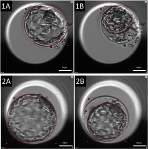
Figure 1. Morphological dynamics of blastocysts illustrating collapse and contraction
The red circle outlines the blastocoel area. (1A, 1B): A blastocyst collapse event, defined as a ≥50% reduction in blastocoel area, from 21,993 μm2 to 9,615 μm2. (2A, 2B): A blastocyst contraction event, characterized by a <50% reduction in blastocoel area, from 30,059 μm2 to 18,291 μm2.
Blastocyst Diameter Measurement Blastocyst diameter (μm) was manually measured approximately 0.5 hours before embryo transfer, following the method described by Almagor et al. (2016). The time points at which 60 blastocysts reached specific diameters were recorded (Figure 2).
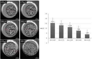
Figure 2. Morphological and morphokinetic characteristics of blastocysts with varying diameters
Left panel: Representative images of blastocysts at different diameters (160μm, 170μm, 180μm, 200μm, 220μm) and at the initiation of blastocyst hatching (tBH). The red line indicates the measured diameter. Right panel: Bar chart comparing the average time (in hours) from reaching specific diameter milestones (t160, t170, t180, t200, t220) to tBH for a cohort of 60 day-5 blastocysts.
Statistical analysis
All analyses were conducted using SPSS Statistics 17 (IBM Inc., NY, USA), with statistical significance set at p<0.05. Continuous variables were compared using the t-test, and categorical variables were compared using the chi-square test or Fisher’s exact test, as appropriate. Multivariable logistic regression was performed to evaluate the independent associations of blastocyst diameter, collapse status, endometrial thickness, age, BMI, FSH, and AMH with live birth. All these variables were entered simultaneously into the model, and adjusted odds ratios (ORs) with 95% confidence intervals (CIs) were calculated. A p-value<0.05 was considered statistically significant. The Cochran-Armitage trend test was applied to assess dose-response relationships between diameter categories and live birth rates within each blastocyst grade.
RESULTS
Patient Characteristics
A total of 476 patients undergoing single blastocyst transfer were included, of whom 174 (36.6%) achieved live birth and 302 (63.4%) did not. As shown in Table 1, maternal age and endometrial thickness significantly differed between the two groups. Patients with live birth were younger (32.2±3.9 vs. 33.3±4.1 years; p=0.006) and had greater endometrial thickness (1.1±0.2 vs. 1.0±0.2 cm; p<0.001). No significant differences were observed in body mass index, basal FSH, AMH levels, infertility duration, or number of aspirated oocytes. The distribution of primary and secondary infertility types was also comparable between groups (p=0.215).
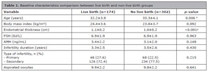
Table 1. Baseline characteristics comparison between live birth and non-live birth groups
a Differences were considered significant when p values<0.05
Blastocyst Development Parameters
Blastocyst characteristics showed clinically significant associations with live birth outcomes (Table 2). The mean diameter of transferred blastocysts was significantly larger in the live birth group than in the non-live birth group (199.83±18.68 vs. 192.34±20.84μm; p<0.001). The incidence of blastocyst collapse was markedly lower in live birth cases (4.6% vs. 12.3%; p=0.006).
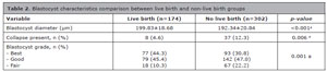
Table 2. Blastocyst characteristics comparison between live birth and non-live birth groups
a Differences were considered significant when p values<0.05
Multivariable Analysis of Factors Associated with Live Birth
To quantify the independent associations of blastocyst parameters and clinical characteristics with live birth, a multivariable logistic regression analysis was performed, adjusting for all variables listed in Table 3. The results showed that collapsed blastocysts had 57% lower odds of live birth compared to non-collapsed blastocysts (OR=0.43, 95% CI: 0.19-0.98; p=0.044). Blastocysts with a diameter >200μm had a 2.49-fold higher likelihood of live birth than those ≤180μm (OR=2.49, 95% CI: 1.46-4.25; p=0.001), while the 180-200μm group also showed significantly higher odds (OR=2.10, 95% CI: 1.20-3.67; p=0.009). Endometrial thickness was strongly associated with live birth (OR=7.31, 95% CI: 2.48-21.52; p<0.001), and increasing maternal age reduced the likelihood of live birth (OR=0.94 per year, 95% CI: 0.89-0.99; p=0.017). Body mass index (OR=1.05, 95% CI: 0.99-1.11; p=0.092), basal FSH (OR=1.03, 95% CI: 0.92-1.16; p=0.570), and AMH (OR=1.04, 95% CI: 0.95-1.15; p=0.387) were not statistically significant in the multivariable model.
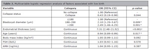
Table 3. Multivariable logistic regression analysis of factors associated with live birth
a Differences were considered significant when p values<0.05
Stratified Analysis by Blastocyst Grade
Given the significant influence of morphological grading on live birth, we further analyzed the effect of diameter within each grade. As shown in Table 4, a consistent diameter-dependent increase in live birth rates was observed across all morphological categories (Best, Good, and Fair), with significant trend p-values (all <0.05). These results indicate that blastocyst diameter provides additive prognostic value beyond conventional morphological assessment.
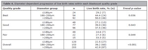
Table 4. Diameter-dependent progression of live birth rates within each blastocyst quality grade
Trend p-values calculated using Cochran-Armitage test for ordered proportions
DISCUSSION
This study demonstrates that blastocyst diameter measured immediately before transfer and the occurrence of blastocyst collapse were independently associated with live birth following SBT. These associations persisted even after controlling for maternal age and endometrial thickness, highlighting their potential as adjuncts to traditional morphological grading.
The strong association between larger blastocyst diameter and improved live birth rates aligns with the concept that the degree of blastocoele expansion reflects functional embryonic competence and developmental potential. Given that blastocyst diameter fluctuates dynamically due to expansion and contraction cycles, our measurement taken approximately 0.5 h before embryo transfer likely captures a state closest to the embryo’s condition at transfer, providing a clinically relevant snapshot. This temporal proximity is crucial, as it may best represent the embryo’s readiness for implantation. Our results are consistent with prior studies reporting that larger blastocyst diameter or area correlates with higher implantation and clinical pregnancy rates (Lagalla et al., 2015; Sciorio et al., 2021; 2025). For instance, Sciorio et al. found that blastocyst width was significantly larger in cycles resulting in clinical pregnancy (Sciorio et al., 2021), while another study reported significantly higher implantation and clinical pregnancy rates for blastocysts with a diameter ≥ 170 µm (Sciorio et al., 2025). Moreover, the graded response we observed, with live birth odds increasing across diameter categories (≤180μm, 180-200μm, >200μm), supports the potential utility of diameter as a continuous variable in embryo assessment models.
Importantly, this diameter-dependent increase in live birth rates persisted even after stratification by conventional morphological grade (Best, Good, Fair), suggesting that diameter provides additive prognostic value within each grade category. This is particularly relevant in clinical scenarios where multiple embryos share similar morphological scores.
In parallel, the significant reduction in live birth odds associated with blastocyst collapse (odds ratio [OR] 0.43) underscores the detrimental impact of this dynamic event. Blastocyst collapse, defined as a transient, substantial reduction in blastocoel volume, is thought to reflect compromised cellular integrity or function (Gazzo et al., 2020; Marcos et al., 2015; Sciorio et al., 2020b). Our findings corroborate previous time-lapse microscopy (TLM) studies linking collapse events to reduced implantation potential (Gazzo et al., 2020; Marcos et al., 2015; Sciorio et al., 2020a;b). Marcos et al. (2015) first systematically described this phenomenon in human embryos and associated it with lower implantation success. Subsequent multicentre studies have confirmed that spontaneously collapsing blastocysts are less likely to implant (Sciorio et al., 2020a;b). Nevertheless, it is worth noting that some evidence suggests collapse should not be evaluated in isolation but rather integrated with other morphokinetic parameters (Bodri et al., 2016).
Beyond these morphological and dynamic features, the clinical implications of our findings must be considered within the broader context of embryo-endometrial synchrony, which is critical for successful implantation (Teh et al., 2016). The transfer of a developmentally competent blastocyst must coincide with the receptive endometrial window, as even minor asynchrony exceeding ±1.5 days can drastically reduce implantation potential (Navot et al., 1991; Teh et al., 2016). Blastocysts transferred on day 5 are generally considered to exhibit higher implantation potential, largely due to better alignment with this window (Shapiro et al., 2008). In this context, blastocyst diameter, which reflects the degree of blastocoele expansion, serves as a tangible, quantifiable marker of embryonic readiness. A larger diameter near the time of transfer likely indicates an embryo that has achieved an advanced developmental stage, thereby enhancing the probability of synchrony with a receptive endometrium, a notion supported by studies identifying expansion as a predictor of live birth (Du et al., 2016).
Our developmental timing analysis (Figure 2) provides further mechanistic insight: early-stage blastocysts (e.g., 160, 170μm) required over 10 hours longer to reach the hatching stage (tBH) compared to their more expanded counterparts (e.g., 220μm). This prolonged timeline implies that such embryos must still undergo substantial expansion and generate sufficient internal pressure to complete zona pellucida escape. Consequently, transferring a smaller-diameter blastocyst risks significant asynchrony, as the embryo may still be in the midst of zona thinning and rupture when the implantation window peaks, thereby compromising implantation success. Thus, diameter not only correlates with developmental competence but also serves as a practical proxy for estimating embryonic temporal alignment with the endometrium.
Several limitations of our study warrant consideration. First, the retrospective design inherently limits causal inference. Second, diameter measurement was performed manually at a single time point; although this captures the state near transfer, it does not fully characterize the dynamic expansion-contraction history of each blastocyst. Automated, continuous morphometric analysis, as employed in some studies (Sciorio et al., 2021; 2025), though laborious (Lagalla et al., 2015), could provide a more comprehensive assessment in the future. Third, our study included only fresh embryo transfer cycles, which may limit the generalizability of our findings to frozen-thawed blastocyst transfer cycles. In frozen cycles, vitrification and warming processes could potentially alter blastocyst morphology, expansion dynamics, and collapse behavior, possibly influencing the predictive value of diameter and collapse for live birth outcomes. Therefore, caution should be exercised when extrapolating our results to cryopreserved embryo transfer settings. Future studies incorporating both fresh and frozen-thawed cycles are warranted to validate whether these morphometric parameters remain equally predictive across different transfer strategies. Fourth, although the sequential culture protocol was strictly standardized, we acknowledge that the in vitro environment itself, including medium renewal, may subtly influence embryo development. While standardization minimizes internal bias, the observed associations may be context-dependent and could vary under different culture systems (e.g., uninterrupted single-step media). Therefore, caution is needed when extrapolating these thresholds to laboratories using substantially different protocols, and multi-center studies are warranted to validate the findings.
In conclusion, even after adjusting for key confounders such as maternal age and endometrial thickness, integrating blastocyst diameter measurement and collapse assessment into standard embryo evaluation protocols provides independent and additive value in predicting live birth outcomes in SBT cycles. These quantitative and dynamic parameters complement traditional morphological grading by capturing subtle differences in embryonic developmental potential, enabling embryologists to make more nuanced selection decisions-especially when choosing between morphologically similar embryos. Ultimately, these refinements may contribute to improved success rates in infertility treatment.
Funding
REFERENCES
Ahlström A, Westin C, Wikland M, Hardarson T. Prediction of live birth in frozen-thawed single blastocyst transfer cycles by pre-freeze and post-thaw morphology. Hum Reprod. 2013;28:1199-209. PMID: 23477908 DOI: 10.1093/humrep/det054
Medline
Almagor M, Harir Y, Fieldust S, Or Y, Shoham Z. Ratio between inner cell mass diameter and blastocyst diameter is correlated with successful pregnancy outcomes of single blastocyst transfers. Fertil Steril. 2016;106:1386-91. PMID: 27567431 DOI: 10.1016/j.fertnstert.2016.08.009
Medline
Ata B, Kalafat E, Somigliana E. A new definition of recurrent implantation failure on the basis of anticipated blastocyst aneuploidy rates across female age. Fertil Steril. 2021;116:1320-7. PMID: 34332750 DOI: 10.1016/j.fertnstert.2021.06.045
Medline
Bodri D, Sugimoto T, Yao Serna J, Kawachiya S, Kato R, Matsumoto T. Blastocyst collapse is not an independent predictor of reduced live birth: a time-lapse study. Fertil Steril. 2016;105:1476-83.e3. PMID: 26940789 DOI: 10.1016/j.fertnstert.2016.02.014
Medline
Bori L, Meseguer F, Valera MA, Galan A, Remohi J, Meseguer M. The higher the score, the better the clinical outcome: retrospective evaluation of automatic embryo grading as a support tool for embryo selection in IVF laboratories. Hum Reprod. 2022;37:1148-60. PMID: 35435210 DOI: 10.1093/humrep/deac066
Medline
Curchoe CL, Bormann C, Hammond E, Salter S, Timlin C, Blankenship Williams L, Gilboa D, Seidman D, Campbell A, Morbeck D. Assuring quality in assisted reproduction laboratories: assessing the performance of ART Compass - a digital ART staff management platform. J Assist Reprod Genet. 2023;40:265-78. PMID: 36637586 DOI: 10.1007/s10815-023-02713-2
Medline
Değer U, Kalı Z, Çağıran F. Endometrial thickness is a good predictor of clinical pregnancy and live birth rates in fresh and frozen-thawed embryo transfer cycles. Eur Rev Med Pharmacol Sci. 2023;27:6351-8. PMID: 37458652 DOI: 10.26355/eurrev_202307_32995
Medline
Desai N, Ploskonka S, Goodman LR, Austin C, Goldberg J, Falcone T. Analysis of embryo morphokinetics, multinucleation and cleavage anomalies using continuous time-lapse monitoring in blastocyst transfer cycles. Reprod Biol Endocrinol. 2014;12:54. PMID: 24951056 DOI: 10.1186/1477-7827-12-54
Medline
Du QY, Wang EY, Huang Y, Guo XY, Xiong YJ, Yu YP, Yao GD, Shi SL, Sun YP. Blastocoele expansion degree predicts live birth after single blastocyst transfer for fresh and vitrified/warmed single blastocyst transfer cycles. Fertil Steril. 2016;105:910-9.e1. PMID: 26776910. DOI: 10.1016/j.fertnstert.2015.12.014
Medline
Fishel S, Campbell A, Montgomery S, Smith R, Nice L, Duffy S, Jenner L, Berrisford K, Kellam L, Smith R, Foad F, Beccles A. Time-lapse imaging algorithms rank human preimplantation embryos according to the probability of live birth. Reprod Biomed Online. 2018;37:304-13. PMID: 30314885 DOI: 10.1016/j.rbmo.2018.05.016
Medline
Fordham DE, Rosentraub D, Polsky AL, Aviram T, Wolf Y, Perl O, Devir A, Rosentraub S, Silver DH, Gold Zamir Y, Bronstein AM, Lara Lara M, Ben Nagi J, Alvarez A, Munné S. Embryologist agreement when assessing blastocyst implantation probability: is data-driven prediction the solution to embryo assessment subjectivity? Hum Reprod. 2022;37:2275-90. PMID: 35944167 DOI: 10.1093/humrep/deac171
Medline
Gardner DK, Lane M. Culture and selection of viable blastocysts: a feasible proposition for human IVF? Hum Reprod Update. 1997;3:367-82. PMID: 9279408. DOI: 10.1093/humupd/3.4.367
Medline
Gardner DK, Schoolcraft WB. Culture and transfer of human blastocysts. Curr Opin Obstet Gynecol. 1999;11:307-11. PMID: 10369209. DOI: 10.1097/00001703-199906000-00013
Medline
Gazzo E, Peña F, Valdéz F, Chung A, Velit M, Ascenzo M, Escudero E. Blastocyst contractions are strongly related with aneuploidy, lower implantation rates, and slow-cleaving embryos: a time lapse study. JBRA Assist Reprod. 2020;24:77-81. PMID: 31524340 DOI: 10.5935/1518-0557.20190053
Medline
Giménez C, Conversa L, Murria L, Meseguer M. Time-lapse imaging: Morphokinetic analysis of in vitro fertilization outcomes. Fertil Steril. 2023;120:218-27. PMID: 37364673 DOI: 10.1016/j.fertnstert.2023.06.015
Medline
Harton GL, Munné S, Surrey M, Grifo J, Kaplan B, McCulloh DH, Griffin DK, Wells D; PGD Practitioners Group. Diminished effect of maternal age on implantation after preimplantation genetic diagnosis with array comparative genomic hybridization. Fertil Steril. 2013;100:1695-703. PMID: 24034939 DOI: 10.1016/j.fertnstert.2013.07.2002
Medline
Immediata V, Patrizio P, Parisen Toldin MR, Morenghi E, Ronchetti C, Cirillo F, Levi-Setti PE. Twenty-one year experience with intrauterine inseminations after controlled ovarian stimulation with gonadotropins: maternal age is the only prognostic factor for success. J Assist Reprod Genet. 2020;37:1195-201. PMID: 32215826 DOI: 10.1007/s10815-020-01733-4
Medline
Lagalla C, Barberi M, Orlando G, Sciajno R, Bonu MA, Borini A. A quantitative approach to blastocyst quality evaluation: morphometric analysis and related IVF outcomes. J Assist Reprod Genet. 2015;32:705-12. PMID: 25854656 DOI: 10.1007/s10815-015-0479-3
Medline
Liu Y, Chapple V, Feenan K, Roberts P, Matson P. Time-lapse deselection model for human day 3 in vitro fertilization embryos: the combination of qualitative and quantitative measures of embryo growth. Fertil Steril. 2016;105:656-62.e1. PMID: 26616439 DOI: 10.1016/j.fertnstert.2015.11.003
Medline
Liu Y, Chapple V, Roberts P, Matson P. Prevalence, consequence, and significance of reverse cleavage by human embryos viewed with the use of the Embryoscope time-lapse video system. Fertil Steril. 2014;102:1295-300.e2. PMID: 25225070 DOI: 10.1016/j.fertnstert.2014.07.1235
Medline
Mahutte N, Hartman M, Meng L, Lanes A, Luo ZC, Liu KE. Optimal endometrial thickness in fresh and frozen-thaw in vitro fertilization cycles: an analysis of live birth rates from 96,000 autologous embryo transfers. Fertil Steril. 2022;117:792-800. PMID: 35109980. DOI: 10.1016/j.fertnstert.2021.12.025.
Medline
Marcos J, Pérez-Albalá S, Mifsud A, Molla M, Landeras J, Meseguer M. Collapse of blastocysts is strongly related to lower implantation success: a time-lapse study. Hum Reprod. 2015;30:2501-8. PMID:26355116. DOI: 10.1093/humrep/dev216
Medline
Navot D, Scott RT, Droesch K, Veeck LL, Liu HC, Rosenwaks Z. The window of embryo transfer and the efficiency of human conception in vitro. Fertil Steril. 1991;55:114-8. PMID: 1986951. DOI: 10.1016/S0015-0282(16)54069-2
Medline
Reig A, Franasiak J, Scott RT Jr, Seli E. The impact of age beyond ploidy: outcome data from 8175 euploid single embryo transfers. J Assist Reprod Genet. 2020;37:595-602. PMID: 32173784 DOI: 10.1007/s10815-019-01661-4
Medline
Ruiz-Alonso M, Blesa D, Díaz-Gimeno P, Gómez E, Fernández-Sánchez M, Carranza F, Carrera J, Vilella F, Pellicer A, Simón C. The endometrial receptivity array for diagnosis and personalized embryo transfer as a treatment for patients with repeated implantation failure. Fertil Steril. 2013;100:818-24. PMID: 23756099 DOI: 10.1016/j.fertnstert.2013.05.004
Medline
Sciorio R, Thong KJ, Pickering SJ. Spontaneous blastocyst collapse as an embryo marker of low pregnancy outcome: A time-lapse study. JBRA Assist Reprod. 2020a;24:34-40. PMID: 31397550 DOI: 10.5935/1518-0557.20190044
Medline
Sciorio R, Herrer Saura R, Thong KJ, Esbert Algam M, Pickering SJ, Meseguer M. Blastocyst collapse as an embryo marker of low implantation potential: a time-lapse multicentre study. Zygote. 2020;28:139-147. PMID: 31928572 DOI: 10.1017/S0967199419000819
Medline
Sciorio R, Thong D, Thong KJ, Pickering SJ. Clinical pregnancy is significantly associated with the blastocyst width and area: a time-lapse study. J Assist Reprod Genet. 2021;38:847-55. PMID: 33471232 DOI: 10.1007/s10815-021-02071-x
Medline
Sciorio R, Greco PF, Tramontano L, Gullo G, Greco E. Morphometric blastocyst assessment: A retrospective study examining the relationship between blastocyst diameter and area and pregnancy outcomes in assisted reproduction technology cycles. J Clin Med. 2025;14:2827. PMID: 40283656 DOI: 10.3390/jcm14082827
Medline
Shapiro BS, Daneshmand ST, Garner FC, Aguirre M, Ross R. Contrasting patterns in in vitro fertilization pregnancy rates among fresh autologous, fresh oocyte donor, and cryopreserved cycles with the use of day 5 or day 6 blastocysts may reflect differences in embryo-endometrium synchrony. Fertil Steril. 2008;89:20-6. PMID: 17224151 DOI: 10.1016/j.fertnstert.2006.08.092
Medline
Teh WT, McBain J, Rogers P. What is the contribution of embryo-endometrial asynchrony to implantation failure? J Assist Reprod Genet. 2016;33:1419-30. PMID: 27480540 DOI: 10.1007/s10815-016-0773-6
Medline
Thompson SM, Onwubalili N, Brown K, Jindal SK, McGovern PG. Blastocyst expansion score and trophectoderm morphology strongly predict successful clinical pregnancy and live birth following elective single embryo blastocyst transfer (eSET): a national study. J Assist Reprod Genet. 2013;30:1577-81. PMID: 24114628 DOI: 10.1007/s10815-013-0100-4
Medline