JBRA Assisted Reproduction 2025;29(Suppl.2 SBRA 2025):22
29th Annual Congress of the SBRA. São Paulo/SP - Brazil, 2025
doi: 10.5935/1518-0557.20263543
P-010. Anti-Müllerian Hormone levels and their association with oocyte maturation rate in controlled ovarian stimulation
JBRA Assisted Reproduction 2025;29(Suppl.2 SBRA 2025):22
Poster Presentation
29th Annual Congress of the SBRA. São Paulo/SP - Brazil, 2025
doi: 10.5935/1518-0557.20263543
P-010. Anti-Müllerian Hormone levels and their association with oocyte maturation rate in controlled ovarian stimulation
1Androlab - Clínica da Fertilidade - Curitiba - PR - Brasil
Objective: While it is established that serum anti-Müllerian hormone (AMH) levels and antral follicle count are predictors of ovarian reserve and outcomes of controlled ovarian stimulation, the specific relationship between AMH levels and oocyte maturation rate remains unclear. Therefore, this study aims to investigate if AMH levels are correlated with the oocyte maturation rate.
Methods: This retrospective cohort study was conducted between January 2021 and July 2025, involving 644 controlled ovarian stimulation cycles at a private fertility clinic in the South of Brazil. The inclusion criteria were patients who had AMH serum levels measured, authorized the use of their data, and underwent controlled ovarian stimulation cycles for either in vitro fertilization (IVF) or oocyte cryopreservation. Serum anti-Müllerian hormone (AMH) levels were categorized into 5 groups. The oocyte maturation rate (proportion of Metaphase II oocytes) and the mean number of oocytes retrieved in each group were analysed.
Results: As shown in Table 1, a clear inverse relationship was observed between AMH levels and the number of oocytes retrieved per cycle. The mean number of oocytes per cycle increased progressively from 3.90 in patients with AMH ≤1 ng/mL to 21.22 in those with AMH >5 ng/mL. Conversely, the mean proportion of mature oocytes (MII) remained relatively stable across AMH categories, ranging from 70.00% to 79.13%. Interestingly, patients with AMH ≤1 ng/mL exhibited the highest MII proportion, despite having the lowest total oocyte yield. There was no statistically significant difference in maturation rates among the groups, reinforcing that diminished ovarian reserve does not necessarily impair oocyte maturation capacity.
Conclusion: Although AMH is strongly associated with the number of oocytes retrieved, the proportion of mature oocytes remained high and stable across all AMH ranges. Notably, even patients with AMH ≤1 ng/mL maintained a good MII rate, indicating that a low ovarian reserve is typically associated with fewer oocytes overall, but not necessarily with a reduced proportion at the MII stage — the maturity level recommended for fertilization or vitrification. This suggests that diminished ovarian reserve primarily affects follicular yield rather than the intrinsic capacity of oocytes to reach the MII stage. These findings can help reassure and motivate such patients during ovarian stimulation, fostering realistic yet optimistic expectations.
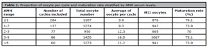
Table 1. Proportion of oocyte per cycle and maturation rate stratified by AMH serum levels.