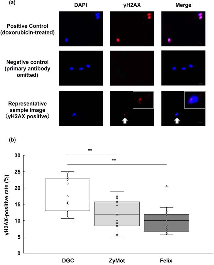
Figure 1. γH2AX immunofluorescence staining and comparison of γH2AX-positive rates among different sperm selection methods(a) Immunofluorescence staining of human sperm nuclei with DAPI and γH2AX. DNA was counterstained with DAPI (blue), and γH2AX immunofluorescence (red) was used to detect nuclear γH2AX signals as a surrogate marker associated with DSB-related DNA damage signaling and chromatin alterations. The positive control (sperm treated with 20 µM doxoru-bicin for 1 h) exhibited clear γH2AX signals, whereas omission of the primary antibody served as a negative control. These controls were used to validate staining performance and were not intended to represent method-specific imag-es. Arrows indicate γH2AX-positive nuclei. Panels from left to right: DAPI, γH2AX, and merged images. Scale bar=5 µm.(b) Comparison of sperm γH2AX-positive rates among different sperm selection methods. Box plots indicate the median, interquartile range (IQR), and individual data points. Overall differences were tested by the Friedman test, followed by Wil-coxon signed-rank tests with Bonferroni correction. Effect sizes are reported as Hodges-Lehmann estimated paired differ-ences (percentage points) with 95% confidence intervals. Asterisks indicate statistically significant differences (**adjusted p<0.01). DAPI, 4',6-diamidino-2-phenylindole.