JBRA Assist. Reprod. 2026;00(0):00-00
ORIGINAL ARTICLE
doi: 10.5935/1518-0557.20260105
Importance of anatomical knowledge in approaches to female fertility
1Pontifical Catholic University of Minas Gerais, School of Medicine, Campus Contagem, Brazil
2University of Jose do Rosario Vellano, School of Medicine, UNIFENAS, Belo Horizonte, Brazil
3College of Natural Science, Michigan State University, Michigan, USA
4Discipline of Anatomy and Human Reproduction, Pontifical Catholic University of Minas Gerais, Brazil
5Human Reproduction Center of the Department of Gynecology, University of São Paulo Medical School, USP, Brazil
CONFLICT OF INTEREST
None.
ABSTRACT
This study aimed to highlight the importance of using cadavers, fresh frozen cadavers, and innovative methodologies such as digital tables and 3D glasses in human anatomy education, particularly in the study of the female pelvis. A total of 900 questionnaires administered between May 2025 and March 2026 were analyzed. Cadavers were the predominant method used by all participants (100%). Fresh frozen cadavers were used by 24%, digital tables by 64%, and 3D glasses by 28%. While anatomical models are popular, cadavers were considered the most effective for studying the female pelvis because of their ability to provide a comprehensive three-dimensional perspective. Practical anatomical sessions also offer emotional preparation for clinical practice.
Keywords: female pelvis, human anatomy, medical education, cadaver dissection, human reproduction
INTRODUCTION
The history of and commitment to understanding the importance of this science for health-related degrees have been fundamental and varied throughout history (Winkelmann, 2007). Since ancient times, different methods have been used to study human anatomy (VanSickle et al., 2022). Some of these methods are listed in this paper. Andrea Vesalius’ work, titled "De Humani Corporis Fabrica," redefined concepts and descriptions, elevating the teaching of human anatomy. This work introduced new study methods and questioned misconceptions. His text proved innovative in anatomical investigations, strongly associating images with agents of anatomical knowledge. He was the first anatomist to dissect the human body (Conroy, 2023).
Anatomical knowledge is essential for the professional practice of anyone in the healthcare field, who deals with the human body throughout their professional life. This includes knowledge of histology, embryology, semiology, and gynecology. Throughout the history of academic studies, human anatomy has refined its centrality, emphasizing its urgency and commitment to quality education in healthcare.
The history of anatomical assessment of the female reproductive tract is interesting and reflects well on the geniuses of reproductive medicine pioneers from the early years of medical practice, focusing on the uterus, fallopian tubes, and peritoneal cavity. This paper aims to highlight the value of cadavers and new methodologies for teaching human anatomy, such as digital tables and 3D glasses (Alvero, 2024; Geampana & Perrotta, 2023). Additionally, it aimed to showcase a practical method that benefits the anatomical study of the female pelvis.
MATERIALS AND METHODS
A descriptive study was conducted based on data collection through the administration of a questionnaire containing exclusively objective questions related to the teachers of human anatomy. The questionnaire was sent via email to the participants between May 2025 and March 2026. Professionals were asked whether they employed, and whether their instructors utilized any technology in the classroom. Finally, the participants were asked about the most favored method for studying the female pelvis. The research followed the Regulatory Norms for Research involving human subjects.
RESULTS
In total, 908 questionnaires were completed. Seven were incomplete and one was excluded. Thus, 900 questionnaires administered between May/ 2025 and March 2026 were analyzed. Cadavers emerged as the predominant practical teaching method most favored by the participants for the study of the female pelvis, accounting for all 900 participants (100%). Fresh frozen cadavers were used by 216 participants (24%). A digital table was used by 576 (64%), and 3D glasses were used by 252 participants (28%) (Table 1).
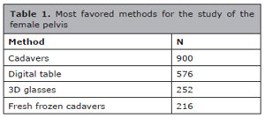
Table 1. Most favored methods for the study of the female pelvis
DISCUSSION
Although technology has revolutionized the world, bringing more dynamism to the population, it seems less conclusive regarding the study of anatomy for healthcare professionals. While anatomical models are available on a wide scale, are considered easy to use, and have good long-term knowledge retention efficacy, they still distort the real spatial understanding of anatomy, making students feel disoriented and frustrated with the methods (Sugand et al., 2010; Yammine & Violato, 2015; den Harder et al., 2022).
Anatomical models offer a three-dimensional representation of the human body or specific organs, allowing students to visualize structures in their real forms and provide a more concrete understanding of anatomical relationships than two-dimensional images in books or screens. Furthermore, the use of anatomical models provides a hands-on experience that helps students develop a deep understanding of body structures in various situations. It serves as a foundation for learning to perform clinical breast examinations during undergraduate studies, which is an important yet complex skill (Collett et al., 2009; Ameer & Peterson, 2025).
Students can explore the location, size, relationship, and function of anatomical structures. These models allow for the repetition and study of different body systems in isolation, which contributes to further enhancing students’ identification skills and spatial memories. Another important factor is that anatomical models are often used as communication tools between healthcare professionals and patients (Asensio Romero et al., 2018; Kavvadia et al., 2023). They assist in explaining medical conditions, surgical procedures, and treatments in a clearer and more visual manner, allowing patients to better understand the procedure and their health conditions, with an emphasis on the study of the female pelvis (Bogomolova et al., 2023).
The digital table is one of the most advanced technologies for 3D anatomy visualization by schools and medical institutions worldwide. It was featured in TEDTalks, PBS, Fuji TV, and various other magazines because of its innovative approach to digital anatomy. The surgical table combined with radiology software and clinical content from methods (Armstrong et al., 2017). This allows users to visualize anatomy exactly as they would on a fresh cadaver. Individual structures are precisely reconstructed, resulting in unprecedented accuracy and dissectible real anatomy in 3D. The anatomy of the female pelvis is presented as a fully interactive experience, in a life-size format.
This technology offers advantages for learning human anatomy beyond what a cadaver can offer. There are no chemicals to handle, no unpleasant odors, no recurring costs for facilities, and no regulations. Additionally, this revolutionary 3D glass system allows users to create various anatomical realities in a matter of seconds. Educators and students can dissect and manipulate anatomical structures while fully immersed in virtual reality. This software was developed using technological advancements adopted from gaming and graphics, as well as medical imaging and modeling methods that have only become available on the market (Lim et al., 2016; O’Reilly et al., 2016).
This application covers all body systems and can be taught using systemic dissection by simply ‘peeling’ away any structure. Each structure is clinically labeled from the smallest ligaments to the nerves and vessels (Hildebrandt, 2010). It is used for patient education and illustrative purposes. A user of the Med version, for example, can fully interact with the anatomy of the ‘shell’ muscles, follow nerve roots, examine skeletal anatomical landmarks, understand muscle insertions and origins, and look at minute details of organs. High-school students in biology classes can better understand various body systems in a fascinating three-dimensional manner. A viewer is first downloaded to the client’s machine and used to dynamically interact with the 3D interactivity with extremely small file sizes.
There is also a discussion about foundational obstetric and midwifery textbooks teaching that clinical pelvimetry and the Caldwell–Moloy classification system can be used to assess the pelvic capacity of pregnant patients. However, the sample on which these techniques were developed betrayed the authors’ bias and did not represent the sample needed to address their interest in obstetric outcomes (VanSickle et al., 2022).
The high cost of digital dissection tables and virtual reality glasses makes them inaccessible to all socioeconomic niches, and the technology cannot replace the sensation of touch provided by studying cadavers, and the lack of tactile feedback. Users often note that digital models may lack the realistic representation of anatomical depth and color, which favors humanization. Anatomical dissection remains a cornerstone in the training of healthcare professionals, providing the necessary foundation for understanding the structures and functions of the human body. In this context, the use of cadavers, especially fresh frozen cadavers allows students to develop observation and dissection skills and experiences that cannot be fully achieved through theoretical studies alone. This hands-on experience enriches academic training and enables a direct connection between theory and practice (Ghosh, 2017).
The dissection of fresh cadavers offers a closer experience of clinical practice. Fresh specimens provide realistic tissue texture and color, unlike the rigidity found in specimens preserved in formaldehyde, which, while useful, does not provide the same level of detail and texture (Cundiff et al., 2001). Research has shown that the use of fresh cadavers can improve students’ confidence in their surgical and clinical skills. This confidence is crucial because many medical procedures require high levels of manual dexterity and anatomical understanding.
In addition to traditional dissection, the introduction of innovative technologies, such as digital tables and 3D glasses, creates a dynamic learning environment. These tools allow students to interactively visualize anatomical structures, thereby facilitating the comprehension of complex concepts. For example, augmented reality can provide an integrated view that allows students to see not only the external anatomy but also internal structures, such as blood vessels and nerves (Briceag et al., 2015). Studies indicate that integrating these technologies is associated with increased student motivation and engagement, thereby contributing to more effective learning.
For instance, in the study of the female pelvis, the combination of dissection with digital technologies enhances the visualization of anatomical nuances and improves student learning. This creates a dynamic learning environment in which students can explore anatomy in multiple dimensions, thereby facilitating the assimilation of complex information. Another significant aspect to consider is the emotional preparation students develop when handling cadavers. Practical experience with cadavers not only serves as a technical learning opportunity, but also sensitizes students to the realities of clinical practice. By dealing with the dead, students are forced to confront feelings related to the fragility of life and the importance of respect for the human body (Chang et al., 2018). Highlighted that exposure to cadavers during training contributed to a deeper academic knowledge. This sensitization is vital for empathetic healthcare professionals who understand the emotional implications of patient care. By developing a deeper connection with human anatomy, students are encouraged to approach patient care with greater humanity.
CONCLUSION
In summary, the combination of fresh and preserved cadavers, with innovative methodologies, such as digital tables and 3D glasses, offers a robust approach for teaching human anatomy. These complementary methods better prepare students to address the challenges of clinical practice. Therefore, anatomy education is not just limited to learning about structures and functions but also involves the development of well-rounded professionals who are technically proficient and empathetic. Anatomical dissection remains an indispensable educational tool that promotes a holistic understanding of anatomy and patient care, and prepares students for future challenges in clinical practice.
REFERENCES
Alvero R. Anatomic fertility assessment: everything old is new again. Fertil Steril. 2024;121:921. PMID: 38703169 DOI: 10.1016/j.fertnstert.2024.04.001
Medline
Ameer MA, Peterson DC. Anatomy, abdomen and pelvis: uterus. In: StatPearls [Internet]. Treasure Island: StatPearls Publishing; 2025. PMID: 29262069
Medline
Armstrong SC, Showell M, Stewart EA, Rebar RW, Vanderpoel S, Farquhar CM. Baseline anatomical assessment of the uterus and ovaries in infertile women: a systematic review of the evidence on which assessment methods are the safest and most effective in terms of improving fertility outcomes. Hum Reprod Update. 2017;23:533-47. PMID: 28903473 DOI: 10.1093/humupd/dmx019
Medline
Asensio Romero L, Asensio Gómez M, Prats-Galino A, Juanes Méndez JA. 3D models of female pelvis structures reconstructed and represented in combination with anatomical and radiological sections. J Med Syst. 2018;42:37. PMID: 29333592 DOI: 10.1007/s10916-018-0891-z
Medline
Bogomolova K, Vorstenbosch MATM, El Messaoudi I, Holla M, Hovius SER, van der Hage JA, Hierck BP. Effect of binocular disparity on learning anatomy with stereoscopic augmented reality visualization: A double-center randomized controlled trial. Anat Sci Educ. 2023;16:87-98. PMID: 34894205 DOI: 10.1002/ase.2164
Medline
Briceag I, Costache A, Purcarea VL, Cergan R, Dumitru M, Briceag I, Sajin M, Ispas AT. Fallopian tubes--literature review of anatomy and etiology in female infertility. J Med Life. 2015;8:129-31. PMID: 25866566
Medline
Chang HJ, Kim HJ, Rhyu IJ, Lee YM, Uhm CS. Emotional experiences of medical students during cadaver dissection and the role of memorial ceremonies: a qualitative study. BMC Med Educ. 2018;18:255. PMID: 30419880 DOI: 10.1186/s12909-018-1358-0
Medline
Collett T, Kirvell D, Nakorn A, McLachlan JC. The role of living models in the teaching of surface anatomy: some experiences from a UK medical school. Med Teach. 2009;31:e90-6. PMID: 19089727 DOI: 10.1080/01421590802516731
Medline
Conroy KR. An otolaryngological tour of Vesalius’ De Humani Corporis Fabrica. J Laryngol Otol. 2023;137:608-13. PMID: 36896601 DOI: 10.1017/S0022215123000257
Medline
Cundiff GW, Weidner AC, Visco AG. Effectiveness of laparoscopic cadaveric dissection in enhancing resident comprehension of pelvic anatomy. J Am Coll Surg. 2001;192:492-7. PMID: 11294406 DOI: 10.1016/S1072-7515(01)00815-8
Medline
den Harder C, van den Eertwegh V, Jongen F, Hageraats E, Nelissen S, Waterval D. Learning the breast examination with Physical Exam Teaching Associates: development and evaluation of the teaching setup. An action research approach. Women Health. 2022;62:502-12. PMID: 35676860 DOI: 10.1080/03630242.2022.2085846
Medline
Geampana A, Perrotta M. Accounting for complexity in healthcare innovation debates: Professional views on the use of new IVF treatments. Health (London). 2023;27:907-23. PMID: 35105230 DOI: 10.1177/13634593221074874
Medline
Ghosh SK. Cadaveric dissection as an educational tool for anatomical sciences in the 21st century. Anat Sci Educ. 2017;10:286-99. PMID: 27574911 DOI: 10.1002/ase.1649
Medline
Hildebrandt S. Lessons to be learned from the history of anatomical teaching in the United States: the example of the University of Michigan. Anat Sci Educ. 2010;3:202-12. PMID: 20648596 DOI: 10.1002/ase.166
Medline
Kavvadia EM, Katsoula I, Angelis S, Filippou D. The Anatomage Table: A Promising Alternative in Anatomy Education. Cureus. 2023;15:e43047. PMID: 37692592 DOI: 10.7759/cureus.43047
Medline
Lim KH, Loo ZY, Goldie SJ, Adams JW, McMenamin PG. Use of 3D printed models in medical education: A randomized control trial comparing 3D prints versus cadaveric materials for learning external cardiac anatomy. Anat Sci Educ. 2016;9:213-21. PMID: 26468636 DOI: 10.1002/ase.1573
Medline
O’Reilly MK, Reese S, Herlihy T, Geoghegan T, Cantwell CP, Feeney RN, Jones JF. Fabrication and assessment of 3D printed anatomical models of the lower limb for anatomical teaching and femoral vessel access training in medicine. Anat Sci Educ. 2016;9:71-9. PMID: 26109268 DOI: 10.1002/ase.1538
Medline
Sugand K, Abrahams P, Khurana A. The anatomy of anatomy: a review for its modernization. Anat Sci Educ. 2010;3:83-93. PMID: 20205265 DOI: 10.1002/ase.139
Medline
VanSickle C, Liese KL, Rutherford JN. Textbook typologies: Challenging the myth of the perfect obstetric pelvis. Anat Rec (Hoboken). 2022;305:952-67. PMID: 35202515 DOI: 10.1002/ar.24880
Medline
Winkelmann A. Anatomical dissection as a teaching method in medical school: a review of the evidence. Med Educ. 2007;41:15-22. PMID: 17209888 DOI: 10.1111/j.1365-2929.2006.02625.x
Medline
Yammine K, Violato C. A meta-analysis of the educational effectiveness of three-dimensional visualization technologies in teaching anatomy. Anat Sci Educ. 2015;8:525-38. PMID: 25557582 DOI: 10.1002/ase.1510
Medline