JBRA Assist. Reprod. 2025;29(Suppl 1):56-56
POSTER PRESENTATION
doi: 10.5935/1518-0557.20250110
P-042. Impact of laboratory factors on the collection of embryonic cell-free DNA (ecfDNA) from spent embryo culture medium (SeCM)
1Center for Human Reproduction Prof. Franco Jr, Ribeirão Preto, Brazil
2Paulista Center for Diagnosis Research and Training, Ribeirao Preto, Brazil
3São Jose do Rio Preto School of Medicine FAMERP, São Jose do Rio Preto, Brazil
Objective: The embryonic cell-free DNA (ecfDNA) released from embryo culture is used to evaluate the genetic information of the embryo and genetically screen for aneuploidy in a noninvasive manner preimplantation. Frequently, a low percentage (8%) of ecfDNA collected from spent embryo culture medium (SeCM) is of embryonic origin, which limits the confidence of the genetic results. In addition, there is no difference in ecfDNA quantity between aneuploid embryos and euploid embryos. Due to the numerous types of ecfDNA collection protocols in IVF laboratories, the objective of this study was to evaluate if the quantity of ecfDNA in SeCM can be affected by laboratory parameters such as culture media volume, culture period, laser zona opening and blastocyst hatching.
Methods: In this study, the ecfDNAs from 153 blastocysts were collected from SeCM, and the effects of 4 important factors (culture media volume, culture duration, laser zona opening, and the presence of a hatched blastocyst) on the quantity of ecfDNA collected were evaluated. The concentrations of ecfDNA from 153 SeCM samples were measured, and the results among the groups were compared. In Group 1, 111 embryos were cultured in 20 µl microdroplets, and 42 were cultured in 15 l microdroplets. In Group 2, 49 embryos were exposed to culture media for ≤48hr, and 104 were exposed for >48 hr. In Group 3, 63 expanded blastocysts collapsed after the application of a single laser pulse, while 90 expanded blastocysts did not. In Group 4, the 84 hatching blastocysts were compared with the 69 nonhatching blastocysts.
Results: The mean ecfDNA concentration collected from SeCM varied from 11.8 to 16.6ng/µl. More ecfDNA was collected from embryos cultured for >48hr than from those cultured for ≤48hr, but there was no difference in the ecfDNA concentration among the other 3 groups (Table 1). Additionally, the quantity of ecfDNA from euploid and aneuploid embryos was not different (15.5±8.8 and 17.2±10.9, respectively; p=0.44).
Conclusion: The quantity of ecfDNA was greater in SeCM from embryos cultured for >48 hr than in those cultured for ≤48 hr but was not affected by the other 3 parameters evaluated or ploidy status. It was not affected by others 3 lab parameters or ploidy status. Culturing embryos for ≥48 h allows the collection of higher ecfDNA concentrations, which can aid in genetic laboratory diagnosis.
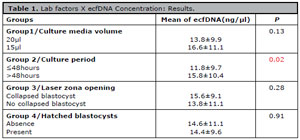
Table 1. Lab factors X ecfDNA Concentration: Results.