JBRA Assist. Reprod 2006;10(2):34-35
RELATO DE CASO
doi: 10.5935/1518-0557.2006.10.2.08
Acral and Renal Malformations Following Intracytoplasmic Sperm Injection
Malformação Acral e Renal Após Injeção Intracitoplasmática de Espermatozóide
aAndrolab - Clínica e Laboratório de Reprodução Humana e Andrologia, Curitiba, Brazil
bDepartment of Human Reproduction - Federal University of Paraná, Brazil
ABSTRACT
This case reports a delivery of a child with limb and renal malformations following intracytoplasmic sperm injection (ICSI). A 29-year-old woman and a 39-year-old man with primary infertility underwent IVF/ICSI-ET therapy cycle. The couple delivered an infant with limb deficience and renal anomalies. When assisted reproductive techniques are used especially ICSI, the risks of genetics abnormalities should be discussed in detail to the couple and sufficient informed consent should be obtained before starting IVF/ICSI-ET procedures.
Key Words: acral, ICSI, malfomation, renal.
INTRODUCTION
Since the publication of the first baby born after IVF and ICSI, was raised the question about the incidence of chromosomal and/or congenital anomalies in these babies. Although no significant risk for congenital malformations after IVF or ICSI (1) has been described. Increased risk for some especific malformations was identified and especifically at ICSI for hypospadias, which seems to be associated with paternal subfertility. We report a child born after ICSI with acral and renal malformations, which includes multiple congenital anomalies. Although the association of limb deficiency and renal anomalies appears well established, concordance of such defects is still relatively rare.
CASE REPORT
A 29-year-old woman with unexplained infertility was stimulated with a combination of a gonadotrophin-releasing hormone agonist and gonadotrophins, oocyte retrieval was carried out and 13 metaphase II oocytes were recovered. Her husband has two children of another relationship, and his semen analysis demonstrated a concentration of 98x106/mL, with 57 % progressive motility and normal morphology.
Gamete preparation and ICSI procedures are described in order to clarify any potential negative impact. The sperm fraction with high motility was isolated by discontinuous Isolate (Irvine Scientific, Santa Ana, USA) gradient separation (90% and 45% layers). The supernatant was removed and the pellet re-suspended in Human Tubal Fluid (HTF, Irvine Scientific) containing 15% of Serum Synthetic Substitute (SSS, Irvine Scientific) and centrifuged at 300g for 10 minutes. The supernatant was removed and the pellet was re-suspended in 0.5 mL of culture medium (HTF + 15% SSS) and incubated in humidified atmosphere of 5% CO2 at 37oC until the moment of injection. Control of fine movement within the injection pipet was provided by a Narishige microinjector (IM-6, Nikon Inc., Garden City, USA) set to the egg-holding and injection micropipettes (Humagen; 10 MPH-120 and 10 MIC, USA, respectively) with inner diameters of 20-25 μm and 4-5 μm, respectively, mounted on a micromanipulator (Narishige, USA) in conjunction with a heated microscope stage, on an inverted microscope (Nikon Inc.). The injection pipet has a 35º. For the procedure, a dish was prepared by adding a 5 μL drop of 10% poli-vinylpyrrolidone (PVP, P-5288, Sigma Chemical) onto the center. Two μL of the sperm sample was added to the center of this drop. Drops of medium, one for each oocyte to be injected, were added around the central PVP drop, and the entire dish was covered with washed mineral oil (M- 8410, Sigma Chemical) that had been equilibrated at 370C, 5% CO2 overnight. The medium for injection consisted of modified HTF medium, supplemented with 0.5 g% BSA (A-4161, Sigma Chemical). These media were maintained warmed at 37ºC. Immediately after dish preparation, oocytes were added to the medium droplets and the dish was placed on the heated stage (37ºC) of the inverted microscope. After alignment of the pipettes within the dish, a motile sperm was identified, immobilized and aspirated into the pipet. With the oocyte held in place by the egg-holding pipet with the polar body at 6 o’clock position, the injection pipet was pushed firmly through the zona pellucida, the cytoplasm was drawn into the injection pipet and then expelled along with the spermatozoon.
The obstetric ultrasound showed at 25 weeks of pregnancy a renal pelvis and collecting system with a moderate dilatation in the left kidney. The right kidney and bladder were of normal size and appearance to the ultrasound. The right forefoot showed a completely fused first and second toe, the third, fourth and fifth toe were fused almost completely. The left foot was normal. The right forehand also showed a completely fused first and second toe, the third, fourth and fifth was fused almost completely (ectrodactyly). The girl was born with a birth weight of 3.000g in the 39th gestational week by cesarean section.
The girl was submitted to a functional surgery at 5 months years old which showed in agreement with the X-ray observations the following acral malformations: (1) on the right hand complete cutaneous syndactyly of the 1st and 2nd fingers; shallow axial cleft between 2nd and 3rd fingers; 3rd and 4th fingers had a complete syndactyly with synostosis of the distal phalanges; depressed longitudinal scar in the mid-palmar area not configuring a lobster claw malformation; no absence of phalanges. (2) left hand showed a pre-axial polydactyly type II of Wassel which consisted of 2 distal phalanges of the first finger; absence of the distal phalange of second finger. (3) right foot showed a transverse phalange in between 1st and 2nd metatarsal-phalangeal joint; shallow axial cleft between 1st and 2nd toe; absence of the distal phalange of second toe. (4) left foot unremarkable. Radiographs of the right hand is shown in figure 1.
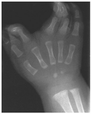
Figure 1. Right hand radiograph. on the right hand complete cutaneous syndactyly of the 1st and 2nd fingers; shallow axial cleft between 2nd and 3rd fingers; 3rd and 4th fingers had a complete syndactyly with synostosis of the distal phalanges; depressed longitudinal scar in the mid-palmar area not configuring a lobster claw malformation; no absence of phalanges.
Renal malformation includes a mild hydronephorosis maybe due to a urethero-pelvic junction stenosis. She showed with a blepharophimosis and at the fundal examination a papilla hypoplasia (optic nerve). Slight nasal hypoplasia is present. Karyotype was analysed and showed a 46, XX baby. She has a normal neurodevelopment to her age. She had a purpuric maucular rash after using vaselin in her first day of life.
DISCUSSION
An association of limb and renal malformations could be interpretated as part of a single developmental field defect, as components of a large number of multiple malformation syndromes and communities of syndrome. Sutcliffe et al (1999) comparing singleton pregnancy conceived naturally or after IVF/ICSI did not find any difference as far as mental development and congenital abnormalities is concerned. Bonduelle et al (2002) comparing almost 3000 babies conceived by IVF or ICSI did show neither an increased risk for major malformations nor to neonatal complications in the ICSI babies. We are actually not sure whether or not exist a link between this malformations and ICSI. As it is a rare malformations, we decide to publish it, in order to help others couples before going through an ICSI therapy. As far as we know this is the first case reporting an acral and renal malformation following an ICSI treatment. The association of an increased risk of malformations in children conceived after ICSI is still speculative, further comparisons are needed before a final conclusion could be drawn.