JBRA Assist. Reprod 2007;11(3):16-19
ARTIGO ORIGINAL
doi: 10.5935/1518-0557.2007.11.3.03
Decrease of the DNA Damage Levels After Sperm Preparation by Layering Method
Redução dos Níveis de Dano no DNA Após Preparo do Sêmen pelo Método de Camada
1Centro de Reprodução Humana Prof. Franco Junior, Ribeirão Preto, SP, Brasil
2Pós-Graduando, Departamento de Ginecologia e Obstetrícia, Faculdade de Medicina de Botucatu - Universidade Estadual de São Paulo-UNESP, Botucatu, SP, Brasil
3Departamento de Ginecologia e Obstetrícia, Faculdade de Medicina de Botucatu - Universidade Estadual de São Paulo-UNESP, Botucatu, SP, Brasil
4Correspondência para: José Gonçalves Franco Junior Centro de Reprodução Humana Prof. Franco Junior Av. Prof. João Fiusa, 689 - CEP 14025-310 Ribeirão Preto - São Paulo - Brasil, Tel/FAX: 55-16-3911.1100, E-mail:
ABSTRACT
The aim of this prospective study was to determine the DNA fragmentation levels before and after sperm preparation by layering method. A total of 78 patients submitted to assisted reproduction technology (ART) for infertility treatment were evaluated. Ejaculated spermatozoa were obtained by masturbation on the day of ART procedure. The evaluation of DNA fragmentation was performed in the fresh semen and after preparation by a layering method, respectively. After washing with PBS, the sperm pellets were smears and then processed for the terminal deoxyribonucleotidyl transferase (TdT)- mediated dUTP nick-end labelling (TUNEL) assay that was performed using a Cell Death Detection Kit with tetramethylrhodamine-labelled dUTP. For quantitative evaluation, 200 spermatozoa in randomly selected areas on microscope slides were evaluated and the percentage of TUNEL positive spermatozoa was determined. If ≥20% of selected sperm were TUNEL positive, the exam was considered abnormal. The mean percentage of DNA sperm fragmentation before sperm preparation was 17±8.3% and after 7.8±6.5% (p<0.0001). The exam was considered normal in 49 patients before preparation and in 73 patients after (p<0.0001). The sperm preparation with a layering method for the ART procedure is effective to select sperm with a significant decrease of the DNA damage.
Palavras-chave: Fragmentação do DNA, sêmen, método de camada, TUNEL, fertilização in vitro/ICSI
RESUMO
O objetivo deste estudo perspectivo foi determinar os níveis de fragmentação DNA antes e depois do preparo do sêmen pelo método de camada. Foram avaliadas amostras de sêmen de 78 pacientes submetidos à reprodução assistida. A analise da fragmentação do DNA foi realizada no esperma antes e após o preparo pelo método de camada, através do teste TUNEL (the terminal deoxyribonucleotidyl transferase (TdT)-mediated dUTP nick-end labelling) utilizando o Kit In Situ Cell Death Detect, com tetramethylrhodamine-labelled dUTP. Para avaliação quantitativa, foram analisados 200 espermatozóides em áreas dos esfregaços selecionadas aleatoriamente e a porcentagem de espermatozóides TUNEL-positivos foi determinada. Se ≥20% da esperma selecionada eram TUNEL-positivo, o exame era considerado anormal. As porcentagens médias de fragmentação do DNA antes e após o preparo do sêmen foram 17±8.3 % e 7.8±6.5 % (p<0.0001), respectivamente. O exame foi considerado normal em 49 pacientes antes do preparo e em 73 pacientes depois do preparo (p<0.0001). Em conclusão, o preparo de esperma pelo método de camada para procedimentos de RA é eficaz para selecionar a esperma com uma redução significativa do dano do DNA.
Key Words: DNA fragmentation, semen, layering method, TUNEL assay, fertilização in vitro /ICSI
INTRODUCTION
The presence of DNA fragmented sperm in human ejaculate is well documented, especially in men with poor semen quality (Muratori et al., 2000). The higher occurrence in subfertile men has led to concerns about using assisted reproductive technology (ART) with these patients because the fertilizing ability of a DNA spermatozoon and the consequences of embryo development are not entirely known. Indeed, some ART protocols override the physiological sperm selection processes which could increase the risk that a DNA damaged sperm participates in fertilization (Muratori et al., 2003). Also some reports have indicated there is a greater risk of major birth defects in children conceived via in vitro fertilization (IVF) and intracytoplasmic sperm injection (ICSI) (Hansen et al., 2002).
One factor of extreme importance in ICSI is sperm integrity. Good quality sperm DNA is essential for the accurate transmission of genetic material to the next generation. Poor DNA may not necessarily prevent fertilization from occurring and genetically damaged human spermatozoa can still form normal pronuclei in oocytes after ICSI (Twigg et al., 1998).
Sperm washing is an integral part of ART but is important that the sperm processing technique is gentle yet enables recovery of a concentrated and highly functional sperm population. Many of the conventional centrifuge techniques used in cell separation are toxic to spermatozoa. Serial centrifugation of semen induces significant reactive oxygen species (ROS) release by spermatozoa and semen leukocytes, resulting in sperm dysfunction (Aitken and Clarkson, 1988; Appasamy et al., 2007).
In an attempt to prevent centrifuge damage and generation of ROS, other sperm preparation methods have been developed using density gradient media or direct swim-up (layering method) from the original sperm sample (Mortimer, 1991). The later is a recommended method for sperm preparation (World Health Organization, 1999).
Sakkas et al. (13) related that when spermatozoa were prepared using the PureSperm technique a significant decrease in DNA damage was observed. However, the swimup technique produced no significant reduction in DNA.
The objective of this study was to examine the effect of standard sperm processing by layering method on human DNA sperm integrity.
MATERIAL AND METHODS
Study participants
Semen samples (one per subject) were obtained from 78 men of an unselected group of couples attending the Center for Human Reproduction Prof. Franco Jr, for the ART program (IVF/ICSI) in 2006.
Sample collection and preparation
Semen samples were collected in sterile containers on the day of IVF/ICSI by masturbation after a period of 2-5 days of sexual abstinence. The liquefied semen samples were divided into two aliquots for evaluation of DNA fragmentation, one for the layering method and other for direct sperm preparation.-Layering method: Briefly, 2 ml of culture medium (modified HTF-10% Human Serum Albumin) was pipetted into a Falcon tube, then 1.0ml of neat sperm was deposited into the bottom of the tube. The tube was placed in an incubator at 37ºC for 1h. After this, the supernatant was removed and divided in two aliquots: the first was used for IVF/ICSI; the other was centrifuged at 200g for 10min and the result pellet was sent for evaluation of sperm DNA fragmentation analysis.-Direct sperm preparation: Liquefied fresh semen was centrifuged at 200g for 10 minutes at room temperature to separate spermatozoa from seminal plasma. The remaining sperm pellet was resuspended with 1ml of phosphate-buffered saline (PBS) and centrifuged at 200g for 10 minutes. The result pellet was sent to sperm DNA fragmentation analysis.
Determination of DNA fragmentation
DNA fragmentation in spermatozoa cells was measured using the terminal deoxyribonucleotidyl transferase-mediated dUTP nick-end labelling (TUNEL) assay which was performed using a Cell Death Detection Kit with tetramethylrhodaminelabelled dUTP (Roche, Monza, Italy). TUNEL identifies single and double stranded DNA breaks by labelling the free 3’-OH termini with modified nucleotides in an enzymatic reaction with terminal deoxynucleotidyl transferase (TdT). TdT polymerises free 3’-OH DNA ends in a template-independent manner, incorporating labelled nucleotides. The remaining sperm pellets (after layering method and after direct preparation) were smeared on microscope slides, air-dried, fixed with 4% paraformaldehyde in PBS at 4ºC for 25min, pH 7.4 and permeated with 0.1% Triton® X-100 (VETEC Química Fina Ltd, Duque de Caxias, Brazil) in 0.1% sodium citrate at 4ºC for 2min. After washing with PBS, the smears were then processed for the TUNEL assay. The TdT-labelled nucleotide mix was added to each slide and incubated in the dark in a humidified atmosphere for 2h at 37°C. After stopping the enzyme reaction, slides were rinsed twice in PBS and then counterstained with Vectashield® Mounting Medium with DAPI (4,6-diamidino-2- phenylindole 1.5µg/ml) (Vector Laboratories, Burlingame, CA, USA). For quantitative evaluation, at least 200 spermatozoa in randomly selected areas on microscope slides were evaluated using a fluorescent microscope. The percentage of TUNEL positive spermatozoa was determined. The number of cells per field stained with DAPI was first counted; in the same field the number of cells with red fluorescence (TUNEL positive) was expressed as a percentage of DNA fragmentation of the total sample (DNA fragmentation index /DFI). Controls were included in every experiment: for the negative control TdT was omitted in the nucleotide mix. Positive controls were generated by pre-incubating the fixed and permeabilized sperm cells using DNase I 1mg/ml (New England Biolabs, Inc, Ipswich, MA, USA) for 30min at 37°C. TUNEL labelling of the positive controls varied between 89-98% of cells. If ≥20% of selected sperm were TUNEL positive (DFI=20), the exam was considered abnormal (Benchaib et al., 2003). The same technician, who was blinded to the identity of the study subjects, performed all exams.
STATISTICAL ANALYSIS
Data are reported as means±SD and were analysed using the InStat 3.0 program for MacIntosh (GraphPad Software, San Diego, CA, USA). The Mann-Whitney test and Chi-square Test were used where appropriate. The level of significance was set at p<0.05
RESULTS
Mean male age was 37.3±5.9 years (range 23-55 years). The 78 samples in the study were classified according to World Health Organization (18) as follows: normozoospermia, 46; oligozoospermia, 1; asthenozoospermia, 12; teratozoospermia, 12, oligoteratozoospermia, 1; asthenoteratozoospermia, 5; and oligosthenoteratozoospermia, 1. Thirty one men had fathered at least a child (or a pregnancy which end in miscarriage), spontaneously or after fertility treatment prior this ART procedure.
Mean DNA sperm fragmentation index percentage before sperm preparation was 17±8.3% and after 7.8±6.5% (p<0.0001, Mann-Whitney test). Figure 1 shows DNA fragmentation index distribution before and after sperm preparation by layering method.
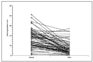
Figure 1. DNA fragmentation before and after sperm preparation by layering method
The exam was considered normal in 49 patients before and in 73 patients after preparation (p <0.0001, Chi-square Test; Table 1).
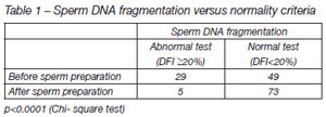
Table 1. Sperm DNA fragmentation versus normality criteria
DISCUSSION
Traditional semen analysis is based on the estimation of sperm concentration, motility and morphology and providing these tests are performed with diligence and adherence to strict guidelines (World Health Organization, 1999), useful prognostic information can be obtained (Tomlinson et al., 1999). However, with an upsurge in the use of assisted reproduction technologies and especially ICSI, the requirements of the diagnostic semen analysis are constantly changing. As assisted reproduction techniques become more diverse, we remove many of the natural selection barriers set in place to ensure that the best spermatozoa are used for fertilization. Semen analysis therefore needs to develop to the point where we can provide patients not only with some idea of prognosis in terms of natural conception, but also whether assisted reproduction treatment will have a successful outcome both in terms of fertilization and in producing a healthy conceptus (Tomlinson et al. 2001).
Defects in sperm genomic material may take the form of condensation or nuclear defects, or sperm chromosomal aneuploidies (Perreault et al., 2003). The causes of these defects have been attributed to disease, drug use, high fever, elevated testicular temperature, air pollution, cigarette smoking, and advanced age. The molecular mechanisms of DNA damage in these diversified conditions is under intense investigation (Agarwal and Allamaneni, 2005). The most important mechanisms under consideration for sperm DNA damage are abnormal chromatin packaging, ROS (Agarwal et al., 2003), and apoptosis (Sakkas et al., 1999). It is likely that multiple mechanisms are involved, based on the clinical diagnosis responsible for DNA damage.
Fragmentation of genomic DNA is one of the hallmarks of apoptosis-the most common form of eukaryotic cell death. Apoptosis, programmed cell death, occurs under normal physiological conditions and proceeds in two main phases. The first is a commitment phase followed by an execution phase which is characterized by a series of stereotypic changes including cell shrinkage, plasma membranedisruption, phosphatidylserine externalization, and condensation and fragmentation of chromatin (Wyllie et al., 1980). A characteristic feature of this process is activation of an endogenous endonuclease which generates numerous DNA strand breaks in chromatin (Wyllie et al., 1980; Santiso et al., 2007).
On the other hand, oxidative damage to sperm DNA does not impede pronucleus formation after microinjection in hamster eggs (Twigg et al., 1998). This observation is rather logical since it is generally assumed that the first development steps on maternal transcripts, thus the paternal expression would normally start at the 6-8 cell stage; it must be remembered that in the majority of cases embryo transfers are performed at day 2 and day 3, i.e. before the complete paternal effect can be expressed.
Benchaib et al. (5, 6) indicate that the proportion of spermwith DNA fragmentation influences the fertilization rate for a threshold value above 10%, and an implantation rate of ICSI-derived embryos. Since no pregnancy was obtained when >20% of selectedsperm were TUNEL positive, this may have a good predictive value in cases of successive failures of implantation for apparently good quality embryos. For this reason, our cut off of normality was < 20% of TUNEL positive.
There is now incontrovertible evidence that centrifugal pelletting of unselected human sperm populations causes irreversible damage to the spermatozoa; this can impair the fertilizing ability of the motile fraction subsequently prepared by swim-up migration. Alternative sperm preparation methods such as direct swim-up (layering method) from liquefied semen (with a subsequent centrifugal washing step) should be used to prepared spermatozoa that will assess or require sperm fertilizing ability (Mortimer 1991). For this reason we decided use the layering method, with a direct swim-up from liquefied semen, and subsequent centrifuged washing step in this study.
One of the confounding problems of relating semen parameters to overall ART outcome is that a certain population of spermatozoa is selected during the semen preparation procedure. It is likely that regardless of the initial sample, a degree of homogenization occurs after sperm preparation. Tomlinson et al. (2001) has shown that simple density gradient centrifugation can enrich the sample both with morphologically normal forms and spermatozoa with improved nuclear integrity. This “normalizing” effect ofdensity gradient preparations may be the reason why pre-preparation sperm parameters had little prognostic value, in terms of fertilization and pregnancy using ART.
Our data demonstrated that of standard sperm processing by layering method is efficient, the mean percentage of DNA sperm fragmentation before sperm preparation was 17±8.3% and after 7.8±6.5% (p<0.0001) and the exam was considered normal in 49 patients before preparation and in 73 patients after (p<0.0001), for selecting sperm with nuclear integrity. However, now is necessary to compare which method is more efficient to select sperm with lower DNA fragmentation.