JBRA Assist. Reprod. 2008;12(3):16-19
ARTIGO ORIGINAL
doi: 10.5935/1518-0557.2008.12.3.03
What’s the signification of nuclear vacuoles in human sperm?
Qual é o significado dos vacúolos nucleares no espermatozóide humano?
1Centro de Reprodução Humana Prof Franco Jr - Ribeirão Preto, SP, Brasil
2Departamento de Ginecologia e Obstetrícia - Faculdade de Medicina de Botucatu - Universidade Estadual Paulista Julio de Mesquita - UNESP - Botucatu, SP, Brasil
3Pós-graduando, Departamento de Ginecologia e Obstetrícia - Faculdade de Medicina de Botucatu - Universidade Estadual Paulista Julio de Mesquita - UNESP - Botucatu, SP, Brasil
4CPDP - Centro Paulista de Diagnóstico e Pesquisa - Ribeirão Preto, SP, Brazil
ABSTRACT
The aim of this study was to determine the extent of DNA fragmentation and the presence of single/denatured or double stranded of DNA in sperm with large nuclear vacuoles (LNV) selected by high-magnification. A total of 30 patients had fresh semen samples prepared by discontinuous concentration gradient. Sperm with normal nucleus (NN) and LNV were selected at 8400x magnification and placed in different slides. DNA fragmentation was determined by TUNEL assay. Denatured and double stranded DNA was identified by acridine orange fluorescence method. The percentage of DNA fragmentation in LNV sperm (29%) was significantly higher (P<0.001) than NN sperm (15.8%). Therefore, cleavage of genomic DNA in low molecular weight DNA fragments (mono and oligonucleosomes), and single strand breaks (nicks) in high molecular weight DNA occur more frequently in LNV. Identically, the percentage denatured stranded DNA in sperm with LNV (67.9%) was significantly higher (P <0.0001) than NN sperm (33%). The high level of denatured DNA in sperm with LNV suggests precocious decondensation and disaggregation of sperm chromatin fibers. Our results support an association between LNV sperm and DNA damage, and the routine selection and injection of morphological motile sperm at high magnification for ICSI. The adverse effect (DNA fragmentation or denaturation) leads to concern particularly about the possibility of iatrogenic transmission of genetic abnormalities.
Keywords: human sperm, nuclear vacuoles, ICSI, DNA fragmentation, denatured stranded DNA
RESUMO
O objetivo deste estudo foi determinar a extensão da fragmentação do DNA e a presença de DNA dupla fita ou fita simples (desnaturada) em espermatozóides com vacúolos nucleares largos (VNL) selecionados pela microscopia de alta magnificação. Um total de 30 pacientes tiveram amostras de esperma fresco preparadas por gradiente descontínuo. Os espermatozóides com núcleo normal (NN) e com VNL foram selecionados a magnificação de 8400x e colocados em laminas diferentes. A fragmentação do DNA foi determinada através do método do TUNEL. O DNA desnaturado (fita simples) e o dupla fita foram identificados pelo método de acridina orange. O percentual de fragmentação do DNA nos espermatozóides VNL (29%) foi significativamente maior (p<0.001) do que nos espermatozóides NN (15.8%). Dessa forma, a quebra do DNA genômico em fragmentos de baixo peso molecular (mono e oligonucleossomos), e quebras em cadeia única (nicks) em DNA com alto peso molecular ocorre mais frequentemente no espermatozóide com VNL. Igualmente, a percentagem de cadeia desnaturada de DNA (fita simples) em espermatozóides com VNL (67.9%) foi significativamente maior (p<0.0001) que em espermatozóides NN (33%). O alto nível de DNA desnaturado em espermatozóides com VNL sugere descondensação e desagregação precoce das fibras da cromatina espermática. Nossos resultadosindicam uma associação entre o espermatozóide com VNL e dano no DNA, além de substanciar o uso da alta magnificação na seleção e injeção dos espermatozóides para ICSI.
Palavras-chave: esperma humano, vacúolos nucleares, ICSI, DNA fragmentação, cadeia desnaturada de DNA
INTRODUCTION
It is known that IVF/ICSI is associated with increased (although relatively low) risk of birth defects and genetic and epigenetic abnormalities in children. To date, it is unclear whether the ICSI procedure or the underlying infertility is responsible for these defects (Cox et al., 2002; Hansen et al., 2002; Ludwig et al., 2005). The risk of birth defects (major and minor) was found to be significantly higher (odds ratio ~ 1.5) in children conceived by IVF/ ICSI than in those naturally conceived. The prevalence of chromosomal abnormalities (de novo abnormalities) was found to significantly higher (1.6%/0.5%) in children conceived by ICSI than naturally (Bonduelle et al,1998). Epigenetics abnormalities, such as errors in DNA methylation, have been linked to certain rare genetic diseases (Beckwith/ Wiedmann and Angelman’s syndromes) and, while still rare, are found in slightly more prevalent in children conceived by IVF/ICSI than naturally (Maher et al., 2003).
Successful human reproduction depends in part on the inherent integrity of sperm DNA. There appears to be a DNA damage threshold beyond which embryo development and subsequent pregnancy outcome are impaired. Clinical evidence now shows that sperm DNA damage is detrimental to reproductive outcomes and that spermatozoa of infertile men posses substantially more DNA damage than do the spermatozoa of fertile men. However, our understanding of the causes of sperm DNA damage and the full impact of this sperm defect on reproductive outcomes in human remains undeveloped (Zini and Libman, 2006).
A specific sperm nuclear alteration is the presence of large vacuoles. Ultramorphological investigation has revealed that this sperm malformation has a clear negative association with natural male fertility potential (Bartoov et al., 1994; Mundy et al., 1994). According to Bartoov et al. (2002) selection of morphological motile sperm at high magnification (MSOME) is the only method to precisely detect large nuclear vacuoles in human sperm for ICSI. Berkovitz et al. (2006a) observed that microinjection sperm (IMSI) with a normal nuclear shape but large vacuoles affects ICSI pregnancy outcome (reduce pregnancy rate and increases early abortion). However, the mechanism behind why large vacuoles impair late embryonic development is not clear. The aim of this study was to determine the presence or absence of DNA damage in sperm with large nuclear vacuoles (LNV) selected by highmagnification and submitted to DNA fragmentation analysis and the presence of denatured/single stranded of DNA by acridine orange fluorescence (AOF).
MATERIALS AND METHODS
Study participants and sperm preparation
Fresh semen samples (one per subject) from 30 patients in an unselected group of couples undergoing infertility investigation and treatment at the Center for Human Reproduction Prof. Franco Junior were prepared by Isolate (Irvine Scientific, USA) discontinuous concentration gradient. The final pellet was resuspended in 0.2 ml modified HTF medium (Irvine). An aliquot of 1µl of sperm cell suspension was transferred to a 5µl microdroplet of modified HTF medium containing 8% polyvinyl pyrrolidone solution (PVP medium Irvine Scientific-USA). This microdroplet was placed in a sterile glass dish (FluoroDishTM-Word Precision Instrument,USA) under sterile paraffin oil (Ovoil-100, Vitrolife, Goteborg, Sweden). The sperm cells, suspended in the microdroplet were placed on a microscope stage above an Uplan Apo x100 oil/1.35 objective lens previously covered by a droplet of immersion oil. In this way, suspended motile sperm cells in the observation droplet could be examined at high-magnification by the inverted microscope (Eclipse TE 2000 U Nikon, Japan) equipped with high-power differential interference contrast optics (DIC/Nomarski). The total calculated magnification was x8400. Sperms with normal nucleus (NN) and sperms with LNV were selected using a micromanipulation system with angled glass micropipets and placed on different slides. Sperm were smeared in a very small area, which was marked on the back of the slides with a glass pen to help find the sperm under the microscope. LNV sperm was defined by the presence of one or more vacuoles occupying ≥50% of sperm head area.
Determination of DNA-fragmentation
DNA fragmentation in spermatozoa cells was measured using the terminal deoxyribonucleotidyl transferasemediated dUTP nick-end labelling (TUNEL) assay which was performed using a In Situ Cell Death Detection Kit with tetramethylrhodamine-labelled dUTP (Roche, Monza, Italy). TUNEL identifies single and double stranded DNA breaks by labelling the free 3’-OH termini with modified nucleotides in an enzymatic reaction with terminal deoxynucleotidyl transferase (TdT). TdT catalyses the polymerization of labeled nucleotides to free 3’-OH DNA ends in a templateindependent manner. Slides with selected sperm were air-dried and then fixed at 4ºC in Carnoy’s solution (methanol/glacial acetic acid, 3:1), and permeabilized with 0.1% Triton X-100 (VETEC Química Fina Ltd, Duque de Caxias, Brazil) in 0.1% sodium citrate at 4ºC for 2min. After washing with phosphate-buffered saline (PBS), the slides were then processed for TUNEL assay. The TdT-labelled nucleotide mix was added to each slide and incubated in the dark in a humidified atmosphere for 2h at 37°C. After stopping the enzyme reaction, slides were rinsed twice in PBS and then counterstained with Vectashield®Mounting Medium with DAPI (4,6-diamidino-2-phenylindole 1.5µg/ml) (Vector Laboratories, Burlingame, CA, USA). The final evaluation was realized using a fluorescent microscope and the percentage of TUNEL positive spermatozoa determined. The number of cells per field stained with DAPI (blue) was first counted; in the same field the numbers of cells with red fluorescence (TUNEL positive) was expressed as a percentage of DNA fragmentation. Controls were included in every experiment: for negative control TdT was omitted in the nucleotide mix. Positive controls were generated by pre-incubating the fixed and permeabilized sperm cells using DNase I 1mg/ml (New England Biolabs, Inc, Ipswich, MA, USA) for 30min at 37°C. TUNEL labelling of positive controls varied between 89-98% of cells. The same technician, blinded to subject identity, performed all exams (Vagnini et al., 2007).
Determination of denatured/single or double stranded DNA by acridine orange fluorescence
Slides with selected sperm were air-dried and then fixed overnight at 4ºC in Carnoy’s solution (methanol/ glacial acetic acid, 3:1). After fixation, the slides were air-dried, stained with AOF staining solution for 5 min and then gently rinsed with distilled water. The AOF staining solution was prepared daily as follows: a mixture of 4 ml 0.1M citric acid and 0.25 ml 0.3M Na2HPO4.7H2O, added to 1 ml 1% AOF stock solution in distilled water. AOF stock solution was stored in the dark at 4ºC for 4 weeks. In order to reduce variation in fluorescence intensity, each stained slide was read immediately after washing. Sperm denatured/single or double stranded DNA was determined under a fluorescence microscope at x400 and x1000 magnification with 450-490nm excitation. Sperm with double-stranded DNA were fluorescent green and those with denatured or single stranded DNA were fluorescent red or yellow. The same technician, blinded to subject identity, performed all AOF procedures.
Sample size and statistical analysis
Sample size was calculated planning a comparison between two proportions control and experimental. Usually, DNA fragmentation and AOF assay both had abnormal values ≤30% in the sperm of the fertile male population. Thus a sample size of 350 sperm in each group has a 80% power to detect an increase of 10% with a significance level alfa of 0.05 (two-tailed).Data were analyzed using InStat version 3.0 (GraphPad Software, San Diego, CA, USA) on a Macintosh computer (Apple Computer Inc., Cupertino, CA). The Fisher’s exact test was used.
RESULTS
Table 1 and 2 show DNA fragmentation and denatured/ double stranded DNA values in sperms with LNV and NN. The percentage of positive DNA fragmentation in LNV sperm was (29%=111/382) significantly higher (P<0.0001) than NN sperm (15.8%=65/410). On the other hand, the percentage of denatured stranded DNA (67.9%=252/371) in LNV sperm was significantly higher (P<0.0001) than in NN sperm (33%=117/354).
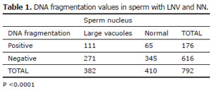
Table 1. DNA fragmentation values in sperm with LNV and NN.
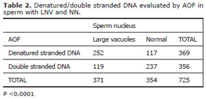
Table 2. Denatured/double stranded DNA evaluated by AOF in sperm with LNV and NN.
DISCUSSION
The accuracy with which morphological normality of spermatozoa for ICSI can be assessed depends on the resolution power of the optical magnification system. Conventionally, ICSI is performed with a x20/x40 objective, resulting in an overall optical magnification of x200 to x400 (De Vos et al., 2003). However, spermatozoa appearing as morphologically normal at this magnification may in fact carry various structural abnormalities that can only be detected with higher optical magnifications, sperm with vacuoles would be not detected in conventional ICSI (Hazout et al., 2006). This is a serious disadvantage, because microinjection of spermatozoa with vacuolated nuclei is clearly associated with low implantation and pregnancy rates, and with early abortion (Berkovitz et al., 2005, 2006a, 2006b). Berkovitz et al. (2006a) suggested that vacuolization of the sperm nucleus reflects some underlying chromosomal or DNA defects but has not showed data confirming this hypothesis.
Evaluation of DNA damage by TUNEL was introduced by Gorczyca et al. (1993) to identify a population of spermatozoa in ejaculate that were believed to be apoptotic. Numerous other studies using the same technique have followed (Sun et al., 1997; Muratori et al., 2000) demonstrating that DNA fragmentation assessed by the TUNEL method was not associated with an apoptosis-like phenomenon in ejaculated spermatozoa and that DNA fragmentation should be considered a sign of defective sperm maturation which probably dates back to the time of DNA packaging. The TUNEL assay is usually described as the method for detecting real DNA damage and providing a direct measurement of DNA breaks in sperm (Li et al., 2006). The first part of our data shows that DNA fragmentation values were significantly higher in sperm nucleus with LNV. Therefore, cleavage of genomic DNA in low molecular weight DNA fragments (mono and oligonucleosomes), and single strand breaks (nicks) in high molecular weight DNA occur in sperm with LNV. Acridine orange staining is an established cytochemical method for determining sperm DNA integrity, allowing the differentiation between normal, double-stranded and abnormal, denatured/single stranded DNA, using the metachromatic properties of the dye (Tejada et al., 1984). Some studies have shown that sperm denatured/singlestranded DNA, detected by AO staining, negatively affects the fertilization process in a classical IVF program (Liu et al., 1994, Hoshi et al., 1996). Increased denatured-stranded DNA in sperm of infertile men after density gradient is linked to results showing fewer embryos suitable for transfer and cryopreservation (Virant-Klun et al., 2002).
The second part of our data showed significantly more denatured DNA in sperm with LNV. This high level of denatured DNA in LNV could mean precocious decondensation and disaggregation of sperm chromatin fibers. Kosower et al. (1992) showed that the color of AOF of sperm nucleus after acetic alcohol treatment is determined by the thiol-disulfide status of DNA-associated protamines. An unwanted high degree of sperm decondensation (disruption of disulphide bridges/red acridine orange fluorescence) can result in asynchronous chromosome condensation and may lead to cytoplasmic fragments in the embryo (Ménézo et al., 2007).
CONCLUSION
Our results support an association between sperm with LNV and DNA damage and the routine use of MSOME/IMSI. The adverse effect (DNA fragmentation or denaturation) leads to concern particularly about the possibility of iatrogenic transmission of genetic abnormalities.