JBRA Assist. Reprod. 2010;14(2):27-31
ARTIGO ORIGINAL
doi: 10.5935/1518-0557.2010.14.2.05
Embryo transfer based on previous uterine length measurement enhances ICSI outcomes when compared to standard ultrasonographic-guided embryo transfer.
A transferência de embriões baseada na medida prévia do comprimento uterino melhora o prognóstico da ICSI quando comparada à transferencia padrão guiada por ultra-sonografia
1Fertility – Assisted Fertilization Center, Av. Brigadeiro Luis Antônio, 4545. São Paulo – SP, Brazil. Zip: 01401-002
2Sapientiae Institute – Educational and Research Center in Assisted Reproduction, Rua Vieira Maciel, 62. São Paulo – SP, Brazil. Zip: 04503-040
3Department of Imaging Diagnosis, São Paulo Federal University. Rua Napoleão de Barros, 421 São Paulo – SP, Brazil. Zip: 04503-040
ABSTRACT
OBJECTIVE: To test the hypothesis that embryo transfer based on previous uterine length measurement is a better method to improve pregnancy rates when compared to the standard ultrasonographic-guided embryo transfer method.
METHODS: Patients undergoing Controlled Ovarian Stimulation (COS) for Intracytoplasmic Sperm Injection (ICSI) cycles were allocated into two groups based on embryo transfer method used. Cycles in which embryo transfer was performed based on previous uterine length measurement (ULM group, n=50) and cycles in which embryo transfer was performed though standard ultrasonographic-guidance (Control group, n=150). The easiness of transfer, implantation and pregnancy rates were compared among the groups using the Student’s t test and Chi-squared or Fisher exact tests as appropriate.
RESULTS: Similar results were found between groups regarding the easiness of transfer. Both implantation (11.9% vs 30.7% for Control and ULM groups respectively, p< 0,001) and pregnancy rates (26.0% vs 54.0% for Control and ULM groups respectively, p< 0,001) were significantly higher in the ULM group.
CONCLUSION: Ultrasound guided embryo transfer based on previous uterine length measurement is useful to increase the pregnancy and implantation rates in patients submitted to COS and ICSI cycles.
KEYWORDS: Assisted Reproduction, Embryo Transfer, Ultrassonography, Implantation, Endometrium
RESUMO
OBJETIVO: Testar a hipótese de que a transferência de embriões guiada por ultra-sonografia e baseada na avaliação prévia do comprimento uterino é melhor para os indices de gravidez quando comparada ao método ultra-sonográfico tradicional.
MÉTODOS: Pacientes sob estimulação ovarian controlada (COS) para ICSI foram alocadas em 2 grupos, baseados no método de transferência utilizado Os grupos incluiram ciclos nos quais a transferência se baseou na avaliação prévia do comprimento uterino. (ULM , n=50) e ciclos nos quais a transferência se deu através do método usual com guia ultra-sonográfica (Grupo controle, n=150). Foram comparadas a facilidade da transferência, a implantação e as taxas de gravidez entre os grupos, através do teste t de Student e do qui-quadrado.
RESULTADOS: Os achados foram similares quanto à faciliadde da transferência. Tanto a implantação (11.9% vs 30.7% para o grupo controle e ULM , respectivamente, p< 0,001) quanto as taxas de gravidez (26.0% vs 54.0% para os controles e ULM, p< 0,001) foram significativamente maiores no grupo de mensuração prévia.
CONCLUSÃO: a transferência de embriões guida por ultra-sonografia, baseada na avaliação prévia do comprimento uterino é útil para aumentar os índices de gravidez e implantação em pacientes submetidas a estimulação controlada para ICSI.
KEYWORDS: Assisted Reproduction, Embryo Transfer, Ultrassonography, Implantation, Endometrium
INTRODUCTION
Since the first successful childbirth resulting from in vitro fertilization (IVF) (Steptoe and Edwards 1978), remarkable improvements were achieved on the treatment of infertile couples through IVF. Throughout the years, the optimization of the several factors known to influence the IVF success rate has been attempted in order to increase pregnancy rate. Embryo transfer (ET) is the last step on the IVF process, and despite major advances achieved on ovarian stimulation protocols, in vitro embryo development and embryo selection, few modifications have been made on the ET technique.
Despite high-quality embryos may be available for transfer, only a small proportion of patients undergoing IVF will ever achieve a pregnancy. In fact, previous studies indicate that up to 80% of the embryos that reaches the uterine cavity fails to implant (Edwards 1995). This may be attributed to multiple factors including embryo implantation potential, endometrial receptivity and the embryo transfer technique itself (Mansour and Aboulghar 2002).
Embryo transfer was originally performed by advancing the transfer catheter until contact with the uterine fundus, then withdrawing the catheter 5–10 mm and expelling the embryos into the uterine cavity. Previous study using ultrasound, however, showed that, during this ‘blind’ technique, the operator is frequently unaware that the catheter is poorly positioned (Strickler et al. 1985). Therefore, transabdominal ultrasound (Leong et al. 1986) and, more recently, vaginal ultrasound guidance (Anderson et al. 2002) have added more consistency to the procedure. The ultrasound guidance during the embryo transfer allows visual monitoring of the process, while navigating embryo placement within the uterine cavity with high precision (Allahbadia et al. 2008).
Besides the ultrasound guidance, Coroleu et al. (2002a) suggested that depth of the embryo insertion into the uterine cavity may influence implantation rates after IVF. For that, the length of the uterine cavity is measured by transvaginal ultrasonography before embryo transfer and based on this measurement, in the moment of transfer, the tip of the catheter is placed 1.5–2 cm below the fundus. Our goal for this study was to test the hyphothesis that embryo transfers based on previous uterine length measurement would improve pregnancy rates in IVF programs when compared to the standard ultrasonographic-guided embryo transfer method.
MATERIALS AND METHODS
Experimental design
This study included 200 patients undergoing intracytoplasmic sperm injection (ICSI) cycles for the first time, in a private assisted fertilization center, during January 2007 to December 2007. The study was approved by the local Institutional Review Board and an written informed consent was obtained, in which patients agreed to share the outcomes of their cycles for research purposes.Cycles were divided into two groups based on embryo transfer method used. Each cycle in which the embryo transfer was based on previous uterine length measurement (ULM group, n=50) was age-matched with three cycles in which embryo transfer was performed though standard ultrasonographic-guidance (Control group, n=150).The easiness of transfer (easy or difficult), presence of blood in the catheter, implantation rate and pregnancy rate were compared among groups.Implantation rate was defined as the total number of gestational sacs with fetal heart activity divided by the total number of embryos transferred. Clinical pregnancy was defined as the presence of a gestational sac with fetal heart activity visualized by ultrasound.
Controlled ovarian stimulation
Controlled ovarian stimulation was achieved by long pituitary down regulation using a gonadotropin-releasing hormone agonist (GnRH, Lupron Kit™, Abbott S.A Societé Française des Laboratoires, Paris, France) followed by ovarian stimulation with recombinant-FSH (Gonal-F®, Serono, Geneve, Switzerland). The follicular dynamic was followed by ultrasound (Echo Camera SSd-500, Aloka®, Tokio, Japan), starting on day 4 of gonadotropin administration. When adequate follicular growth and serum estradiol levels were observed, recombinant human chorionic gonadotrophin (r-hCG, Ovidrel™, Serono, Geneve, Switzerland) was administered to trigger final follicular maturation. Oocytes were collected 35 hours after hCG administration by transvaginal ultrasound ovum pick-up.
Preparation of oocytes and intracytoplasmic sperm injection
After retrieval, oocytes were incubated in culture medium (G-MOPS™-V1, Vitrolife, Kungsbacka, Sweden) covered with mineral oil (Ovoil™, Vitrolife, Kungsbacka, Sweden) at 37°C and 6% CO2 for 3 hours. Cumulus cells were removed with a 30 s exposure to Hepes-buffered medium containing 80 IU/mL hyaluronidase (Irvine Scientific, Santa Ana, USA), after which coronal cells were manually removed using a finely drawn glass Pasteur pipette (Humagen Fertility Diagnostics, Charlottesville, Virginia, USA). Sperm injection was carried out on the heated stage (37°C) of an inverted microscope (Eclipse TE 300; Nikon®, Tokyo, Japan) 40 hours after hCG trigger for MII stage retrieved oocytes or after 24 hours in culture for immature retrieved oocytes that had undergone nuclear maturation.
Assessment of fertilization and embryo quality
Fertilization was assessed 18 hours after ICSI, and normal fertilization was declared when two clearly distinct pronuclei were present. Embryo quality was evaluated under an inverted microscope (Eclipse TE 300; Nikon®, Tokyo, Japan). The following parameters were recorded: (i) the number of blastomeres, (ii) the fragmentation percentage, (iii) variation in blastomere symmetry, (iv) the presence of multinucleation and (v) defects in the zona pellucida and the cytoplasm. High-quality embryos were defined as those having all of the following characteristics: either 4 to 6 cells on the second day or 8 to 10 cells on the third day of development, less than 15% fragmentation, symmetric blastomeres, absence of multinucleation, colorless cytoplasm with moderate granulation with no inclusions, absence of perivitelline space granularity and absence of zona pellucida dysmorphism.
Embryo Transfer
Two to three days after oocyte recovery, from one to three high-quality embryos per patient were transferred depending on patient’s age. When no high-quality embryos were available for transfer, patient was excluded from the study.For embryo transfer, patients with full bladder were placed in the lithotomy position and the cervix was exposed using a bivalve speculum. The mucus in the cervical canal was removed with a cotton swab and the exocervix was cleaned with a phosphate-buffered saline (PBS) solution (Dulbecco’s PBS solution; Irvine Scientific, Santa Anna, California, USA). For patients in ULM group, during controlled ovarian stimulation, the distance from the external cervical os to the fundal endometrial surface was measured by transvaginal ultrasonograhy and this length was used for embryo replacement with the tip of the catheter achieving the half of the endometrial lumen (Figure 1).
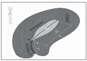
Figure 1. Schematic view of the embryo transfer site for patients in ULM groups
For patients in Control group, during embryo transfer, abdominal ultrasound was performed to visualize the full length of the endometrium, the cervix and the uterine fundus. Under visualization, the tip of the catheter was positioned aiming to place the expelled embryo in the middle of the cavity length (Sallam 2005), according to the clinicians and sonographers’ judgment. For ULM group the embryos’ replacement was based on the previous uterus length measurement.For all patients, a soft embryo transfer catheter (Wallace®, Smiths Medical International, Hythe, UK) was used. During the catheter insertion phase, the outer catheter did not touch the vaginal walls to avoid transferring any infection inside the uterus. As the outer sheath of the catheter was inserted into the cervix, a nurse encouraged the patient to notify the clinician upon feeling the catheter or period like pain. If that occurred, even by a slight amount, sheath insertion was postponed until the discomfort subsided.When the clinician was satisfied that the outer catheter was in the correct position, the embryologist loaded the embryos and brought the inner catheter into the transfer room. The clinician inserted the inner catheter into the outer catheter and the embryos were gently expelled.The catheter was carefully removed immediately after transfer and then checked under a stereomicroscope to ensure that all embryos had been transferred. At the end of the procedure, patients remained resting in bed for 30 min. All embryo transfer procedures were performed by the same person.
Statistical analysis
Results are expressed as mean ± standard deviation (SD) for numeric variables and proportions (%) for categorical variables. Mean values were compared by Student’s t test, and proportions were compared by the Chi-squared or Fisher exact test, where appropriate.Power of test analysis was performed using the program Analyst of the software SAS System for Windows (SAS Institute Inc., Cary, NC, USA).
RESULTS
Power of test analysis performed on implantation rates revealed a value of 0.88 based on a two-sided test. The causes of infertility were equally distributed among the groups (Table 1) No differences were found between groups on female age; number of aspirated follicles, number of MII retrieved oocytes, number of transferred embryos and endometrium thickness (Table 2).
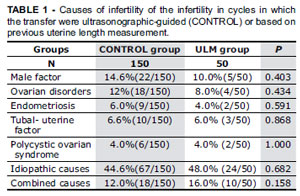
TABLE 1. Causes of infertility of the infertility in cycles in which the transfer were ultrasonographic-guided (CONTROL) or based on previous uterine length measurement.
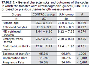
TABLE 2. General characteristics and outcomes of the cycles in which the transfer were ultrasonographic-guided (CONTROL) or based on previous uterine length measurement.
The embryo transfers were classified as easy or difficult and the groups were equal regarding the presence of blood on the catheter (4.7% vs 4.3% for Control and ULM groups respectively, p = 1.000). and easiness of transfer (95.3% vs 96.0% for Control and ULM groups respectively, p = 1.000). The transfer was considered difficult when the assistance of a more rigid catheter was required to successfully accomplish the embryo transfer.
Both the implantation (11.9% vs 30.7% for Control and ULM groups respectively, p < 0,001) and the pregnancy rates (25.3% vs 54.0% for Control and ULM groups respectively, p < 0,001) were significantly higher in the ULM group.
The total number of gestational sacs visualized by ultrasound. and the total number of embryos transferred for Control and ULM groups were respectively 43 vs. 37 and 361 vs. 120.
The incidence of multiple pregnancies were 3 twins and 1 triplets for control group and 10 twins and no triplets for ULM group.
DISCUSSION
Embryo transfer is a crucial tool on assisted reproduction treatment. Here, we tested the hypothesis that embryo transfers based on previous uterine length measurement would increase ICSI outcomes when compared to the standard ultrasonographic-guided embryo transfer method. Results of the present study indicate that the embryo transfer based on previous uterine length measurement do not result on an easier transfer; however increased pregnancy and implantation rates can be achieved using this technique.
Clinical perception of the embryo transfer difficulty is a commonly used marker for the optimum performance of embryo transfer. However, although previous studies indicate that a traumatic embryo transfer technique may result in damage to the endometrium (Cevrioglu et al. 2006), the impact of easiness or difficulty of embryo transfer on subsequent pregnancy rates is controversial. While, decreased pregnancy rates has been reported with difficult embryo transfer (Englert et al. 1986; Mansour et al. 1990), such significant detrimental effect has not been noted by others (Tur-Kaspa et al. 1998; De Placido et al. 2002; Silberstein et al. 2004).
In the present study, although embryo transfers based on previous uterine length measurement did not affect the difficulty of embryo transfer, a significant impact on the clinical outcomes could be observed. In fact, in our trial, difficult embryo transfers were observed in less than five percent of the cases, independently on the embryo transfer method. Therefore it would be surprising to find any improvement with the embryo transfers based on previous uterine length measurement. The increased pregnancy and implantation rates observed in our study, when embryo transfers based on previous uterine length measurement were performed instead of standard ultrasonographic-guided embryo transfer, raise the questions on whether the ultrasonographic guidance may avoid injury to the endometrium.
Some potential advantages of the ultrasonographic guidance have been reported. The placement of soft catheters and the positioning of the tip is facilitated (Allahbadia et al., 2008). Some authors argue that the real benefit of ultrasound guidance lies in the ability to increase the clinical appreciation of the pelvic anatomy during transfer (Abou-Setta 2007).
In fact, previous studies indicate that ultrasonographicguided embryo transfers are easier if compared to the clinical manipulation alone, however the superiority of this technique is still under debate. While some studies show an improvement on IVF outcomes (Coroleu et al. 2000; Matorras et al. 2002; Li et al. 2005), others demonstrate a lack of differences (Coroleu et al. 2002b; Garcia-Velasco et al. 2002; Isaza et al. 2002; Flisser et al. 2006; Kosmas et al. 2007).
Even though ultrasonography may confirm the position of the catheter within the uterus, it may not ease the entry into the uterine cavity when negotiating a tortuous endocervical canal, retroverted uterus and obese patients, when the transduction visualization in difficult. The speculum may also interfere with visualizing the initial catheter placement by scattering or interfering with the acoustic signal. Visualizing the entire catheter track may not always be possible; aligning the canal and endometrial stripe within the narrow acoustic window of the transducer may be technically difficult if the route traversed by the catheter falls wide of this path (Flisser et al., 2006). Moreover, a recent meta analysis conducted to determine whether ultrasound-guided embryo transfer improved clinical pregnancy rates and live birth rates in assisted conception did not demonstrate a difference (Drakeley et al. 2008).
Conversely, in another meta-analysis, it was concluded that the use of transabdominal ultrasound to guide catheter placement during embryo transfer may be beneficial and should become routine for all patients .(Abou-Setta et al. 2007). Instead, in our trial we demonstrated that embryo transfer based on previous uterine length measurement could potentially improve implantation rate, avoiding the ultrasound guidance available during the embryo transfer.
Nevertheless, some catheters have an additional feature, due to an echodense tip or echogenicity extending along the catheter. It was hypothesized that these echogenic catheters could minimize disruption of the endometrium with improvement in implantation rates (Letterie et al. 1999). A prospective, randomized study concluded that the use of these echodense catheters simplifies ultrasound-guided embryo transfer and the need to move the catheter for identification, but no differences in success rates were observed when compared with non-echodense catheters (Karande et al. 2002). Moreover, Coroleu at el (2006) reported similar pregnancy rates among groups with embryo replacement through echodense and nonechodense catheters. Thus, even with the advent of echogenic catheters, pregnancy rates do not seem to be higher with the use of ultrasound-guided embryo transfer. Previous studies already investigated the influence of transfer distance from the fundus on clinical outcomes. Oliveira et al., (2004) demonstrated that the pregnancy rate is significantly affected by the transfer distance from the uterus fundus. Frankfurter et al. (2004) showed that both pregnancy and implantation rates are positively affected by directing embryo placement to the lower to middle uterine segment. Coroleu et al.(2002a) concluded that applying the fixed distance of 15–20 mm away from the fundus might optimize the results of embryo transfers.
In a previous study designed to determine the location of the physiological embryo implantation site using a three dimensional transvaginal ultrasound, results indicated that the part of the endometrium suitable for human blastocyst implantation, under physiological conditions, is located at the uterine fundus, especially near the uterotubal junction (Minami et al. 2003). Conversely, Frankfurter et al (2003; 2004) suggested that higher pregnancy rate could be achieved when the site of embryo replacement was more distant from the uterine fundus, from the middle to lower segments of the uterus compared to the upper segment. Moreover, through a logistic regression analysis, Pope et al. (2004) demonstrated that for each additional millimeter that embryos are deposited away from the fundus, as noted by abdominal ultrasound, the odds of clinical pregnancy increased by 11% (Pope et al. 2004).
In our trial, the embryos were deposited in the middle of the cavity length, and, in comparison with the standard ultrasonographic-guided embryo transfer, a significant improve on implantation and pregnancy rates was observed. These findings suggest that the endometrial location may provide a more favorable region for embryo deposition and the embryo transfer based on previous uterine length measurement may be useful to determine the depth beyond which catheter insertion should occur.
CONCLUSION
In conclusion, our findings suggest that embryo replacement in the middle of the cavity length can significantly improve pregnancy and implantation rates, with no additional costs, and therefore embryo transfer based on previous uterine length measurement should be performed during the follicular growth control in controlled ovarian stimulated cycles.
ACKNOWLEDGMENTS
The authors thank Marcílio Nichi, PhD for the critical reading and evaluation of this manuscript.