JBRA Assist. Reprod. 2010;14(4):29-32
ARTIGO ORIGINAL
doi: 10.5935/1518-0557.2010.14.4.05
Motile sperm binding to egg yolk membrane is an easy and feasible function test for acrosome integrity
Ligação de espermatozóides à membrana vitelínica de ovos de galinha: teste funcional simples e fácil para verificação da integridade acrossomal
1Huntington Medicina Reprodutiva, São Paulo Brasil.
2Departamento de Ginecologia da Universidade Federal de São Paulo, São Paulo, Brasil.
3Disciplina de Ginecologia da Faculdade de Medicina da Universidade de São Paulo, São Paulo, Brasil.
This investigation was performed at Huntington Medicina Reprodutiva.
ABSTRACT
Objective: Acrosomal integrity is a feature necessary to the correct interaction of the spermatozoon to the oocyte. The assessment of functional integrity of the acrosome is a useful tool for evaluation of male fertility before assisted reproduction treatments. We aimed to validate a perivitelinic binding test(PBT) for human sperm.
Methods: Perivitelinic membranes were washed in PBS for yolk removal and squares of 1cm2 were obtained for the incubation of 1mL of Hepes diluted sperm (105sptz/mL) for 2 hours at 37ºC. Membranes were washed in PBS for the removal of unbound sperm and they were mounted onto glass slides for count of motile sperm bound(MSB) to the membrane. Acrosome integrity was also assessed in spear smears stained with fast green/Bengal rose for computation of Pearson’s correlation coefficient to MSB. Validation of PBT comprised linear regression of percent of MSB in fresh sperm samples diluted with dead sperm samples (semen plunged into N2L and thawed five times) at the dilution rate of 0:1; 1:4; 1:1; 3:4; 1:0. Pearson’s correlation coefficient between MSB and intact acrosome of fresh samples was calculated. Significance was attained at 0.05.
Results: Percent of MSB was correlated with acrosome integrity (r=0.65;p=0.04). Furthermore, MSB was also correlated to the dilution rate(r=0.927;p<0.001) and they featured a significant linear relationship (%MSB=0.09 + 0.7730 dilution rate; p-value <0.001).
Conclusion: Motile sperm binding to egg yolk membrane is an easy and feasible test for the assessment of functional integrity of acrosome. This is a low cost tool for evaluation of semen before IUI and IVF.
Keywords: acrosome, semen analysis, acrosome reaction
RESUMO
Objetivo: A integridade funcional do acrossoma é necessária para a ligação espermático-oocitária e para a fertilização. A avaliação da sua integridade funcional é uma ferramenta importante na avaliação da fertilidade masculina antes do tratamento de infertilidade. Nosso objetivo foi validar o teste de ligação de espermatozóide à membrana perivitelínica de ovo.
Métodos: As membranas perivitelínicas foram lavadas em PBS três vezes para remover o vitelo e cortadas em quadrados de um centímetro. O teste foi realizado pela incubação de 1 mL de espermatozóides (105sptz/mL) com as membranas perivitelínicas por 2 horas a 37ºC e posterior lavagem em PBS. As membranas foram montadas entre lamina e lamínula e a motilidade avaliada em microscópio óptico (200x) em três campos diferentes. A integridade acrossomal também foi avaliada em esfregaços corados por meio do método fast green/Bengal rose para cálculo do coeficiente de correlação de Pearson com o teste em questão. A validação foi realizada por meio de regressão linear entre a porcentagem de espermatozóides móveis em amostras frescas diluídas com amostras contendo apenas espermatozóides mortos obtidas após 5 mergulhos/descongelamentos em N2L nas seguintes proporções: 0:1;1:4;1:1;3:4;1:0.
Resultados: A porcentagem de espermatozóides móveis ligados foi correlacionada com a integridade acrossomal avaliada por fast green: bengal rose (r=0,65;p=0,04) e com a taxa de diluição com espermatozóides móveis (0,927;p<0,001). Foi observada uma relação linear significativa (% de espermatozóides ligados=0,09 + 0,7730 diluição; p <0,001) entre o teste de ligação de espermatozóide à membrana perivitelínica de ovo e a taxa de diluição com os espermatozóides móveis
Conclusão: O teste de ligação de espermatozóide à membrana perivitelínica de ovo é um teste funcional de integridade acrossomal fácil, exeqüível e barato.
Palavras-chave: acrossoma, análise do sêmen, reação acrossômica
INTRODUCTION
Traditional sperm analysis according to the criteria established by the World Health Organization (WHO) is part of the investigation of infertile couples, and it aims to recognize potentially fertile conditions (WHO, 2010). If the analysis does not reach the normal criteria, assisted reproduction techniques could be used to overcome the male deficiency using their sperm or irreversible conditions that might indicate the use of donor’s sperm (WHO, 2006). The parameters defining a normal ejaculate were initially published by WHO in 1980 (Lewis, 2007) and they have been subjected to several changes in the past 30 years with five updates (Joffe, 2010). However, all editions are still based on spermatozoa parameters such as sperm concentration, motility and morphology as the core of the evaluation. Functional tests are still grouped as experimental tests and have not indicated for basic workup (Eliasson, 2010; Lamb, 2010).
The definition of normal semen parameters was based after sperm analysis of men who fathered pregnancies that took 12 months or less to conceive; notwithstanding, ejaculate volume, sperm concentration, motility and morphology are not able to predict the time to pregnancy, the ability to conceive and, more importantly, neither to distinguish fertile and infertile males (Joffe, 2010; Lamb, 2010). The crucial question is to determine the semen capable to contain adequate amounts of spermatozoa and undergoes a series of cellular and biochemical changes, allowing to deliver the male genome inside the oocyte. These subtle spermatozoal changes could be better assessed by sperm function tests. In last two decades, several methods were developed trying to measure normal and abnormal sperm function.
The ability of spermatozoa to interact with the zona pellucida is a crucial step during in vivo fertilization process and might be a predictor to determine the standard in vitro fertilization treatment. The sperm-zona interaction occurs through the binding of the spermatozoa with the zona pellucida 3 protein (ZP3) accomplished by a receptor located at acrosome (Yanagimachi, 1994). This interaction can be evaluated through hemizona assay (Coddington et al., 1991; Lanzendorf et al., 1992; Oehninger et al., 1992; Yogev et al., 1995; Janssen et al., 1997; Oehninger et al., 1997); however, human zona pellucida is a rare and difficult material to be routinely spent on sperm function tests.
Poultry perivitelinic membrane has a considerable homology to ZP3 (Waclawek et al., 1998) and allows the sperm binding in several mammalian species (Barbato et al., 1998; Amann et al., 1999a). In fact, commercial tests employing protein extract from egg yolk membrane were already validated to human sperm populations (Amann et al., 1999b). Assessment of sperm binding to poultry perivitelinic membrane can be prepared in house and might consist in a cheap, easy and feasible function test to assess male fertility. The aim of this work was to validate an in house sperm binding to perivitelinic binding test (PBT).
MATERIALS AND METHODS
Preparation of perivitelinic membranes
Fresh chicken eggs were washed with neutral detergent and sanitized with 70% ethylic alcohol for 5 minutes. Egg shells were gently opened and albumen was drained. Vitelinic sac was opened with scissor and the yolk was dumped for isolation of the perivitelinic membrane. Each perivitelinic membrane was subjected to 4 washes in 7 mL of PBS for 5 minutes under mild agitation for complete removal of yolk. Membranes were transferred to 60mm dishes and they were spread to allow the section of squares of 1cm2. Membranes were kept in PBS until use.
Perivitelinic binding test (PBT)
Freshly ejaculated samples from 5 donors were liquefied for 30 minutes at 37ºC and they were submitted to regular sperm analysis following WHO’s criteria. Samples were diluted in HEPES to obtain the test concentration of 105 sptz/mL (Barbato et al., 1998).Squares of perivitelinic membranes were spread on the bottom of a two well dish and covered with the sperm solution. Incubation was carried out for 2 hours at 37ºC and it was followed by 2 washes in PBS to remove unbound sperm. Membranes were mounted between glass slide and coverslips for submission to light microscope under 200X magnification. Sperm counts were performed in 3 different fields and the result was expressed by the percent of motile sperm bound (Barbato et al., 1998).
Validation of PBT and statistical analysis
Validation of PBT comprised two steps: i) calculation of Pearson’s correlation coefficient; and ii) determination of linear regression between percent of motile bound sperm and percent of dead sperm dilute in the sample.
i) Aliquots of freshly ejaculated samples were also prepared by fast-green/Bengal rose method for the assessment of acrosome integrity and comparison with the percent of motile bound sperm. Briefy, 5µL of semen and 5µL of a fast-green/Bengal rose solution were merged onto a glass slide, smeared and let to dry. Preparations were submitted to light microscopy under 1000X magnification for the identification of sperm with intact acrosome (stained in purple). Percent of sperm with intact acrosome in fresh samples was determined after the observation of 200 cells and Pearson’s correlation coefficient with percent of motile sperm bound to the perivitelinic membrane was computed (Pope et al., 1991).Additionally we performed a power analysis for Pearson’s correlation and linear regression to determine if the sample size yielded reliable results. Significance was attained at p<0.05 for all statistical tests.
ii) Subsets of the freshly ejaculate samples were submitted to 5 cycles of snap freezing in liquid nitrogen and thaw at 37ºC without addition of cryoprotectants. Samples for validation were prepared by the dilution of dead sperm into fresh samples according to the following dilution rates: 0:1(live:dead;0% live); 1:4(25%live); 2:4(50%live); 3:4(75%live); 1:0(100%live). Stepwise regression between the percent of motile sperm bound to the membrane and percent of dead sperm added to the sample was calculated.
RESULTS
Ejaculates from 5 donors were submitted to standard sperm assessment and the results were demonstrated according to the WHO laboratory manual for semen analysis (Table 1).
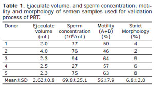
Table 1. Ejaculate volume, and sperm concentration, motility and morphology of semen samples used for validation process of PBT.
Fresh samples submitted to fast green/Bengal rose staining showed 72.8±7.8 % of acrosome integrity. Furthermore, determinations of acrosome integrity with fast green/Bengal rose were positively correlated to the percent of motile sperm bound to the perivitelinic membrane (Table 2; r=0.65; p=0.04).
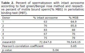
Table 2. Percent of spermatozoon with intact acrosome according to fast green/Bengal rose method and respective percent of motile bound sperm(% MBS) in perivitelinic binding test (PBT).
Five cycles of snap freezing in liquid nitrogen and thaw at 37ºC without addition of cryoprotectants were enough to virtually vanish motile sperm in all semen samples. The percent of motile sperm bound to the perivitelinic membrane in samples prepared for validation was significantly correlated to the expected rate of live sperm (0.927; p<0.001). Additionally, there was a linear relationship between the observed percent of motile sperm bound (MSB) and the dilution rate (Table 3; Figure 1) (%MSB=0.09 + 0.7730 dilution rate; p-value <0.001). The observed MSB was also correlated to the percent of motile sperm observed in each diluted sample (r=0.97; p=0.004) (Table 4). There was a linear relationship between the observed percent of motile sperm bound (MSB) and the percent of motile sperm observed in each diluted sample (%MSB = 6,24 + 1,48 percent of motile sperm observed in each diluted sample; p=0.004).
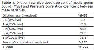
Table 3. Dilution rate (live:dead), percent of motile sperm bound (MSB) and Pearson's correlation coefficient between these variables.
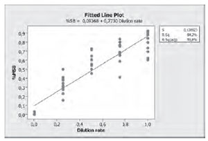
Figures
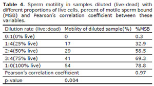
Table 4. Sperm motility in samples diluted (live:dead) with different proportions of live cells, percent of motile sperm bound (MSB) and Pearson's correlation coefficient between these variables.
DISCUSSION
To produce the fertilization, mammalian sperm has to interact with the zona pellucida, initiating a series of morphological and biochemical changes which will culminate with sperm penetration into the oocyte. Consequently, the loss sperm ability to interact to the zona pellucida might exert a deleterious impact on male fertility. Infections of the accessory glands, varicocele among others are recognized factors impairing male fertility through the diminishment of the competence of the sperm to bind to the zona pellucida and fuse with the oolema (Comhaire et al., 1999; Villanueva-Diaz et al., 1999).
Although the interaction between the gametes seems to be a crucial determination, the "gold-standard" in the evaluation of the male factor continue to be the semen analyses. The sperm-zona interaction might be elucidated through human hemizona binding assay, test developed during the late 1980's. Several prospective tests recognized that hemizona binding assay has a positive predictive result for intrauterine insemination and in vitro fertilization treatments (Franken et al., 1989a; Franken et al., 1989b; Oehninger et al., 1989; Oehninger et al., 1992; Franken et al., 1993; Oehninger et al., 1997; Arslan et al., 2006; FrankenOehninger, 2006). However, the obtainment of zona pellucida relies on the efforts to isolate material from immature oocyte retrieved during in vitro fertilization procedures or even from ovarian tissues (Arslan et al., 2006). Unfortunately, the test depends on a scarce and rare material. Alternative materials, providing similar results were developed to assess sperm function evaluation and among then chicken's perivitelinic membrane were indicated.
Barbato et al. (1998) demonstrated the ability of several mammalian sperms to bind to perivitelinic membrane of chicken's egg and proposed its use to detect subfertile males in several species including humans. Additionally, Amann et al.(1999b) showed that proteins of the perivitelinic membrane of chicken's egg adsorbed to plastic dishes are suitable in the identification of sperm with acrosome function preserved after cryopreservation. In our study, we choose to validate the use of minimally processed perivitelinic membranes by several reasons: it is timeless and low material consuming; it might be easily included in semen evaluation workup, could differentiate the male factor to predict the choice to in vivo to in vitro procedures. Staining of semen samples with fast green/Bengal rose for production of smears provides useful information on acrosome integrity (Pope et al., 1991; Valle et al., 2008). The percent of sperm with intact acrosome detected by this staining was positively correlated with the percent of MSB. Preparation and reading of PBT is longer than staining semen samples with fast green/Bengal rose (Pope et al., 1991; Barbato et al., 1998), but has an additional gain in providing information on the sperm motility status as well as, the intact acrosome. This simple correlation between these two evaluations could be considered enough for validation; nevertheless, we decided to assess if the proportion of MSB would be kept in different concentrations of dead sperm.
Cycles of snap freezing/thawing without cryoprotectants provided samples rich of dead sperm that were diluted to our fresh samples to produce decreasing concentrations of live sperm. Our results indicated that the percent of MSB are highly correlated to the proportion of live sperm contained in a sample and they presented a linear relationship; moreover, MSB was correlated to the percent of motile sperm in each sample with different proportions of live cells. Thus, PBT can be considered sensible and specific for the detection of MSB, even for samples with low proportion of motile sperm able to bind to PM.
Taken together, the correlations of MSB with the results of fast green/Bengal rose, the proportion of live sperm and percent of motile sperm contained in samples were enough to validate PBT and its ability to faithfully identify sperm with acrosome intact whilst indicate the proportion of cells with preserved motility.
In summary, PBT seems to be a validated, cheap, easy and feasible test for the assessment of sperm function; moreover, this test indicates the ability of motile sperm to interact to the zona pellucida. This is low cost tool might benefit patients undergoing fertility screening before intrauterine insemination and standard in vitro fertilization; however, clinical data to validate the extensiveness of these benefits should be the objective of future studies.
Joffe M. Semen quality analysis and the idea of normal fertility. Asian J Androl. 2010;12:79-82.
Lewis SE. Is sperm evaluation useful in predicting human fertility? Reproduction. 2007;134:31-40.