JBRA Assist. Reprod. 2011;15(1):24-27
ARTIGO ORIGINAL
doi: 10.5935/1518-0557.2011.15.1.04
Use of microarrays Comparative Genome Hybridization for preimplantation genetic screening of poor prognosis patients
O uso da Hibridação Genômica Comparativa por arrays para o screening cromossômico preimplantacional em pacientes com baixo prognóstico reprodutivo
1Genesis Genetics Brasil, São Paulo, Brasil
2Huntington Centro de Medicina Reprodutiva, São Paulo, Brasil Este trabalho foi realizado no laboratório Genesis Genetics Brasil
RESUMO
Introdução: Muitos embriões gerados in vitro apresentam alterações cromossômicas e, assim, baixo potencial de desenvolverem gestações viáveis. A metodologia mais frequentemente usada no diagnóstico genetico préimplantacional envolve a biópsia embrionária no terceiro dia do desenvolvimento embrionário, seguido por hibridação fluorescente in situ (FISH) para análise de 5 a 12 cromossomos. Neste trabalho relatamos uma nova estratégia para screening completo de aneuploidias cromossômicas em embriões gerados por Fertilização in vitro para pacientes com baixo prognóstico reprodutivo, a Hibridação Genômica Comparativa por microarrays (aCGH).
Materiais e Métodos: Um total de 23 embriões provenientes de 5 casais com histórico de falhas sucessivas de Fertilização in vitro e perdas gestacionais foram biopsiados no terceiro ou quinto dia do desenvolvimento embrionário. O screening cromossômico foi realizado pela técnica de aCGH.
Resultados: A aCGH apresentou uma taxa de eficiência de 95,6%. No total, 47,8% dos embriões analisados eram euplóides. Nove embriões foram transferidos para o útero materno sem vitrificação prévia, resultando em 75% (6/8) de taxa de implantação (presença de saco gestacional) e 50% de fetos.
Conclusão: A metodologia proposta neste trabalho para a análise completa das aneuploidias embrionárias supera a maioria das limitações ténicas anteriormente observadas na realização do screening genético preimplantacional e pode, finalmenteme, favorecer as taxas gestacionais em casais com baixo prognóstico reprodutivo.
Palavras-chave: aCGH, PGS, implantação embrionária, aneuploidia, taxa gestacional
ABSTRACT
Introduction: Many embryos produced in vitro contain chromosomal abnormalities and have little potential for forming a viable pregnancy. The most commonly used method for preimplantation genetic diagnosis involves embryo biopsy on day 3 of development, followed by fluorescence in-situ hybridization analysis of 5-12 chromosomes. Here we report a new strategy for comprehensive chromosome screening in IVF embryos for poor reproductive prognosis patients, the Comparative Genome Hybridization by microarrays (aCGH).
Matherial and Methods: A totol of 23 embryos, from 5 couples undergoing IFV treatment with history of unsuccessful attempts (>2 attempts) and miscarriages, were examined by aCGH after blastomere or trophectoderm biopsies.
Results: The aCGH showed an efficiency of 95.6%. A total of 47.8% of embryos were euploid. Nine embryos were transferred to uterus without previous vitrification, reveling that 75% (6/8) of them produced fetal sac snd 50% of transferred embryos produced a fetus.
Conclusion: The full chromosome complement screening method described overcomes the majority of the problems that limited earlier aneuploidy screening techniques and may finally allow preimplantation genetic screening to improve the implantation rates in poor prognosis patients.
Key-words: aCGH, PGS, embryo implantation, aneuploidy, pregnancy rate
INTRODUCTION
In vitro fertilization (IVF) treatments typically involve the production of multiple embryos. However, the viability of individual embryos is highly variable. Even among a cohort of sibling embryos competence can vary greatly. The challenge for IVF clinics is to correctly identify the most viable embryos and prioritize them for transfer to the uterus.
One of the most powerful methods for improving IVF efficiency is embryo selection based on morphologic and developmental characteristics; even so, the implantation potential of human embryos produced in vitro remains low. One reason is that the majority of human embryos produced in vitro are chromosomally abnormal (1-9). A study involving over 6000 embryos (2) showed that chromosome abnormalities were widespread regardless of maternal age and morphology. For example, only 42% of the best embryos, according to morphology, in patients 35 to 37 years of age were chromosomally normal, and abnormalities increased with age and poorer embryo developmental characteristics (2). Although morphology is clearly correlated with euploidy, its use in selecting embryos for replacement can at best only improve euploidy by a few percentage points. Wells et al. (10) have indicated that over 40% of blastocysts are still chromosomally abnormal and that most monosomies can reach the blastocyst stage.
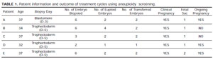
Table 1. Patient information and outcome of treatment cycles using aneuploidy screening.
Because morphology alone can only slightly improve the probability of selecting a chromosomally normal embryo for transfer, Preimplantation Genetic Screening (PGS) in combination with other selection techniques may provide a benefit by accurately identifying euploid embryos, thus leading to improved ART (Assisted Reproductive Technology) outcomes. Despite large studies indicating the advantages of aneuploidy screening, confusion and controversy persist concerning the benefits of PGS in part because of the differing results of studies that have used widely varying procedures, some more effective than others.
Here we report clinical application of a novel aneuploidy screening strategy, the Comparative Genome Hyubridization by microarrays (aCGH). The method was applied both in day-3 embryos and in blastocyst stage, 2-3 days later than traditional methods of PGS. Additionally, the procedure involves screening of the entire chromosome complement, rather than the limited chromosome assessment typically used for the purpose of PGS. We suggest that this approach may finally achieve a high clinical potential, improving the pregnancy rate in patients with a history of failed IVF attempts or/and spontaneous abortions.
MATHERIALS AND METHODS
Patient Details and Biopsy
Data presented were derived from 5 couples undergoing assisted reproductive treatment. The average maternal age was 35.4 years (range 32-37years). The chromosome screening was offered to these patients, not for advanced reproductive age, but due to their history of unsuccessful IVF attempts (>2 attempts) and miscarriages.There are 2 particularly difficult cases: (A) patient with 32 years old who experienced two miscarriages after IVF treatment, in third IVF attempt it was included aneuploidy screening for 9 chromosomes and the patient failed to get pregnant; (B) patient with 37 years old who experienced 11 FIV attempts and more than two pregnancy losses.Four of these patients had their embryos biopsied on day-5 during blastocyst stage and transferred after the aCGH analysis, on day-6, without vitrification. One patient had her embryos biopsied on day-3 and the euploid embryos were transferred to uterus on day-5. CGH results were available for 22 of the 23 embryos biopsied.
Comparative Genome Hybridization microarray (aCGH)
Each sample biopsied (trophectoderm and blastomeres) was washed in sterile phosphate-buffered saline and transferred to a microcentrifuge tube in 2ul of a lysis solution. To generate the ~1 mg of DNA required for aCGH analysis, the biopsied cells were lysed and the whole genome amplified using degenerate oligonucleotide-primed polymerase chain reaction. Amplified DNA was labeled with a green fluorescent molecule (Spectrum Green-dCTP, G&E Healthcare). Similarly, DNA from a chromosomally normal individual was labeled with red fluorescence (Spectrum Red-dCTP, G&E Healthcare). The green (embryo) and red (normal reference) DNAs were mixed together and simultaneously hybridized on a BAC - array platform able to cover all the 24 chromosomes. Scanned images were analyzed and quantified, and whole chromosomal copy number ratios were reported using the arrayanalysis software (Fig 1).
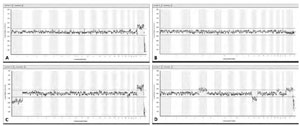
Figure 1. A: Example of a normal female embryo analyzed from a trophectoderm biopsy by aCGH. B: Example of a normal male embryo analyzed from a trophectoderm biopsy by aCGH. C: aCGH analysis of an embryo with a karyotype: 45,XX,-1. D: aCGH analysis of an embryo with a karyotype: 47,XX,+5,-13,+19.
RESULTS
The data described here were derived from 5 patients. These patients produced a total of 23 embryos. Seventeen of them were biopsied on blastocyst stage and 6 of them on day-3. Biopsy was successful for 100% of embryos, presenting DNA enough for subsequent aCGH analysis. A total of 22 embryos were successfully assessed using aCGH (95.6% diagnostic efficiency), 1 biopsied sample had degraded DNA that did not enable the analysis.
Of these, 11(47,8%) were diagnosed as euploid and 9 were transferred to uterus immediately after the aCGH report, with no vitrification.
All the patients showed at least 1 euploid embryo. One patient had only one embryo transferred and the other 4 patients had two embryos transferred.
The 5 patients become pregnant. The proportion of transferred embryos that successfully implanted was evaluated by ultrasound 6 weeks after transfer, revealing that 75% (6/8) of embryos transferred after aCGH analysis produced a fetal sac. Later ultrasound screening to assess fetal heartbeat, showed that 50% embryos transferred produced a fetus. These results are summarized in Table 1.
DISCUSSION
Preimplantation Genetic Screening was developed to try and improve pregnancy rates in certain groups of patients undergoing IVF procedures owing to infertility. Reported indications for PGS include patients with advanced maternal age, repeated miscarriage, repeated implantation failure, and severe male factor infertility (11).
PGS was first reported by Verlinsky et al. (12) and Munne et al. (13) in the analysis of polar bodies. Since these first reports, there have been numerous papers on the use of PGS; however, as with many new technologies brought into the IVF clinic, there has been little evidence to show that PGS increases delivery rates. There are now at least 11 randomized controlled trials (RCTs) applied to both good- (14-18) and poor- (19-23) prognosis patients. None of these trials have shown that PGS improved the delivery rate compared with a control group.
All of these studies used Fluorescent in situ Hybridization (FISH) based techniques to study 5-12 chromosomes, and almost all were applied to cleavage-stage embryos, except Jansen et al. (16), who performed trophectoderm biopsy. Despite the controversy surrounding the use of PGS, there is a universal agreement that even embryos reaching the best morphologic scores often fail to achieve implantation or do not produce a live birth, and those with poor scores sometimes succeed in producing a child. In many cases, the underlying cause of implantation failure or spontaneous pregnancy loss is the presence of numerical chromosomal abnormalities (aneuploidy).
A lot has been discussed about PGS testing of 24 chromosomes and recent studies (24) have renewed hope that new techniques and a fresh look at the moment of biopsy (moving away from cleavage stage where mosaicism is rampant) may finally show the usefulness of aneuploidy screening.
The present study displayed a high diagnostic efficiency, with comprehensive aneuploidy screening results obtained from 95.6% of embryos. The high implantation rate reported here may be indicative of improved embryo selection due to full chromosome screening on either cleavage stage embryos or blastocysts.
It has often been argued that the transfer of more than one embryo is advisable for patients of advanced maternal age or those with a history of multiple unsuccessful IVF treatments. Our data show that single embryo transfer after CGH screening would yield implantation rate (i.e., the probability of an embryo selected for transfer forming a clinical pregnancy) of 50% per blastocyst transfer compared with ~33% in cycles without screening published in ESHRE - ART fact sheet (www.eshre.com). It is probable that implantation and pregnancy rates would be higher in patients without a history of failed IVF attempts. In this report, 75% (6/8) of embryos transferred after aCGH analysis produced a fetal sac, suggesting that single embryo transfer should still be considered for such patients, at least for those capable of producing chromosomally normal blastocysts.
To conclude, although randomized controlled trails are still needed, this report has demonstrated the usefulness of aneuploidy screening by aCGH technique, increasing the pregnancy rate of poor prognosis patients.