JBRA Assist. Reprod. 2011;15(3):26-28
ARTIGO ORIGINAL
doi: 10.5935/1518-0557.2011.15.3.06
Preimplantation Genetic Diagnosis (PGD)- FISH versus CGH
Diagnóstico genético pré-implantacional (PGD)- FISH versus CGH
Reproferty, São José dos Campos- SP, Brazil
ABSTRACT
Objective: To determine embryos viability in preimplantation genetic diagnosis by Fluorescent In Situ Hybridization or Comparative Genomic Hybridization.
Materials and methods: Were biopsied 57 embryos from patients with clinical cases and similar age. Of these, 13 had DNA degraded/ absent, 17 were analyzed by Comparative Genomic Hybridization, 18 by Fluorescent In Situ Hybridization with 11 probes and 9 with 9 probes. We analyzed the percentage of aneuploids results that could have been obtained by each technique.
Preliminary Results: Among embryos diagnosed with aneuploidy by Fluorescent In Situ Hybridization with 9 probes, 60% could be diagnosed using 5 or 7 probes. The results of the method with 11 probes indicated that 80% of embryos with aneuploidy could have been diagnosed by 5 probes, 86.7% through the use of 7 probes and 100% with 9 probes. In relation to Comparative Genomic Hybridization, could have been diagnosed 53.8% of aneuploid embryos by 5 probes, 69,2% by 7 probes and 84,6% using 9 or 11 probes.
Conclusions: Diagnosis by Comparative Genomic Hybridization provides more accurate results. Its indication for patients with recurrent implantation failure and maternal advanced age should be considered because allowing the selection of best embryos. However, our data demonstrate that diagnosis by Fluorescent In Situ Hybridization may be sufficient to identify the most cases of aneuploidies, without need for further investment. Is important emphasize that the sample size is still small, so the study will continue to obtain more conclusive results.
Key words: spontaneous abortion, FISH, CGH, aneuploidy
Objetivos: Determinar a viabilidade de embriões através do diagnóstico genético pré implantacional por hibridização in situ fluorescente ou hibridização genômica comparativa.
Material e métodos: Foram realizadas biopsias de 57 embriões de pacientes com casos clínicos e idades similares. Desses, 13 apresentaram ausência ou degradação de DNA, 17 foram analisados pela hibridização genômica comparativa, 18 por hibridização in situ fluorescente com 11 sondas e 9 com 9 sondas. Foram analisados os porcentuais dos resultados de aneuploidias obtidos por cada técnica.
Resultados preliminares: Dentre os embriões diagnosticados com aneuploidia por hibridização in situ fluorescente com 9 sondas, 60% poderiam ser diagnosticados usando 5 ou 7 sondas. Os resultados dos métodos com 11 sondas, indicam que 80% dos embriões com aneuploidia poderiam ser diagnosticados com 5 sondas, 86,7% através do uso de 7 sondas e 100% com 9 sondas. Em relação à hibridização genômica comparativa, poderiam ser diagnosticados 53,8% das aneuploidias nos embriões por 5 sondas, 69,2% por 7 sondas e 84,6% usando-se 9 ou 11 sondas.
Conclusões: O uso do diagnóstico por hibridização genômica comparativa oferece resultados mais precisos. Sua indicação para pacientes com falhas recorrentes na implantação e idade materna avançada deve ser considerada, por permitir a seleção dos melhores embriões. Entretanto, os dados demonstram que o diagnóstico por hibridização in situ fluorescente pode ser suficiente para identificar a maior parte dos casos de aneuploidias, sem a necessidade de maiores investimentos. Deve-se enfatizar que o tamanho das amostras é bem pequeno, deste modo, o estudo deve continuar para obtermos resultados mais conclusivos.
Palavras-chaves: aborto espontâneo, FISH, CGH, aneuploidia.
INTRODUCTION
The Pre-implantation Genetic Diagnosis (PGD) is indicated for patients undergoing treatment for Assisted Reproduction (AR) and that fall into at least one of the following cases (Wilton, 2005): (I) recurrent failure of implantation, ie failure in more than three cycles of RA in which good embryos were transferred, (II) women over 35 years, due to the high incidence of chromosomal abnormalities derived from meiotic alterations, such as non-disjunction, which occur with advancing maternal age (Margalioth et al., 2006; Andersen et al., 2000; Romero-Maldonado et al., 2002), (III) patients with recurrent spontaneous abortions.
The chromosomal anomalies have an important contribution in spontaneous abortions and recurrent implantation failure in cases in vivo and in vitro. Its incidence reaches including embryos with good morphological quality and high rates of development (Voullaire et al., 2002; Margalioth et al., 2006). It is estimated that 70% of the normal embryo are in fact aneuploid and about 37% of trisomic embryos reach the blastocyst stage (Sandalinas et al., 2001).
The chromosomal numeric abnormalities are the most common in samples from spontaneous abortions: 50 to 60% of trisomy, 20 to 25% poliploidy and 15 to 25% monosomy of the chromosome X. The genetic factor is related to the occurrence of abortion and its subsequent repetition. Fetal chromosomal abnormalities are the cause of 50-60% of spontaneous abortions occurring during the first trimester of pregnancy (Munné et al., 2002).
The most plausible explanation for the high incidence of chromosomal abnormalities in cases of spontaneous abortions is that the most chromosomal damages results in a disorganized development of the conceptus, which is incompatible with uterine survival and birth. According to literature, the degree of selection among different chromosomal defects is highly variable. For example, in trisomy of 21, responsible for Down Syndrome, about a third of the fetuses surviving to term gestation, whereas in cases of triploidy, ie in which all pairs have an extra chromosome, less than 1 % reach the third trimester of development (Rodini et al., 2004).
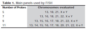
Table 1. Main panels used by FISHnumber of Probes
To complement the selection of embryos based on morphology and thereby improve the success rates of pregnancy, one of the techniques available for PGD it is the Fluorescent In Situ Hybridization (FISH) performed on biopsied cells from embryos during cleavage stage (Gutierrez-Mateo et al., 2004). This method uses specific probes for chromosomes related to aneuploidy commonly found in newborns and fetuses spontaneously aborted. The number of chromosomes evaluated, as well as the cost of examination per embryo, varies with the amount of probe used. The numbers of probes most used are 5, 7, 9 and 11. The chromosomes evaluated according to each panel of probes are shown in Table1.
Althought imply increases in success rates of RA, the use of FISH is limited by the small number of chromosomes analyzed simultaneously and reduced accuracy when many probes are combined (Abdelhadi et al., 2003). Some studies indicate that 25-30% of chromosomal abnormalities remain undiagnosed using FISH, which entails transferring aneuploid embryos incorrectly diagnosed as normal (Sandalinas et al., 2001).
By allowing complete chromosome analysis and then detect the aneuploidies that are unable to be diagnosed by a set of FISH probes, the Comparative Genomic Hybridization (CGH) is a promising alternative (Wilton et al., 2005; Gutierréz-Mateo et al., 2004). This is a molecular cytogenetic technique that can be applied to individual cells in interphase and allows the simultaneous analysis of each chromosome (Voullaire et al., 1999; Wells et al., 1999). However, it presents high costs and its benefits need to be properly assessed before his indication for the patient.
The objective of this study was to determine whether the more complex techniques, and consequently more expensive, are necessarily the most suitable for PGD in patients under AR treatment. We compared the results of aneuploidy in embryos subjected to the methodologies by CGH and FISH with 5, 7, 9 and 11 probes.
MATERIALS AND METHODS
This is an ongoing prospective study. Biopsies were performed on third day of developing embryos of 57 patients with similar clinical cases and mean age of 35 years. The blastomeres of each embryo were sent to a laboratory expert in PGD. Of these, 13 had DNA degraded or absent, 17 were analyzed by CGH, 18 by FISH with 11 probes and 9 by FISH with 9 probes.
For each method, we analyzed results of aneuploidy which could have been obtained using a smaller number of probes. For example, an embryo diagnosed by CGH with trisomy on chromosome 17 could have been properly diagnosed as aneuploid with the use of FISH with a panel of nine probes, since it has the probe to detect this chromosome. However, using 5 or 7 probes, this aneuploidy wouldn’t be detected. On the other hand, a monosomy of chromosome 2, for example, only can be detected by CGH (see Table 1). The data obtained allowed to establish a comparison between the methodologies’ viability based on the percentage of diagnoses that can be obtained by less expensive techniques.
PRELIMINARY RESULTS
Among the embryos analyzed by CGH, two were considered euploid, ie, the karyotype with exactly 23 chromosomes pairs. The analysis by FISH with 9 and 11 probes found 4 and 3 embryos without alteration, respectively. Of the 44 embryos analyzed, 35 (78.5%) were diagnosed with aneuploidy. Among the aneuploid embryos analyzed by FISH with 9 probes, 60% could be diagnosed using 5 or 7 probes. The results of the method with 11 probes indicate that 80% of embryos with aneuploidy could have been diagnosed with only 5 probes, 86.7% by 7 probes and 100% through the use of 9 probes. In relation to CGH, could have been diagnosed 53.8% of aneuploid embryos using 5 probes, 62.9% through the use of 7 probes and 84.6% using 9 or 11 probes.
Moreover, 4 embryos (11.4%) would only be correctly diagnosed as aneuploid by CGH, because the abnormalities found are related to chromosomes that aren’t analyzed by the probes commonly used in FISH, and these were trisomies of chromosomes 10 and 5, monosomy of chromosomes 2 and 4 and deletion of the short arm of chromosome 1.
CONCLUSIONS
It is known that CGH allows to obtain more accurate results and its indication for patients with recurrent implantation failure and advanced maternal age should be considered because allowing the selection of best embryos and thus increase the chances of successful treatment (Romero-Maldonado et al., 2002). However, our data show that the FISH with few probes may be sufficient to identify more than half cases of aneuploid embryos, avoiding the investment in more expensive techniques. This is explained by the fact that the chromosomes selected for investigation by FISH are precisely those with a higher frequency of chromosomal anomalies and, thus, are directly involved with the major genetic diseases and causes of spontaneous abortions (Andersen et al., 2000; Rodini et al., 2004).
It is important to emphasize that in cases in which FISH did not detect changes, the diagnosis of aneuploidy may not necessarily be excluded, since abnormalities may occur in chromosomes non-assessed by the probes used. In these cases, the CGH is the only methodology available of detecting all aneuploidies and its indication is mainly recommended for patients with advanced maternal age.
The use of the techniques of PGD is a useful tool to identify embryos with high implantation potential (Fragouli et al., 2010) and should be approached with the couple for a decision on do or not to do embryo biopsy after being informed about the risks of technique.
It should be noted that the sample size is still small, so the study will continue to obtain more conclusive results.