JBRA Assist. Reprod. 2011;15(3):29-31
ARTIGO ORIGINAL
doi: 10.5935/1518-0557.2011.15.3.07
Array-Comparative Genome Hybridization: what expect of a full aneuploidy screening in embryos produced in vitro
Hibridação Genômica Comparativa por arrays: o que esperar da análise completa de aneuploidias em embriões produzidos in vtro
1Genesis Genetics Brasil, São Paulo, SP, Brazil
2Genesis Genetics Institute, Detroit, MI, USA
This study was done in Genesis Genetics Brasil, supervised by Pericles Assad Hassun Filho.
ABSTRACT
Objective: Aneuploidy is the principal genetic factor affecting reproductive success in humans. A great number of morphologically normal embryos either do not implant or spontaneously abort early in pregnancy because their chromosome number deviates from the normal diploid 46.
Material and methods: The present study used comparative genomic hybridization by microarrays (aCGH), a molecular cytogenetic method that allows simultaneous enumeration of all chromosomes to establish a review of a full aneuploidies screening in FIV embryos.
Results: The present study illustrates that aneuploidy can affect any chromosome with a slightly higher prevalence of monosomies, suggesting that good quality embryos revealed that even the most severe chromosomal abnormalities (e.g. monosomy, imbalance affecting the largest chromosomes and aneuploidy affecting multiple chromosomes) were often capable of surviving up to the blastocyst stage.
Key-words: CGH, arrays, aneuploidy, embryos, implantation rate, PGD
RESUMOObjectivo: Aneuploidia é o principal fator genético que afeta o sucesso reprodutivo em humanos. Um grande número de embriões humanos morfologicamente normais ou não implantam ou são abortados espontaneamente no início da gravidez porque o seu número de cromossomos difere da norma diplóide de 46.
Material e métodos: O presente estudo usou a hibridização genômica comparativa por microarrays (aCGH), um método de citogenética molecular que permite a contagem simultânea de todos os cromossomos para estabelecer a revisão de um rastreio completo de aneuploidias em embriões de FIV.
Resultados: O presente estudo mostra que aneuploidia pode afetar qualquer cromossomo, com uma prevalência ligeiramente maior de monossomias. Mesmo com as anormalidades cromossômicas mais graves (por exemplo, monossomia e aneuploidia), embriões foram muitas vezes capazes de sobreviver até o estágio de blastocisto.
Palavras-chave: CGH, matrizes, aneuploidia, embriões, taxa de implantação, PGD
INTRODUCTION
Aneuploidy is one of the main causes behind low pregnancy rates in humans (Obradors et al., 2010). Chromosome abnormalities are found in early cleavage stage embryos and first trimester, spontaneously aborted fetuses (Boue et al., 1985; Munne et al., 1995). More than 70% of all In Vitro Fertilization (IVF) embryos fail to implant, and only 35% - 40% of all cycles succeed after an IVF procedure. It is believed that the single major cause of IVF failure is the quality of harvested eggs, and up to 75% of such eggs are aneuploid (Fishel et al., 2010).
Chromosomal abnormalities arising after fertilization, at the cleavage stage, have also been the subject of cytogenetic study. The technique routinely used for aneuploidy screening in IVF embryos is fluorescence in situ hybridization (FISH), which is a fast and easy method to perform. While this approach has provided a useful insight into aneuploidy during early human development, most chromosomes remain untested and the cytogenetic assessment is therefore incomplete, once the most common FISH diagnosis analysis from 5 to 12 chromosomes in one cell.
While many studies have investigated chromosomes in cleavage stage embryos, the cytogenetics of human blastocysts (5 or 6 days post-fertilization) has received comparatively little attention.
Recently, there have been efforts to provide a more detailed characterization of blastocyst cytogenetics, using methods such as Comparative Genome Hybridization (CGH), a molecular cytogenetic method that allows simultaneous enumeration of all chromosomes (Fraagouli et al., 2008). From the clinical perspective, there has been increasing interest in screening blastocyst-stage embryos for chromosomal abnormalities, with a view to detecting and preferentially transferring euploid normal embryos during IVF cycles. Early clinical data suggest that an approach combining blastocyst biopsy and comprehensive chromosome screening using CGH or microarray-CGH (aCGH) may represent the optimal approach for preimplantation genetic screening (PGS) (Fragouli et al., 2011).
The aim of the present study is to report an overview of the chromosomal condition of 172 embryos of patients undergoing routine assisted reproduction treatment (ART), using the aCGH technique.
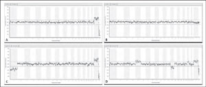
Figure 1. A: Example of a normal female embryo analyzed from a trophectoderm biopsy by aCGH. B: Example of a normal male embryo analyzed from a trophectoderm biopsy by aCGH. C: aCGH analysis of an embryo with a karyotype: 45,XX,-1. D: aCGH analysis of an embryo with a karyotype: 47,XX,+5,-13,+19.
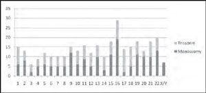
Figure 2. Presence of aneuploidies affecting all the chromosomes with a slightly higher prevalence of monosomies mainly those involving the chromosome 16.
MATHERIAL AND METHODS
Patients Details and Biopsy
Data presented were derived from 53 patients undergoing assisted reproductive treatment. The average maternal age was 38 (30-45) years. The chromosome screening was offered to these patients for advanced reproductive age, history of unsuccessful IVF attempts (>2 attempts) and miscarriages.Forty six of these patients had their embryos biopsied on day-5 during blastocyst stage and transferred after the aCGH analysis, on day-6, without vitrification. Seven patients had her embryos biopsied on day-3 and the euploid embryos were transferred to uterus on day-5. Array-CGH results were available for 161 of the 172 embryos biopsied. Eleven biopsied samples had degraded DNA that did not enable the analysis.
Comparative Genome Hybridization microarray (aCGH)
Each sample biopsied (trophectoderm and blastomeres) was washed in sterile phosphate-buffered saline and transferred to a microcentrifuge tube in 2ul of a lysis solution. To generate the ~1 mg of DNA required for aCGH analysis, the biopsied cells were lysed and the whole genome amplified using degenerate oligonucleotide-primed polymerase chain reaction. Amplified DNA was labeled with a green fluorescent molecule (Spectrum Green-dCTP, G&E Healthcare). Similarly, DNA from a chromosomally normal individual was labeled with red fluorescence (Spectrum Red-dCTP, G&E Healthcare). The green (embryo) and red (normal reference) DNAs were mixed together and simultaneously hybridized on a BAC - array platform able to cover all the 24 chromosomes. Scanned images were analyzed and quantified, and whole chromosomal copy number ratios were reported using the arrayanalysis software (Figure 1).
RESULTS
Of the embryos tested, 68,3% were found to be chromosomally abnormal (110 out of 161). A wide variety of aneuploidies was detected. Indeed, it was found that any chromosome can be affected by aneuploidy in IVF embryos (Figure 2). Monosomies and trisomies represented almost equally, with a slightly higher prevalence of monosomies mainly those involving the chromosome 16. Sixteen of the abnormal embryos were a consequence of chromosome breakage, resulting in partial losses or gains of chromosomal material, most of which affected the larger chromosomes (chromosomes 1-11 and chromosome X). Although the majority of embryos could be incompletely diagnosed by FISH for 9 chromosomes (13, 15, 16, 17, 18, 21, 22, X and Y), 37 of them (23%) would be classified as completely normal and probably transferred. Complex abnormalities (more than 3 aneuploidies in the same embryo) were seen in 32 embryos. As expected, a strong association between advancing maternal age and aneuploidy was observed. The number of normal embryos obtained from patients 30-37 (mean 34.1) years old were compared with those from patients 38-45 (mean 41) years old and we found out that the number of normal embryos in the first group is 2 fold higher than the second group (34/17).
DISCUSSION
Earlier studies have reported associations between aneuploidy and several distinct abnormal morphologies (Bielansk et al., 2002; Munne et al., 2006; Moayeri et al., 2008). However, in most cases the correlations have been weak, and no morphologic marker permitting accurate differentiation of euploid embryos from those with a lethal chromosome abnormality has yet been identified. This failure may simply demonstrate that cleavage stage morphology is a poor guide to aneuploidy.
The present study illustrates that aneuploidy can affect any chromosome (Figure 2). It is therefore inevitable that some embryos from earlier studies, with abnormalities affecting untested chromosomes, were wrongly categorized as euploid, like was seen in ~23% of all analyzed embryos in this report, that could be misdiagnosed by FISH 9 chromosomes and then transferred to uterus and fail to implant. In the present study, aCGH analysis was used, allowing all of the chromosomes to be accurately assessed. Furthermore, the majority of the embryos tested was based on the analysis of several cells rather than just one, an approach that theoretically reduces the risk of misclassification due to mosaicism (Fragouli et al., 2008). Around 70% of all embryos analyzed (68,3%) were chromosomally abnormal. A strong association between maternal age and aneuploidy was observed, decreasing the number of normal embryos from 34 in patients aged 31-37 (mean 34.1) years to 17 in the embryos of women aged 38-47 (mean 41) years.
With few exceptions, abnormalities affecting the largest chromosomes, monosomies, and abnormalities affecting multiple chromosomes simultaneously are not observed during prenatal testing. This suggests that such ‘‘severe’’ anomalies are lethal before the end of the first trimester of pregnancy, although the precise point at which affected embryos succumb remains to be defined. In contrast to our data, almost totally obtained from trophectoderm biopsies, one earlier study suggested that most monosomies are incapable of forming viable blastocysts (Sandalinas et al., 2001). A detailed evaluation of all the chromosomes in 158 good quality blastocysts revealed that even the most severe chromosomal abnormalities (e.g. monosomy, imbalance affecting the largest chromosomes and aneuploidy affecting multiple chromosomes) were often capable of surviving up to the blastocyst stage (Fragouli et al., 2008).
In conclusion, our results are in agreement with a previous investigation of more than 2000 embryos (personal communication with Genesis Genetics Institute) that found the same proportion of monosomies and trisomies involving all the chromosomal complement and confirm that even though some women of advanced reproductive age can produce embryos capable of becoming blastocysts, they have an elevated risk of carrying chromosome errors compared with embryos from younger women.
Boue A, Boue J, Gropp A. Cytogenetics of pregnancy wastage. Adv Hum Genet. 1985;14:1-57