JBRA Assist. Reprod. 2000;04(01):19-22
ARTIGO ORIGINAL
doi: 10.5935/1518-0557.2000.4.1.04
Accuracy of hysterosalpingography and hysteroscopy for diagnosis of intrauterine lesions in infertile patients in an assisted fertilization program
Acurácia da histerossalpingografia e histeroscopia para diag nóstico de lesões intrauterinas em pacientes de um programa de fertilização assistida
Hospital de Clínicas de Porto Alegre, Setor de Reprodução Assistida, Universidade Federal do Rio G rande do Sul
Abstract
To determine the accuracy of hysterosalpingography with relation to hysteroscopy for detection of intrauterine abnormalities in in fertile patients. Design: Cross-sectional study. Setting: University hospital. Subjects: 375 women of an assisted fertilization program. Intervention: Patients were submitted to hysteroscopy and hysterosalpingography prior to intrauterine insemination or in vitro fertilization. Main outcome measures: Hysterosalpingographic and hysteroscopic findings. Results: Hysteroscopy proved satisfactory in 95.2% of cases. Two-hundred and twenty-two patients (62.2%) had a normal hystero scopic evaluation. The most prevalent hysteroscopiç findings were : cervical stenosis (n=46); chronic endometritis (n=23); synechiae (n=23); endometrial hyperplasia (n= 19); polyps (n=17); and myomas (n= 10). The comparison of histeroscopic and histerosalpingographic findings revealed a sensitivity of 52.3% and a specificity of 85.1 % for hysterosalpingography; the positive and negative predictive values were 65.7% and 76.5%, respectively. Conclusions: Hysterosalpingography is not sufficiently accurate or sensitive for screening pathologies of the endometrial cavity in infertile patients. Hysteroscopy should be performed in all patients before IVF and arti ficial insemination.
Key words: hystero scopy, A RT, in fertility, hysterosalpingography
Resumo
Determinar a acurácia da histerossalpingografia em relação a histeroscopia na detecção das anormalidades uterinas em pacientes com infertilidade. O estudo foi realizado em um hospital universitário com 375 mulheres do programa de fertilização, onde as pacientes foram submetidas à histeroscopia e histerossalpingografia antes à inseminação artificial (IA) oufertilização "in vitro " (FIV). A histeroscopia provou ser satisfatória em 95. 2% dos casos. Cerca de 222 (62. 0%) apresentaram uma histeroscopia normal. Os maiores achados foram estenose cervical; (n =46), endometrites crónicas (n =23); sinéquia (n =23); hiperplasia de endométrio (n =19); pólipos (n =17) e miomas (n =10). A comparação da histeroscopia com a histerossalpingografia revelou sensibilidade em 52. 3% e especificidade em 85. 1% das histerossalpingografias. Os valores positivos e negativos foram 65. 7% e 76. 5% respectivamente. Em conclusão, a histerossalpingografia não apresenta uma acurácia suficiente ou uma sensibilidade para a seleção das patologias da cavidade endometrial em pacientes inférteis. A histeroscopia deverá ser procedimento de rotina em pacientes antes de realizar FIV ou IA.
Introduction
Most health care institutions consider hysteroscopy (HSC) as the gold standard for diagnosing intrauterine abnormalities. Moreover, since HSC is a tolerahle and safe test, it can be carried out in outpatient settings, without anesthesia. (Ayuida et al. 1997 and Gubbini et al. 1996). During the last few years, many authors have recommended the routine use of HSC for assessing the uterine cavity of infertile patients, especially of those who will be submitted to intrauterine insemination (IUI), or to in vitro fertilization with embryo transfer (IVF- ET). La Sala et al. (1998), Malhota et al. (1997), Cravello et al. (1997), Shamma et al. (1992) and Valle et al. (1991) demonstrated that in 18 to 43% of the cases studied, other methods were not effective for the detection of intracavitary abnormalities.
The actual role of anatomical anomalies in infertility has not been established. It is known that in spite of such anomalies, many women do impregnate and do get through the gestation period, without finding out about the problem. However, many studies have shown that the majority of women in this situation have difficulties in carrying out the gestation. Shamma et al. (1992) observed differences of up to 29% in the pregnancy rate of patients with abnormal hysteroscopic findings when compared to infertile patients with a normal hysteroscopic evaluation. In another study, Goldemberg et al. (1991), observed a 19% prevalence of intrauterine anomalies in patients who had been previously submitted to IVF and who did not gestate.
Currently, hysterosalpingography (HSG) IS routinely performed as part of most fertility investigation protocols throughout the world for examination of the uterine cavity, fallopian tubes and peritoneal cavity. However, a screening method for intrauterine abnormalities must have an adequate and established accuracy, which might not be case of HSG. Therefore, it is important to raise the question of whether HSG, or perhaps HSC, should be indicated to ali patients being submitted to insemination procedures or to embryo transfer. The objective of the present study was to assess the accuracy of HSG with relation to HSC for investigation of intrauterine abnormalities in a group of patients of an assisted fertilization program.
Subjects and Methods
The study population included 375 women of an assisted fertilization program at Hospital de Clínicas de Porto Alegre (Teaching Hospital, Universidade Fedéral do Rio Grande do Sul). Patients up to 38 years of age with indication for IUI and IVF, submitted to HSC between January 1996 and August 1998, and who agreed to participate in the study, were enrolled.
All patients were submitted to diagnostic HSC on an outpatient basis and without preoperative anesthesia. Ali exams were performed by the sarne physician (JSLCF), using a 4 mm Hamou Optics II hysteroscope (Karl-Storz, Germany). Carbon dioxide was used for uterine distention.
Hysteroscopic and hysterosalpingographic findings were analyzed in order to calculate the accuracy of HSG in relation to HSC. Only HSG examinations carried out in our hospital were included in the analysis (249 patients). HSG was carried out and evaluated by the sarne physician (JSLCF). In addition, the prevalence of diagnoses obtained with each method was determined.
To determine the accuracy of HSG, sensitivity, specificity, positive and negative predictive values were calculated in relation to the HSC diagnosis.
The Epi-Info 6.04 statistical package was employed for data analysis.
Results
The age average of our patients was 30 years, with a standard deviation (SD) of 6.7, and a range of 18 to 38 years. HSC proved satisfactory in 95.2% of ali cases, and there were no complications after this examination was performed. Two-hundred and six patients (55%) presented primary infertility, and 169 patients (45%) presented secondary infertility.
Two-hundred and twenty-two patients (62.2%) had a normal hysteroscopic evaluation. Abnormalities were found in 135 (37.8%) women. The most prevalent hysteroscopic findings were: cervical stenosis (46 cases); chronic endometritis (23 cases); synechiae (23 cases); endometrial hyperplasia (19 cases); polyps (17 cases); and myomas (10 cases). Malformation was observed in only 6 cases (figure 1).
Twenty-four patients (6.4%) presented two associated diagnosis. From these, 6 (1.6%) presented cervical stenosis and were resubmitted to HSC. The HSG results of 249 patients were analyzed in our study, out of which 179 were normal (72%). Synechiae, non-specific uterine filling defects, and cervical stenosis were the most frequent findings. The sensitivity of HSG in relation to HSC findings was 52.3%, with a specificity of 85.1%, and positive and negative predictive values of 65.7% arid 76.5%, respectively (Table 1).
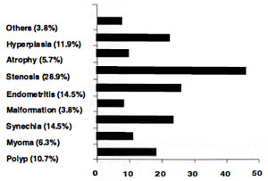
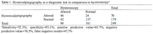
Table 1. Hysteroslpingography as a diagnostic test in comparison to hysteroscopya
Discussion
The prevalence of intrauterine anomalies diagnosed by HSC in our population was 37.8%, similar to the results reported by Ayida et al. (36%) (1997), the prevalence was lower in studies by La Sala et al. (1998) and by Maneschi et al. (1995) (18% and 10%, respectively). Still, other studies reported an even higher prevalence than what we found (48%, 43%, 45%, and 49%). Gubbini et. al. (1996), Malhotra et al. (1997), Shamma et al. (1992) e Nagele et al. (1996).
The most frequent finding in our study was cervical stenosis, fo llowed by chronic endometritis and synechiae. In a similar study, Malhotra et al. (1997) found synechiae in 25% ofcases; myomatosis in 9.4%; atrophy in 9.4%; and malformation in 6. 1 % of cases. This difference in result may have been caused by differences in the criteria employed in each study, as well as by differences in the population.
Chronic endometritis was a significant finding, which was visually diagnosed in 6. 1 % of cases (without biopsy). In a study by Cravello et al., (1997) the authors obtained the following results with patients in IVF programs: 22% had chronic endometritis; 14% had unexplained infertility; and 23.6% had frequent abortions. Those authors observed that the correlation between hysteroscopic and histologic findings was only 35%.
In the 1950s, Noyes et al. had already described the presence of lymphocytes in the endometrial stroma as one of the diagnostic cri teria for age-dating the endometrium during the second stage ofthe menstrual cycle. The characterization of endometritis would require the presence of other cellular elements of difficult anatomopathological diagnosis.
The prevalence of endometrial hyperplasia (5%) was considered high for the age of our patients, suggesting an association with chronic anovulation.
In our study, the sensitivity of HSG with relation to HSC was 52.3%, with a specificity of 85.1 %. In comparison to our results, Wang et al. (1995) reported higher sensitivity and specificity (87% and 91 %, respectively), whereas Malhotra et al .(1997) found lower sensitivity and specificity values (47% and of 69%, respectively, with 52.6% offalse negative results). The rate of false negative results in our sample was 47.7%. This shows that almost half of the patients submitted to HSG had some sort of intrauterine pathology which was not diagnosed by this examination.
Although it would be difficult to determine to what extent the diagnosed anomalies affected our patients infertility, the detection of such anomalies has been shown to be important for the reproductive prognosis. For example, a study Shamma et al. (1992) regarding the pregnancy rate of in fertile patients and its association with the presence of intrauterine pathologies (diagnosed by HSC), did observe that the pregnancy rate per cycle was 37.5% in patients with normal HSC results, as opposed to 8.3% for patients with abnormal HSC results. Moreover, in a study with patients submitted to at least two failed IVF attempts, La Sala et al. (1998) observed, through HSC, that 18% had endouterine abnormalities.
Screening exams must be sensitive enough to reduce the number of false negative results. In our study, we observed that HSG is a method that does not fulfill this criterion; it is not accurate enough for diagnosing pathologies of the endometrial cavity in infertile patients.
Because there is a high prevalence ofuterine anomalies in patients of assisted fertilization programs, and also because such anomalies affect pregnancy rates, it is essential that HSC become a routine procedure prior to IVF and artificial insemination.
Valle R. F. - Hysteroscopy. Current Opinion in Obstetrics and Gynecology 3: 422-426, 1991.