JBRA Assist. Reprod. 2001;05(03):102-105
ARTIGO ORIGINAL
doi: 10.5935/1518-0557.2001.5.3.03
Assisted hatching helps in selected patients with demonstrable abnormalities in the zona pellucida of their eggs
Assisted hatching ajuda na seleção de pacientes com anormalidades na zona pelucida de seus embriões
Abstract
This study was about the use of assisted hatching (AH) for helps in selected patients with demonstrable abnormalities in the zona pellucida of their eggs. We used a simple mechanical process involving ICSI injection pipette. The objective was determinate whether AH was of value in selected patients. A group of 75 patients IVF/ICSI, that multi pie IVF failure, aged 38 or over or patients with embryos with thick or descoloured zona pellucida (ZP). The results showed, that live birth rates were significantly better in the study group than in the age matched control group. The live pregnancy rates assisted hatching seems to ouer an advantage, was statistically significant. On frozen embryo no advantage accured to the hatching study group.. In conclusion the real benefict of AH may simply be the facilitation of early extrusion of the embryo though the ZP and its subsequent interaction with the endometrium maximising the opportunity for implantation.
Key words: assisted hatching, ICSI, IVF, multiple IVF/ICSI failure
Resumo
O estudo refere-se ao uso do assisted hatching (AH), para ajudar na seleção de pacientes com anormalidades demonstradas em zona pelucida de seus oócitos. Fizemos uso do mecanismo simples involvendo a pipeta de injeção para ICSI. O objetivo foi determinar quando o AH, era de grande valor para seleção das pacientes. Um grupo de 75 pacientes do programa de FlV/ICSI que apresentava múltiplas falhas de implantação e com idade acima de 38 anos ou pacientes com embriões que tenham espessamento ou descoloração em zona pelucida. Os resultados mostram que a taxa de nascimentos são signiticante melhores que as do grupo controle. As taxas de gravidez quando usado AH em embriões a fresco oferecem a vantagem de serem estatisticamente significante enquanto em embriões congelados não apresentam vantagens com o uso do AH. Em conclusão o real benefício do AH é simplesmente a facilitação da extrusão do embrião para fora da zona pelucida e subsequentemente a interação com o endométrio, maximizando assim a oportunidade de implantação
Unitermos: hatching assistido, ICSI, FIV, múltiplas falhas de implantação, implantação
Introduction
About one in four blastocysts derived from in vitro fertilized ovarian oocytes will hatch (Fehilly et al., 1985; Lindenberg, 1989), suggesting that human blastocysts are capable of hatching in the absence of uterine Iysins, but also demonstrating that zonae are often resistent to dissolution. Blastocyst formation and hatching can be increased by culturing human embryos on monolayers of reproductive tract cells (Lindenberg, 1989), which supports the idea that many in-vitro fertilized embryos may be deficient in the amount of zona Iysins needed to intiate and complete hatching. Bearing this in mind, it is thought that hatching can be promoted by opening the zona pellucida shortly before replacement (Malter & Cohen, 1989; Cohen et al., 1990). Much has been written about assisted hatching (see Brinsden et al., 1999) and it has been argued that the greatest benefit from assisted hatching (AH) might be derived by women aged 38 and over.
Assisted hatching may help because even when an embryo becomes a blastocyst, it may be unable to hatch because, even though it develops into an "expanded blastocyst", the zona pellucida does not break. This would leave a potentially viable embryo trapped inside the zona. This scenario may be caused by the zona pellucida being too thick or too hard. In such cases, where it is thought that there is problem with hatching, a procedure resulting in the opening up of a hole(s) of some description into the zona pellucida (assisted hatching), might help. In this study, we used a simple mechanical process involving an ICSI injection pipette. It differs from Cohen's (Malter & Cohen, 1989; Cohen et al., 1990) original method insofar as the abrasion process is omitted, whereby the ICSI injection needle is used to introduce just two small openings (small "perforations" about 7-1511- in diameter) in the zona by means of a simple "skewer" action. The aim was to determine whether AH was of value in selected patients over 35 years of age, who had endured previous multiple fail ure with IVF.
A further factor was examined during this study. Tucker et al.(1991) proposed that AH might benefit frozen embryo replacement (FER) cycles. An additional study group consisting of FER cycles was established to determine whether AH was useful for these patients.
Materials and Methods
Methods and criteria for screening embryos for assisted hatching Patients over 38 years of age and with multiple IVF failure were chosen to participate in the study. Additionall y, the zona pell ucida of all emhryos were routinely examined under a microscope containing a graticule, enabling ZP thickness to be assessed aml recorded. Furthermore, the ZP was assessed for disc ol ouration (either brown or yellow). Any patient exhibiting thick (>15 microns) or discol oured ZP were offered AH.
Patients
For the stud y 75 IVF/ICSI patients were recruited (multipie IVF failures aged 38 or over or patients with embryos with thick or discoloured ZP). Each study patient was also matched with next consecutive case as a control and age matched control. The average ages for study, control and age matched controls were as follows: study 37 years, control 33 years and age matched control 37 years.
A further study group of was established, consisting of 50 patients receiving FER. Ali these patients received embryos subjected to AH. Each study patient was matched to a control patient, not having AH. Average ages were: study 35 years, control 35 years.
Sperm preparation
Sperm were prepared using Percoll substitute provided by Fertipro (Belgium) by means of a discontinuous gradient of 45 and 90%, centrifuging for 20 minutes at 600g. Pellets were retrieved and washed in the appropriate medium and then resuspended in fresh medium at concentrations of about 1 million per ml.
IVF procedures
Ovarian stimulation was done using buserelin (Suprefact, Hoechst) in conjunction with Gonal-F (Serono, UK). Egg collection was carried out by transvaginally directed ultrasound. Oocytes were col lected and washed, after which they were placed in Ferticult medium. The routine protocol for IVF has been previously published Lee & Carvalho (1999). Insemination was carried out using 50-100000 sperm per ml. Pronuclei check was done 18 to 24 hours later after which embryos were placed in new dishes containing fresh medium (equilibrated for 24 hours in the incubator before use (using sarne medium as lhe initial culture). Forty two to 48 hours after egg collection, embryos were checked, after which three embryos (where available) were chosen for embryo transfer. For day 3 embryo transfer, embryos were placed in G3 for the final 24 hours of culture.
ICSI
Patients were advised to have ICSI treatment if they had previously undergone IVF which resulted in failed or poor fertilisation rates, or where the semen parameters were too poor for standard IVF treatment. Semen was assessed according to count, motility, progression and abnormal forms. Cases where the total motile count was less than 3 x L06; less than 5% motile sperm; if progression was very poor and/or < 5% abnormal forms; or a combination of the above; were treated with ICSI.
Eggs were treated with 80 iu/ml Hyaluronidase (Medicult, Denmark; Microm, Oxford; Scandinavian IVF Science, Hunter Scientific, Essex) for a maximum ofthree m inutes. The cumulus and corona were then removed by repeated passage through a Cook micropipetter and 140 micron Cook pipettes (Cook, Herts). The eggs were then washed well in fresh medium and then placed into dishes for ICSI. The eggs were placed in 20ul droplets of culture medium (covered in mineral oil) in Falcon 1006 dishes. Dishes were filled with 6-8 eggs at a time and also contained two droplets of polyvinylpyrrolidine (PVP) to act as reservoirs for the sperm.
ICSI was done using a Narishige setup mounted on a Nikon inverted microscope. Holding and injection pipettes were provided by Cook (Herts)or Humter Scientific (Essex). For cryptozoospermia, sperm was prepared using Sil-Select. The resulting pellet was spread out in one large droplet of medium under oil in Falcon 1006 dish. Motile sperm swim to the edge where they were 'harvested' for use. For percutaneous or micro-epididymal sperm aspiration, Sil-Select preparation was performed. The resulting pel let was washed and then placed in a 250µl eppendorf. The pellet was layered with approximately 25µl medium on top and allowed to swim up for an hour or soo With testicular biopsy (no Sil-Select preparation required), tissue was minced with green gauge injection needles (Becton Dickinson, Oxford) and then pl aced in a large 500ul droplet ofmedium under oil in a Falcon L006 dish. The droplet was then scrutinised carefully for up to 3 hours in order to obtain sperm for microinjection.
Freezing and thawing methods
Surplus embryos were frozen after IVF or ICSI. A commercial formulation (Scandinavian IVF Science, Hunter Scienti fic, Saffron Walden) was used to freeze and thaw. Brietly, embryos were washed in freeze I and then placed into freeze 2 for 15 minutes, then freeze 3 for 15 minutes, then loaded into a straw (Embryo freezing straw, Rocket of London, Watford) (1-3 at a time). The dish is originally at 37 C, but allowed to cool during the freezi ng process, so when the straw is filled, the embryos are at room temperature. The straw(s) were then placed in a KRYO 10 Series III automated freezer (Planar Products, Berkshire) and the standard on board embryo freezing programme was useo. Seeding was done manually after the temperature has been at - 7°C for 5 mino After the programme was complete, the straws were quickly placed into storage under liquid nitrogen in an MVE (Planar Products, Berkshire) liquid ni trogen holding canister until needed for frozen embryo replacement(FER)
For thawing, straws were removed from the MVE canister (Planar Products, Berkshire) and allowed to thaw at room temperature. The embryos were then expelled into thaw I and washed, then placed into thaw 2 for 5 minutes, then thaw 3 for 5 minutes, then into thaw 4 and washed before placing into a culture dish for preincubation prior to embryo transfer. During the thaw, the dish starts at room temperature, is then placed on a heated plate at 37C until the embryos are moved into a culture dish and placed into the incubator.
Assisted hatching method
A simple mechanical process involving an ICSI injection pipette was used to introduce two small "perforations" (7-15µl) into the zona. The embryos are placed into an ICSI dish (Falcon 1006, Becton Dickinson, Oxford) in a 100 µl drop of culture medium, which is covered in mineral oil (Microm, Oxford). A holding pipette (Cook, Herts) is used to steady and hold the embryo, whilst an ICSI needle is used to lance the zona either at the north of south pole (see Figure I). It differs from Cohen's (Malter & Cohen, 1989; Cohen et al., 1990) original method insofar as the abrasion process is omitted, whereby the ICSI injection needle is used to introduce two small openings (small "perforations" about 7-15µl in diameter) in the zona by means of a simple "skewer" action.
Results
Results for assisted hatching.
Results shown in Table I show that live birth rates (study group-23% vs control group - 18%, age matched control group- 10%; p<0.04 Fisher's Test) were significantly better in the study group than in the age matched control group. When looking at the live pregnancy rates assisted hatching seems to offer an advantage (AH-23% vs age matched controls- 10%), this difference was statistically significant (Fisher's Test, Instat, Graphpad, San Diego, USA).
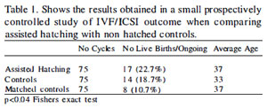
Table 1. Shows the results obtained in a small prospectively control led study of IVF/ICSI outcome when comparing assisted hatching with non hatched controls.
In the secondary study to examine the effect of AH on frozen embryos (see Table 2), no advantage accrued to the hatching study group.
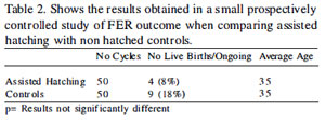
Table 2. Shows the results obtained in a small prospectively controlled study of FER outcome when comparing assisted hatching with non hatched controls.
Figure 1. Showing the assisted hatching method used in this study. (I) denotes the ICSI needle.
Discussion
Hatching was offered selectively; usually only after 3 failed IVF cycles or where the female partner is over 38 years old. Additionally, indications such as thick, discoloured or disfigured zonas were used as criteria for those having AH.
Control cases and age matched controls done at the sarne time as the AH cases have been paired to provide further data for comparison. The tive birthrate for AH patients was significantly higher (22.7% vs 10.7%) than for the matched controls. It is possible that zona hardness is reflected by thickness(> 15 microns), the presence of discoloration, unusually opacity and even by the presence of large numbers of sperm attached to the ZP.
Assisted hatching is a simple procedure, but it has been suggested that it may have profound consequences for embryonic development. For instance, microorganisms, vi ruses and cytotoxins present in the insemination suspension, could invade and infect the embryo via the artificially produced gap in the zona. Blastomeres may be lost through large holes, possibly causing embryonic death or vesiculation (Talansky and Gordon, 1988). Immune cell invasion through gaps in the zona may cause embryonic death, prior to the formation of tight junctions (Willadsen, 1979). No such problems were observed in this study and furthermore, no precautions such as prophylactic antibiotic therapy was given to any patient following embryo replacement after hatching. Moreover, only small perforations were made in this study.
Tucker et al., 1991 suggested that FER embryos might fare better if hatched. To examine this possibility, we additionally carried out a small study to see whether this might be the case. Surprisingly, we saw no such advantage. If anything the study group fared less well than the controls.
The real benefit of AH may simply be the facil itation of early extrusion of the embryo through the ZP and its subsequent interaction with the endometrium, maximising the opportunity for implantation. Assisted hatching is not very time consuming, simple to perform and does not require the use of new culture media or additives. Risks of damage from micromanipulation are minimal (for > 200 embryos micromanipulated none of the blastomeres were damaged). In particular, we have concentrated on thick and abnormal zonas, which Cohen et al., 1989 and Wright et al., 1990 showed had a poorer chance of implantation.
The results obtained in this study suggest that for selected patients AH is a worthwhile procedure.