JBRA Assist. Reprod. 2004; 08(02):7-20
ARTIGO ORIGINAL
doi: 10.5935/1518-0557.2004.8.2.02
Preimplantation Genetic Diagnosis
Centre for Medical Genetics, Medical School and University Hospital of the Dutch-speaking Brussels Free University (Vrije Universiteit Brussel, VUB)
Abstract
Preimplantation genetic diagnosis (PGD) is a very early form of prenatal diagnosis. Oocytes or preimplantation embryos are obtained in vitro and are genetically analysed, after which only those embryos that are judged to be free of the genetic defect under consideration are transferred. Initially, PGD was developed to help couples at risk for monogenic diseases.
Key words: Technologies used in PGD, Outcome of PGD, Accuracy of the diagnosis, pregnancy outcome.
Introduction
Preimplantation genetic diagnosis (PGD) is a very early form of prenatal diagnosis. Oocytes or preimplantation embryos are obtained in vitro and are genetically analyzed, after which only those embryos that are judged to be free of the genetic defect under consideration are transferred. To this end, polar bodies are removed from oocytes, or blastomeres (either at the cleavage stage or at the blastocyst stage) are removed from preimplantation embryos, and these cells are used for the genetic diagnosis. Initially, PGD was developed to help couples at risk for monogenic diseases. The only technique available to analyze single cells (polar bodies or blastomeres) was PCR, which was used either to sex the embryos (Handyside et al., 1990) or to detect specific mutations such as the ΔF508 mutation in cystic fibrosis (Handyside et al., 1992, Verlinsky et al., 1992). Later, fluorescent in situ hybridization (FISH) was downscaled to the single cell level (Griffin et al., 1992) and this opened possibilities to analyze embryos at the chromosomal level: either, again, for sexing (Harper et al., 1994), or for chromosomal aberrations such as Robertsonian or reciprocal translocations (Conn et al., 1998), or, finally for aneuploidy screening (Verlinsky and Kuliev, 1996).
Indications for PGD
Monogenic Diseases
Not surprisingly, most PGD cycles for monogenic diseases have been performed for the most frequent monogenic diseases. These are also more or less the monogenic diseases for which most prenatal diagnoses are performed. Examples are of course most importantly cystic fibrosis, spinal muscular atrophy, βglobinopathies, Duchenne’s muscular dystrophy, Fragile X disease, myotonic dystrophy and Huntington’s disease. Cystic fibrosis was the first monogenic disease for which a specific diagnosis at the single cell level was performed, analyzing the most frequent ΔF508 mutation (Handyside et al., 1992), and several reports using slightly different approaches have been published since (Verlinsky et al., 1992, Liu et al., 1993, Moutou and Viville, 1999, Goossens et al., 2000). More recently, multiplex approaches designed for analysis of single cells, and analyzing a CF mutation with one or more linked markers (Strom et al. 1998, Moutou et al., 2002, Goossens et al., 2003), or a panel of linked markers, have been described (Dreesen et al., 2000, Eftedal et al., 2001, Vrettou et al., 2002). Similarly, strategies have been developed for PGD for spinal muscular atrophy, both based on analysis of the most prevalent mutation (a large deletion encompassing several exons, Dreesen et al., 1998, Fallon et al., 1999, Moutou et al., 2001) and on the multiplex analysis of the mutation and linked markers (Moutou et al., 2003). Mutations in the β-globin gene, both leading to sickle cell anemia and β-thalassemia have received a considerable attention from the PGD community, because of their high frequency in certain populations. Because so many different mutations can be found in the β-globin gene (only one of them leading to sickle cell anemia, the others leading to various types of β-thalassemia), the strategies developed for PGD are different from e.g. SMA and aim to detect as many different mutations as possible with the same assay. Examples of this are given by Xu et al. (1999, on PGD for sickle cell anemia), Kanavakis et al. (1999, using DGGE for PGD for β-thalassemia), Kuliev et al. (1999, on polar body biopsy for PGD for βthalassemia), De Rycke et al. (2000, on PGD for sickle cell anemia and β-thalassemia using fluorescent PCR), and most recently Jiao et al. (2003, using PEP for PGD for β-thalassemia).Patients at risk for transmitting an autosomal dominant disease have always been particularly interested in PGD because of the high recurrence risk. Examples of this abound in the literature, and again the more frequent diseases have received the most attention. We (Sermon et al., 1999) and others (Harton et al.,1996, Blaszczyk et al., 1998) have described several different approaches for Marfan’s disease. PGD for diseases caused by dynamic mutations have also been reported. Noteworthy here are myotonic dystrophy type 1 (DM1) (Sermon et al. 2001, Pyiamongkol et al. 2001, Dean et al. 2001) and Huntington’s disease (Sermon et al, 1998, Sermon et al., 2001, Stern et al., 2002).Sexing with FISH has been most frequently used for X-linked diseases such as Duchenne’s muscular dystrophy, hemophilia A and B, retinitis pigmentosa, and others (Staessen et al., 1999), but more and more specific DNA diagnoses are developed and used. The advantages of a specific DNA diagnosis are important: firstly, healthy male embryos are not discarded and secondly, female carriers can be identified and, according to the patient’s wishes and the centre’s policy, are then not selected for transfer. Examples of tests developed for the specific diagnosis of an X-linked disease are given by Hussey et al. (1999), Ray et al. (2001) and Girardet et al. (2003) for Duchenne’s muscular dystrophy. Sermon et al. (1999) and Apessos et al. (2001) described protocols for PGD for Fragile X syndrome. Because girls who are carriers of Fragile X can be affected, only a specific DNA analysis can be used for PGD.For more examples of diseases for which PGD has been performed and the appropriate references, the reader is referred to Sermon et al. (2004).
Chromosomal Aberrations
Reciprocal translocations are characterized by the exchange of fragments between chromosomes, while in Robertsonian translocations a whole acrocentric chromosome is translocated to another one through centromeric fusion. Normal carriers of these translocations are at risk to have children with congenital anomalies and mental retardation due to chromosomal imbalances or more frequently suffer from recurrent miscarriages or infertility (especially if the male is a carrier). This explains the large interest this group of patients has shown for PGD. The first reports (Munné et al., 1998) described the use of probes that were custom-designed for one specific translocation usually occurring in only one family, which is why the application of this approach remained limited. However, it was only since the widespread availability of fluorescent probes in different colours (Conn et al., 1999, Van Assche et al., 1999, Coonen et al., 2000), that it has been possible to propose PGD to these patients in more than a handful of highly specialized centres. Examples of reports on larger series are Munné et al. (2000) and Pickering et al. (2003).
Aneuploidy Screening
It has been a well-established fact that human embryos carry cytogenetic abnormalities in high proportions: using classical karyotyping to investigate embryos, between 23 and 80% of embryos were found to be aneuploid, the latter number found in embryos of poor quality (Zenzes et al., 1992, Pellestor et al., 1994). More detailed information has become available after the advent of FISH. Earlier reports by Delhanty et al. (1993), Harper et al. (1995) and Munné et al. (1993) showed a whole range of abnormalities, such as monosomies, trisomies, triploidies and combined abnormalities. These authors also reported that abnormally developing embryos showed abnormalities in as much as 70% of the embryos, even if only five chromosomes (X, Y, 13, 18 and 21) were analyzed. In a more recent report on a large number of non-viable cleavage stage embryos, Marquez et al. (2000) could show that aneuploidy (from 1.4% in patients between 20 and 34 years to 52.4% in patients between 40 and 47) increases with maternal age, while polyploidy and mosaicism are related to poor embryo morphology. Several authors suggested that, considering the high rate of abnormalities in preimplantation embryos, together with the higher risk for fetal aneuploidy at an advanced maternal age and the fact that 50-60% of all spontaneous abortions from clinically recognized pregnancies carry an abnormal karyotype (Boué et al., 1985), embryo selection based on chromosome complement would improve IVF results in groups of patients with poor outcome, as well as avoid the birth of babies with chromosomal defects. The obvious patient groups for whom PGD-AS could be beneficial are patients with advanced maternal age (Verlinsky et al., 1999, Munné et al., 2003), repetitive implantation failure after IVF (Gianaroli et al., 1999) and recurrent miscarriage not due to translocations (Wilton et al., 2002, Rubio et al., 2003). Comparative Genomic Hybridisation (CGH, Voullaire et al. 2000, Wells and Delhanty. 2000, Wilton et al., 2001, and Wells et al., 2002) has been used in PGD-AS: the important advantage of CGH over FISH, is that a whole karyotype is obtained. In this way, abnormalities are found in embryos which would have been missed by FISH. Wilton et al. (2003) estimate that FISH for five or nine chromosomes would have missed 38% and 25% of the abnormal blastomeres, respectively. However, the complexity of the CGH, as well as the time currently needed to obtain a karyotype (5 days) explain why for the time being, CGH is not so widely applied as FISH. PGD-AS using FISH has now become widely applied in the patient groups mentioned higher, because of the relative ease of the technique and the large potential patient group. However, the evaluation of the benefit of these treatments awaits the results of large prospective trials, because the PGD-AS data in these studies are not compared to a suitable control group (Wilton et al., 2002). Large multicentre, randomized studies are currently undertaken and will allow in the future to evaluate the efficiency of PGD-AS and to delineate patient groups who will most benefit from PGD-AS.
Technologies Used in PGD
Assisted Reproductive Technology
Intracytoplasmic sperm injection (ICSI) is usually preferred over regular IVF, because the risk for unexpected fertilization failure is reduced. Moreover, when PCR is used for diagnosis, the presence of sperm stuck to the zona pellucida after IVF represent an important source of contamination (Liebaers et al., 1998).
Biopsy Techniques: Pros and Cons
Polar body biopsy
Just before fertilization, a normal oocyte is at the metaphase II stage of the meiosis, i.e. it has extruded the first polar body (PBI). The second polar body (PBII) is then extruded after normal fertilization. These two PBs have no further function in the embryonic development, and can thus be retrieved for analysis. The genetic content of the oocyte is the mirror image of the genetic content of the polar bodies and can thus indirectly be deduced (Verlinsky et al., 1992 and 1999). The advantages of PB biopsy are self-evident: the embryo proper is not touched and there are thus no detrimental effects of the decrease of embryonic mass as in blastomere biopsy. Another important argument in favour is ethical. Indeed, it is possible to biopsy and analyze the two PBs before syngamy of the male and female pronucleus, and thus before what is legally regarded in several countries, e.g. Germany, as the beginning of life. A third advantage is that the difficulties raised by mosaicism in the embryo are avoided. The important disadvantages have led most centres to prefer cleavage stage embryo biopsy. Firstly, only the maternal contribution is analyzed, thus the technique is not applicable in autosomal dominant diseases or translocations where the father is carrier. It is also not applicable for sexing. Secondly, if the analysis is to be finished before syngamy, it leaves very little time to complete the diagnosis. Conversely, if ethics are not an issue, more time is available for analysis than in cleavage stage biopsy.Technically, PB biopsy is quite straightforward. The zona pellucida (ZP) is breached using either mechanical slitting with a fine needle or laser technology. The chemical breaching of the ZP is not a valid alternative as the Acidic Tyrode’s solution used for this purpose damages the oocyte. After hatching, a small diameter pipette is introduced into the hole and the two polar bodies are removed (Verlinsky et al., 1999).
Cleavage stage biopsy
Cleavage stage biopsy is the most widely spread technique for obtaining embryonic material for PGD (ESHRE PGD Consortium, 2002). The biopsy is performed at the morning of day 3 when the embryo is normally at the eight-cell stage. One or two cells are retrieved, and it is generally agreed that the biopsy of embryos below the six-cell stage is of limited benefit. Whether the biopsy of two cells would significantly impair the implantation potential of the embryo is still under debate, although some studies seem to indicate that good quality embryos easily recover from the removal of one quarter of their cell mass (Van de Velde et al., 2000, Parriego et al., 2003). However, there seems to be a consensus that for some applications (e.g. PGD for autosomal dominant diseases) the risk for misdiagnosis on one cell is too important and in these cases two cells should be biopsied, while this risk is often considered much smaller in other applications (e.g. PGD-AS) and here, biopsy of one cell is sufficient (De Vos and Van Steirteghem, 2001).Several techniques have been used to breach the ZP and to make the blastomeres accessible. The ZP can be opened mechanically, like for polar body biopsy. Usually, two perpendicular slits are made with a needle, giving rise to flaps in the ZP that can be lifted to allow the introduction of the biopsy pipette (Cieslak et al., 1999). The chemical opening is the most widespread method. A thin stream of Acidic Tyrode’s (AT) solution (pH 2.2) is applied with a drilling pipette of about 10-12 µm to the ZP to dissolve it. This technique requires some skill, as the AT can lyse the cells immediately under the ZP. Lately, the opening of the ZP using a non-contact diode infrared laser has found entrance in the PGD lab. Two or exceptionally three pulses of 5-8ms and with a wavelength of 1.48 µm are applied at a safe distance (more than 8 µm from the nearest blastomere) (De Vos and Van Steirteghem, 2001). We (Joris et al., 2003) have found that the number of cells lysed after zona hatching is significantly reduced using the laser as compared to AT, and that the time needed to biopsy the cell(s) is also significantly reduced. Moreover, the laser does not have a detrimental effect on the further development of the embryos.It was shown by Dumoulin et al. (1998) that the use of Ca++ and Mg++-free medium to de-compact the embryos did not have a detrimental effect on the development of the embryo. This is why most centres performing PGD at the cleavage stage incubate the embryos in Ca++ and Mg++-free medium, either only before biopsy, or during the whole biopsy procedure (ESHRE PGD Consortium Steering Committee, 2002).For the biopsy itself, several techniques have been developed and applied with changing success. Biopsy by flow displacement (where a flow of fluid is applied through one opening in the zona to blow out the blastomere through another hole) or extrusion (where pressure is applied to the embryo to push a blastomere through the opening in the ZP) are just mentioned for completeness. The most widely used method is described extensively in Joris et al. (2003). The embryo is held with a holding pipette (100 µm outer, 25-30 µm inner diameter) through the application of a slight negative pressure. A blunt biopsy pipette (inner diameter of 35-40 µm) is introduced through the hole in the ZP and the blastomere is gently aspirated. The blastomere is usually not aspirated completely into the pipette, but is “grabbed” partially and pulled through the hole in the ZP (Figure 1).
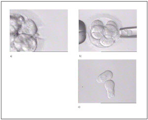
Fig. 1. Example of an embryo biopsy. Panel a) shows the hole in the zona pellucida after application of the laser for zona hatching. Panel b) shows how one blastomere is gently aspirated outside the embryo. Panel c) shows two biopsied blastomeres with a clear nucleus.
Single Cell PCR
The polymerase chain reaction (PCR) is the only method that allows the analysis of the DNA from one single cell, i.e. 6 pg of DNA. Since the first report on PGD, the methods of analysis of the PCR products have evolved from electrophoresis on simple agarose gels (Handyside et al., 1990), over fragment analysis on automated sequencers (Sermon et al., 1998), to minisequencing (Fiorentino et al., 2003) and real-time PCR (Rice et al., 2002). Although the refinement of the analysis methods has increased the efficiency and accuracy tremendously, there are still a number of pitfalls that are inherent to single cell PCR and that still cause incorrect diagnosis. These are: (1) the (lack of) specificity of the PCR, (2) contamination with DNA extraneous to the analysed cell, and (3) allele drop-out.
Specificity of the PCR
Because Taq DNA polymerase incorporates mistakes, smears and a-specific products appear when a large number of cycles are performed for single cell PCR. In nested PCR, which efficiently solves this problem, a first PCR round is performed, followed by a second PCR round in which a small amount of the first round PCR product serves as a template and primers are used that amplify a fragment inside the first fragment. A-specific fragments are thus not amplified in the second PCR round, and a clear, pure PCR product is obtained (Holding et al., 1989). Later, the number of PCR cycles necessary to obtain enough PCR product for analysis was significantly reduced with the introduction of fluorescent PCR. Not only is this method much more sensitive than analysis on agarose gels (approximately 1000 x), but the resolution of fragments is also much greater (Lissens and Sermon, 1997). The introduction of DNA polymerases with a high proofreading activity, alone or in mixtures with regular Taq polymerase, has reduced the number of cycles necessary for a sufficient yield even more (Sermon et al., 1998). Finally, the need for the tedious fine-tuning of PCR protocols for single cell analysis was significantly reduced with the introduction of minisequencing techniques as applied for instance in the SNaPshot kit from Applera (Fiorentino et al., 2003).
Contamination with extraneous DNA
The introduction of one molecule of foreign DNA in the PCR tube along with the cell that is to be analyzed can lead to a wrong diagnosis. A number of measures need to be taken to avoid and/or detect contamination (Lissens and Sermon, 1997):
1. Granulosa cells are meticulously removed from the oocytes and fertilisation is achieved through ICSI to avoid contamination with sperm.
2. The pre-PCR area and the post-PCR area need to be strictly separated.
3. The PCR reactions are set up in a laminar flow, that is fully equipped with dedicated pipettes, filtered tips and UV light.
4. For each sample, a blank containing all PCR components except DNA should be run. A more efficient way to detect contamination is the use of linked or unlinked polymorphic markers, amplified in duplex with the locus of interest.
Allele drop-out
Allele drop-out (ADO) is defined as the non-amplification of one allele when starting from a single cell. It can thus only be detected in a heterozygous cell. ADO can lead to serious misdiagnoses, e.g. in the detection of mutations in autosomal dominant diseases (Navidi and Arnheim, 1991). Supposedly, ADO has led to two misdiagnoses in CF (Harper and Handyside, 1994, Verlinsky, 1996). A first efficient way to significantly reduce ADO is to use fluorescent PCR. Together with the use of more efficient DNA polymerases, fluorescent PCR has been the most important breakthrough so far to reduce the risk for ADO (Sermon et al., 1998).ADO cannot be reduced to zero, so detection methods have been devised. The most important is the multiplex PCR of the mutation together with linked markers, or a set of linked markers and has now become the golden standard in PGD for monogenic diseases (Lewis et al., 2001). Usually, microsatellites are used as linked markers because they are highly polymorphic and can thus be used in several families (Rechitsky et al., 1999, Dreesen et al., 2000, Apessos et al., 2001, Dean et al., 2001, Pyiamongkol et al., 2001, Goossens et al., 2003, Moutou et al., 2003). Several new and promising techniques have emerged that will facilitate the development of new diagnostic tests. Minisequencing has been advocated as a means to reliably detect mutations and single nucleotide polymorphisms (SNPs) at the single cell level without a tedious pre-clinical work-up (Fiorentino et al., 2003).Another newly introduced method in single cell PCR, is real-time PCR, which has the advantage that PCR fragments are analyzed as they are formed, and not after a complete PCR programme. It has been used by several authors with a low rate of ADO (Rice et al., 2002, Pierce et al. 2000). Finally, microarrays are currently pervading all branches of molecular biology (Syvänen, 1999). A combination of microsequencing and microarray technology would be applied: the array would be covered in oligonucleotides that anneal specifically to PCR fragments just adjacent to the SNP to be analyzed. As in minisequencing, the complementary nucleotide would be added in (Kurg et al., 2000).
Single cell cytogenetics
Fluorescence in situ hybridization
In fluorescence in situ hybridization (FISH), fluorescent probes carrying distinct fluorochromes for different chromosomes, are hybridized to cell nuclei spread either in metaphase or in interphase. When embryos are analyzed, the cells are usually in interphase. Careful choosing of the type and location of the probes allows not only enumeration of chromosomes, as in sexing and PGD-AS, but also the diagnosis of chromosome imbalances in structural abnormalities.Two methods are currently in use for the fixation of the blastomeres. The first method is derived from the Tarkowsky method to fix embryos and uses acetic acid and methanol as a fixative. In the second method the blastomere is spread on a glass slide in spreading solution (0.1 N hydrochloric acid, 0.01% Tween 20). Some authors claim that the modified Tarkowski method yields better spread nuclei, so that the different fluorescent dots are better separated and more easily enumerated (Velilla et al., 2002). The Coonen method is easier to use, and gives reasonable spreading results (Staessen et al., 1999), even when five different chromosomes are analyzed concurrently, in the labs that are more experienced with this method (Staessen et al., 2003).Fluorescent probes are commercially available. For more widespread uses, such as sexing and PGDAS, kits are available, containing directly labeled probes for X and Y, 13, 18, and 21 (MultiVysion PGT kit from Vysis) or 13, 16, 18, 21 and 22 (MultiVysion PB kit from Vysis). These chromosomes were chosen either because they are present in liveborn trisomies (trisomies 13, 18 and 21) or because they are frequently present in miscarriages (16 and 22). Because the number of fluorochromes available is limited, the number of chromosomes that are analyzed can be increased either through ratio labelling and computerized analysis, or/and through the application of two and three hybridization rounds. Munné et al. (2003) showed that in this way, up to nine different chromosomes (X, Y, 13, 15, 16, 17, 18, 21 and 21) could be analyzed for aneuploidy screening, and these authors reported an increase in implantation rate, as compared to the implantation rate obtained after analysis with five probes.For structural abnormalities, a judicious choice of probes is made depending on the chromosomes involved and the breakpoints present. The first cases were performed using probes that specifically delineate the translocation breakpoints (Munné et al., 1998, Munné et al. 2000). A more general approach was described by Conn et al. (1999), taking advantage of the availability of centromeric and subtelomeric probes for each chromosome. For Robertsonian translocations, a combination of one locus-specific probe centromeric to the breakpoint of one of the chromosomes involved and one on the telomere of the other chromosome involved, is applied. Three FISH probes are used for reciprocal translocations: two on the centromeric side of the breakpoints of the chromosomes involved, and a third one on one of the telomeres on the other side of the breakpoint (Figure 2). The two or three probes used must each carry a different fluorochrome, and it must be possible to use them together in one assay (Scriven et al., 2000).
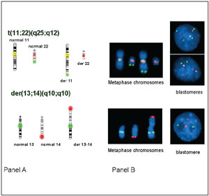
Fig. 2. An example of the probes chosen for a reciprocal translocation (t(11,22), panel a) and for a Robertsonian translocation (t(13; 14), panel b). The results are shown schematically, on lymphocyte metaphase spreads, and on interphase spreads of blastomeres.
Comparative Genomic Hybridization
Comparative genomic hybridization (CGH) was developed to characterize the often complex chromosomal rearrangements present in cancer tissue. Total genomic DNA from the cells to be analyzed (e.g. the cancer cells) is labeled with a green fluorochrome, while a normal DNA sample is labeled with another, e.g. red, fluorochrome. Both labeled DNA samples are mixed and hybridized to a normal metaphase. If the test sample contains more of a certain sequence, e.g. in a duplication, then that sequence will show up more green after computerized analysis of the metaphase. Conversely, if the test sample contains less of a certain sequence (e.g. a deletion), this sequence will be more red on analysis. CGH has been downscaled to the single cell level by introducing a first step of whole genome amplification (WGA) of the DNA in the single blastomere (Voullaire et al., 2000; Wells and Delhanty, 2000; Wilton et al., 2001; Wells et al., 2002). At this point, CGH at the single cell level still requires several days for analysis, which is why the groups that have presented clinical application of CGH in PGD either had to recur to polar body analysis (Wells et al., 2002), or to cryopreservation of the embryos (Voullaire et al., 2000, Wilton et al., 2002). Both authors however foresee that the introduction of microarrays to replace the metaphase spread would significantly reduce the time necessary for the CGH analysis, and bring it back well within time to transfer embryos before day 5.
Outcome of PGD
Accuracy of the Diagnosis
One misdiagnosis for sexing and two for CF are mentioned in early reports (Harper and Handyside, 1994, Lissens and Sermon, 1997), and these were mainly due to the low efficiency of single cell PCR. Further technical developments, i.e. FISH for sexing and multiplex fluorescent PCR, have ruled out the re-occurrence of this type of errors. Munné et al. (1999) reported one misdiagnosis (trisomy 21 after aneuploidy screening) on a total of 57 pregnancies. These authors estimate that the misdiagnosis rate after biopsy of one blastomere was around 7% in a large series; nearly 6% of the embryos were misdiagnosed due to mosaicism in the embryo (Munné et al., 2003). Pickering et al. reported one misdiagnosis for spinal muscular atrophy (SMA) out of 18 ongoing pregnancies. The third report of the ESHRE PGD Consortium (2002) mentions eight misdiagnoses: five after PCR (two for sexing, one each for Duchenne’s muscular dystrophy, βthalassemia, CF and myotonic dystrophy) and three after FISH (one each for social sexing, translocation (11;22) and a trisomy 21 after PGD-AS). This gives a figure of 8/265 (3%) if only those fetal sacs for which a control had been carried out are counted. The identification of these misdiagnoses and the reasons why they occurred has lead to the initiation of the drawing up of guidelines for PGD within ESHRE.
Pregnancy Outcome
The European Society for Human Reproduction and Embryology (ESHRE) PGD Consortium has reported on PGD cycles and their outcome (ESHRE PGD Consortium, 2002).For structural chromosomal abnormalities (amongst which translocations), 368 cycles led to 62 clinical pregnancies (17%). For sexing, 254 cycles led to 41 pregnancies (16%). For monogenic diseases, 575 cycles led to 119 (21%) pregnancies. These numbers are lower than can be expected in a regular IVF cycle, but it must be taken into account that a large cohort of embryos is diagnosed as affected or abnormal. Especially in patients carrying reciprocal translocations, as many as 80% of the embryos carry an unbalanced karyotype (Munné et al., 1998, Van Assche et al., 1999, Pickering et al., 2003). This contrasts with the results for PGD-AS: of the 799 cycles reported, 199 or 25% led to a clinical pregnancy in patient groups with a poor prognosis (advanced maternal age, repetitive IVF failure and recurrent miscarriages).The most important reason for morbidity and mortality in the pregnancies conceived after PGD is multiplicity. In this, and many other aspects such as birth weight and congenital malformations, children born after PGD are comparable to children born after ICSI (Bonduelle et al., 2002).The International Working Group reported that more than 3000 clinical PGD cycles had been applied by mid-2001, resulting in around a 24% pregnancy rate (The International Working Group, 2002). Close to 700 children have been born following these pregnancies and 4.9% of these were reported to show abnormalities. Taking the four most active groups world wide together, a total of 2774 PGD cycles, resulting in 2265 transfers and 652 clinical pregnancies (29%) were mentioned. Here too, it was stressed that outcome of pregnancy was comparable to IVF populations.Another large cohort of children, which were not included in the ESHRE PGD Consortium report, were reported by Strom et al. (2000). A total of 109 infants were described and here too, the conclusions were that children born after PGD are very comparable with children born after ICSI, and that PGD is a safe method to avoid the birth of children with genetic defects.