JBRA Assist. Reprod. 2020;24(3):241-244
ORIGINAL ARTICLE
doi: 10.5935/1518-0557.20190080
Predicting The Developmental Potential Of Cleavage Stage Embryos Based On Oxygen Consumption Rate In FET Cycles
1Chongqing Key Laboratory of Human Embryo Engineering, Chongqing Reproductive and Genetics Institute, Chongqing Health Center for Women and Children. Chongqing, China
2Department of Histology and Embryology, Laboratory of Stem Cells and Tissue Engineering, Chongqing Medical University. Chongqing, China
CONFLICT OF INTEREST
None declared.
ABSTRACT
Objective: To study the value of oxygen consumption (OC) as a predictor of the developmental potential of D3 embryos in frozen embryo transfer (FET) cycles.
Methods: This observational study included 148 patients undergoing FET cycles with two embryos transferred per cycle. OC rates were examined by scanning electrochemical microscopy before embryo transfer. Implantation, clinical pregnancy, miscarriage, and live birth rates were calculated.
Results: A total of 296 embryos were transferred in 148 cycles, or two embryos per cycle. The embryos were divided into three groups based on OC: Group A included the cases in which the OC rate of each of the two transferred embryos was greater than 3.0 fmol/s; Group B included the cases in which the OC rate of one of the embryos was greater than 3.0 fmol/s and the OC rate of the other embryo was less than 3.0 fmol/s; and Group C included the cases in which the OC rates of the two embryos were less than 3.0 fmol/s. Higher live birth rates and lower miscarriage rates were observed in Group A (p<0.05).
Conclusions: Our data suggest that OC is positively correlated with embryo developmental potential. Therefore, measuring the OC of human embryos may be useful in embryo assessment.
Keywords: oxygen consumption, FET, scanning electrochemical microscopy (SCEM), implantation rate
INTRODUCTION
Accurate assessment of developmentally competent embryos is crucial for the success of ART treatment. For decades, embryo quality assessment based on morphological evaluation was used to predict implantation and pregnancy rates to some degree (Gardner et al., 2004). Numerous alternatives to morphological assessment have been proposed to assess the quality of embryos, including methods based on spent embryo culture medium, cell cycle timing, morphological evaluation by time-lapse systems (TLSs), and PGS (Twisk et al., 2006; Armstrong et al., 2015). The above technologies have provided important insight into early embryo development and the arbitrary nature of embryo selection; however, none has become standard practice, and an early prediction of clinically relevant, quality embryos before transfer still remains challenging (Kurosawa et
al., 2016).
Embryo evaluation based on oxygen metabolism or consumption rate has been attempted via objective and quantitative methods (Rodgaard et al., 2015). Oxygen consumption rate has been recently considered a metabolic indicator that reflects the activity of mitochondrial oxidative phosphorylation (OXPHOS), by which adenosine triphosphate (ATP) is synthesized. Animal experiments have described a correlation between embryo OC and developmental potential. Using a non-invasive method, Shiku et al. (2001) succeeded at determining oxygen consumption by individual oocytes and embryos using a measurement system based on scanning electrochemical microscopy (SECM) that enables the evaluation of mitochondrial activity in oocytes and embryos. Tejera et al. (2016) showed that OC is a marker for human oocyte competence. To our knowledge, few observational studies have looked into OC as a marker to predict human embryo developmental potential.
This study aimed to measure embryo OC patterns before frozen-thawed embryo transfer (FET) cycles and to explore the correlation between OC rate and embryo developmental potential.
MATERIALS AND METHODS
Patient and embryo selection
The study included embryos warmed between January 2015 and May 2015 at the Chongqing Reproductive and Genetics Institute. All survived and two embryos were transferred per patient in the first FET cycle. The patients were aged ≤ 37 years and had been diagnosed with tubal factor infertility. They had undergone insemination by IVF and had ≥3 D3 embryos available. No fresh embryo transfer at oocyte retrieval (OR) was included. Patients with positive preimplantation genetic diagnosis and transfers with donated gametes were excluded. All procedures were performed according to the Declaration of Helsinki on medical research involving human subjects. The Ethics Committee of the Chongqing Obstetrics and Gynecology Hospital approved the study (certificate no. RGI-ZA201408). The embryos included in the study were divided into three groups based on OC rate: Group A included the cases in which the OC rate of each of the two transferred embryos was greater than 3.0 fmol/s; Group B included the cases in which the OC rate of one of the embryos was greater than 3.0 fmol/s and the OC rate of the other embryo was less than 3.0 fmol/s; and Group C included the cases in which the OC rates of the two embryos were less than 3.0 fmol/s.
Fresh cycle, embryo cryopreservation by vitrification
In fresh cycles, ovarian stimulation and luteal supplementation were performed according to our standard practice (Ye et al., 2009). A standard insemination protocol was used for all patients. In sum, 39-40 hours after hCG administration, 10,000-15,000 spermatozoa per microdrop were added to G-IVF medium pre-equilibrated overnight. The oocyte was then added to the microdrop, and co-cultured with sperm at 37°C in 5% O2 and 6% CO2 in an incubator. The day of oocyte retrieval was defined as Day 0. Before 9 am on Day 1, the oocytes were observed after the removal of granular cells, and fertilization was confirmed by the presence of two pronuclei (PN) and two polar bodies. Embryo quality was evaluated using a morphological scoring system, which accounts for the regularity of blastomeres, degree of fragmentation, and embryo microscopic appearance, as previously described (Xiong et al., 2011). The embryos were cryopreserved on Day 3. The Cryotop method for embryo vitrification was as described by Kuwayama (2007). After vitrification, the Cryotop devices were plunged directly into liquid nitrogen. The embryos were further stored in vapor phase nitrogen containers.
FET cycles
Warmed embryos were transferred in natural cycles, stimulated cycles (gonadotropin or clomiphene citrate), or hormone replacement cycles as described previously (Yamanaka et al., 2011). The embryos were transferred on Day 4. In order to warm the embryos, the Cryotop was removed from LN2 storage and immediately plunged into thawing solution (TS; 1.0 M sucrose in TCM199+20% SSS) at 37°C. After 1 min, the embryos were transferred to a dilution solution (DS; 0.5 M sucrose in TCM199+20% SSS) at room temperature. After 3 min, they were transferred to a washing solution (WS; TCM199+20% SSS) for 5 min twice. Subsequently, the embryos were transferred to G2.5 (Vitrolife, Sweden) medium containing 5% human serum albumin cultured overnight before transfer. Embryos with more than 50% intact blastomeres were recorded as survivors. The tools and solutions used in embryo freezing and thawing were procured from Kitazato (Kitazato BioPharma Co., Japan).
Evaluation of embryo oxygen consumption rates and embryo transfer
After overnight culture (20-24h), the oxygen consumption rate of individual embryos was quantified on Day 4 before transfer with a modified SECM measuring system described in the literature (Yamanaka et al., 2011). In sum, the two embryos of each patient were transferred into a well filled with modified TCM199+20% SSS medium. The embryos sank to the bottom of the cone-shaped microwell and remained at its lowest point, each microwell containing one embryo. A platinum microdisk electrode was loaded into 5 ml of modified TCM199+20% SSS medium, and its tip potential was maintained at -0.6 V versus Ag/AgCl with a potentiostat to monitor local oxygen concentration. The microelectrode scanned along the z-axis from the edge of each embryo, and the oxygen consumption rate was calculated with custom software based on spherical diffusion theory. Oxygen levels were measured in about two minutes. After oxygen consumption rates were measured, the two embryos were returned to culture medium and then transferred to the patients. Clinical pregnancy was established when a gestational sac was detected by transvaginal ultrasound 28 days after embryo transfer.
Statistical analysis
The data sets were analyzed on software package Stata (Stata 10.0 version, STATA Inc., College Station, TX). Patient data were expressed as mean ± SD for purposes of comparison. Quantitative data were compared using the t-test. Categorical variables were analyzed via the chi-square test. A p value<0.05 was considered to be statistically significant.
RESULTS
A total of 296 embryos from 148 transfer cycles met the inclusion criteria. One hundred and seventy embryos from 85 cycles were included in Group A; 72 embryos from 36 cycles were included in Group B; and 54 embryos from 27 cycles were included in Group C. Table 1 describes patient demographic and baseline characteristics. The demographic characteristics of the patients submitted to frozen-thawed embryos transfers were not significantly different in terms of age, body mass index (BMI), number of oocytes retrieved, number of available embryos, mean blastomere number, or endometrial thickness.
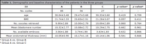
Table 1. Demographic and baseline characteristics of the patients in the three groups
Table 2 shows implantation, clinical pregnancy, miscarriage, and live birth rates. The three groups were not significantly different in terms of implantation, pregnancy, or miscarriage rates (50.00% vs. 45.83% vs. 40.74, p>0.05; 67.06% vs. 50.00% vs. 51.85%, p>0.05; 4.07% vs. 11.11% vs. 14.81%, p>0.05). However, Group A yielded significantly higher live birth rates than Groups B and C (56.47% vs. 36.11% vs. 33.33%, p<0.05).
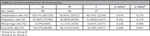
Table 2. Clinical outcomes from the three groups
DISCUSSION
Oxygen consumption rate has been recently considered a metabolic indicator that reflects the activity of mitochondrial oxidative phosphorylation (OXPHOS), by which adenosine triphosphate (ATP) is synthesized (Kurosawa et al., 2016). Therefore, mitochondrial dysfunctions may lead to inferior embryo development and lower pregnancy rates from IVF cycles. According to the literature, respiration increases indicate mitochondrial development from the morula to the blastocyst stage in most animal embryos, while embryos with high oxygen consumption rates provide for good viability and freezing ability. In addition, it has been established that embryos with above average respiration rates also yield higher pregnancy rates. These results demonstrate that oxygen consumption is a good parameter to assess embryo quality.
OC rate detected by SECM has often been regarded as a better indicator of overall metabolic activity (Tejera et al., 2011). It has also been demonstrated that oxygen consumption is related to oocyte fertilization capability, which is consistent with previous data regarding higher ATP turnover in oocytes (Ruvolo et al., 2013). Yamanaka et al. (2011) found that the OC rate might be used to select vitrified-warmed blastocysts with high developmental potential. Yoshida et al. (2013) found that when the oxygen consumption rate increased in a group of thawing embryos, their rate of blastocyst development and freezing ability for cryopreservation increased. Tejera et al. (2012) found that OC rates during embryo development were positively associated with implantation potential and embryo quality; therefore, OC measurements during embryo culture - performed almost immediately after embryo transfer - were deemed to add value to the process of selecting viable embryos.
This observational study was designed to test how effectively OC may be used as a predictor of embryo developmental potential, and to investigate the relationship between OC and pregnancy outcomes. Our study was based solely on FET cycles. The OC rates of each of the 296 embryos included in the study were calculated before embryo transfer. The embryos were divided into three groups based on OC: Group A included the cases in which the OC rate of each of the two transferred embryos was greater than 3.0 fmol/s; Group B included the cases in which the OC rate of one of the embryos was greater than 3.0 fmol/s and the OC rate of the other embryo was less than 3.0 fmol/s; and Group C included the cases in which the OC rates of the two embryos were less than 3.0 fmol/s. The three groups were compared for implantation, clinical pregnancy, miscarriage, and live birth rates. Transfers of two embryos with OC rates greater than 3.0 fmol/s yielded higher live birth rates, a finding that situates the OC rate as a reliable predictor of human cleavage-stage embryo developmental potential.
OC showed a positive correlation with embryo developmental potential. Embryo OC measurement based on SECM enabled the assessment of respiration of single human embryos. Our findings and current literature on the subject indicates that this might become an alternate method for embryo selection in IVF laboratories. Randomized controlled trials are required to further validate the existing evidence.
Authors’ roles
Han Shubiao and Huang Guoning designed the study. Hou Jiying participated in the experiment. Zhang Xiaodong analyzed the data. Han Shubiao and Zhang Xiaodong wrote the paper.
Funding
This study was supported by Chongqing basic and frontier research project, China (cstc2016jcy JAX0007); Foundation of the Chinese Medical Association of Clinical Medicine Research, China (No. 16020350651); and the Basic and Prospective Foundation of Yuzhong District, Chongqing province, China (No. 20150141)
REFERENCES
Armstrong S, Arroll N, Cree LM, Jordan V, Farquhar C. Time-lapse systems for embryo incubation and assessment in assisted reproduction. Cochrane Database Syst Rev. 2015;2:CD011320.
Medline Crossref
Gardner DK, Surrey E, Minjarez D, Leitz A, Stevens J, Schoolcraft WB. Single blastocyst transfer: a prospective randomized trial. Fertil Steril. 2004;81:551-5.
Medline Crossref
Kurosawa H, Utsunomiya H, Shiga N, Takahashi A, Ihara M, Ishibashi M, Nishimoto M, Watanabe Z, Abe H, Kumagai J, Terada Y, Igarashi H, Takahashi T, Fukui A, Suganuma R, Tachibana M, Yaegashi N. Development of a new clinically applicable device for embryo evaluation which measures embryo oxygen consumption. Hum Reprod. 2016;31:2321-30.
Medline Crossref
Kuwayama M. Highly efficient vitrification for cryopreservation of human oocytes and embryos: the Cryotop method. Theriogenology. 2007;67:73-80.
Medline Crossref
Rodgaard T, Heegaard PM, Callesen H. Non-invasive assessment of in-vitro embryo quality to improve transfer success. Reprod Biomed Online. 2015;31:585-92.
Medline Crossref
Ruvolo G, Fattouh RR, Bosco L, Brucculeri AM, Cittadini E. New molecular markers for the evaluation of gamete quality. J Assist Reprod Genet. 2013;30:207-12.
Medline Crossref
Shiku H, Shiraishi T, Ohya H, Matsue T, Abe H, Hoshi H, Kobayashi M. Oxygen consumption of single bovine embryos probed by scanning electrochemical microscopy. Anal Chem. 2001;73:3751-8.
Medline Crossref
Tejera A, Herrero J, de Los Santos MJ, Garrido N, Ramsing N, Meseguer M. Oxygen consumption is a quality marker for human oocyte competence conditioned by ovarian stimulation regimens. Fertil Steril. 2011;96:618-23.e2.
Medline Crossref
Tejera A, Herrero J, Viloria T, Romero JL, Gamiz P, Meseguer M. Time-dependent O2 consumption patterns determined optimal time ranges for selecting viable human embryos. Fertil Steril. 2012;98:849-57.e1-3.
Medline Crossref
Tejera A, Castelló D, de Los Santos JM, Pellicer A, Remohi J, Meseguer M. Combination of metabolism measurement and a time-lapse system provides an embryo selection method based on oxygen uptake and chronology of cytokinesis timing. Fertil Steril. 2016;106:119-26. e2.
Medline Crossref
Twisk M, Mastenbroek S, van Wely M, Heineman MJ, Van der Veen F, Repping S. Preimplantation genetic screening for abnormal number of chromosomes (aneuploidies) in in vitro fertilisation or intracytoplasmic sperm injection. Cochrane Database Syst Rev. 2006;(1):CD005291.
Medline Crossref
Xiong S, Han W, Liu JX, Zhang XD, Liu WW, Liu H, Huang GN. Effects of cumulus cells removal after 6 h co-incubation of gametes on the outcomes of human IVF. J Assist Reprod Genet. 2011;28:1205-11.
Medline Crossref
Yamanaka M, Hashimoto S, Amo A, Ito-Sasaki T, Abe H, Morimoto Y. Developmental assessment of human vitrified-warmed blastocysts based on oxygen consumption. Hum Reprod. 2011;26:3366-71.
Medline Crossref
Ye H, Huang GN, Zeng PH, Pei L. IVF/ICSI outcomes between cycles with luteal estradiol (E2) pre-treatment before GnRH antagonist protocol and standard long GnRH agonist protocol: a prospective and randomized study. J Assist Reprod Genet. 2009;26:105-11.
Medline Crossref
Yoshida H, Abe H, Arima T. Quality evaluation of IVM embryo and imprinting genes of IVM babies. J Assist Reprod Genet. 2013;30:221-5.
Medline Crossref