JBRA Assist. Reprod. 2020;24(3):316-323
ORIGINAL ARTICLE
doi: 10.5935/1518-0557.20200009
Effects of Vitamin D on Apoptosis and Quality of Sperm in Asthenozoospermia
1Cellular and Molecular Research Center, Ahvaz Jundishapur University of Medical Sciences, Ahvaz, Iran
2Department of Anatomical Sciences, Faculty of Medicine, Ahvaz Jundishapur University of Medical Sciences,Ahvaz, Iran
3Fertility, Infertility and Perinatology Center, Imam Khomeini Hospital, Ahvaz Jundishapur University of Medical Sciences, Ahvaz, Iran
4Department of anatomical sciences, Abadan School of Medical Sciences, Abadan, Iran
CONFLICT OF INTEREST
None of the contributing authors has any conflict of interest.
Part of this study was presented in 9th European Congress of Andrology 21 - 23 September 2016 Rotterdam, the Netherlands
ABSTRACT
Objective: Vitamin D receptor (VDR) is expressed in human spermatozoa. However, the role
of vitamin D (VD) in human male reproduction has not yet been clarified. In
this study, effects of VD on sperm parameters and its apoptosis in
asthenozoospermic and healthy men were evaluated.
Methods: The study was carried out on discharged semen samples of 80 asthenozoospermic
and healthy men. The samples were divided into control and experimental
groups (received 20 µMol of VD). This study assessed sperm motility using
the Makler chamber, their morphology by Diff quick, apoptosis and necrosis
by Annexin-V and TUNEL assays, and their chromatin integrity was assessed by
Aniline blue and Toluidine blue staining, according to WHO guidelines.
Results: The results revealed that: 1) the total number of motile sperms was increased
by VD in both groups, but it was only significant in the asthenozoospermia
group. 2) The progressive motility was increased with significant difference
in both groups. 3) Morphology of sperm did not show any changes due to VD in
any of the groups. 4) Early apoptosis and necrosis of sperms were reduced in
both groups, but the results of late apoptosis showed no statistical
difference in these groups. 5) The percentage of positive toluidine blue was
significantly decreased after using VD in the asthenozoospermia group.
Conclusion: VD could improve motility, early apoptosis, and sperm necrosis, especially in
asthenozoospermic men and it could be used for therapeutic
opportunities.
Keywords: apoptosis, asthenozoospermia, sperm motility, sperm morphology, vitamin D
INTRODUCTION
Infertility is a common disorder with significant medical, psychosocial, and economic
aspects (Benyamini et al.,
2005), which has had a worldwide increase, and now approximately 1/6 of
the couples are struggling with it (Thonneau et al., 1991). The male factor infertility remains a
significant problem contributing to 50% of the cases visiting infertility clinics
(Schulte et al., 2010).
Asthenozoospermia is a common cause of male infertility, in which motile sperm is
less than 40% and progressive motile sperm is less than 32% (WHO, 2010), which could even be 0% (absolute immotile) (Ortega et al., 2011). Sperm
motility is extremely important for migration from the vagina to the fallopian
tubes, penetration of the cumulus oophorus, and for processes involved in
fertilization (Ortega et al.,
2011); thus asthenozoospermia has a poor fertility prognosis (Beauchamp et al., 1984). Some
studies showed that apoptosis could affect sperm motility and lead to poor sperm
motility and asthenozoospermia (Moradian Fard et al., 2019).
Apoptosis is physiologically-programmed cell death, which is different from necrosis
and affects cells without any related inflammation in the surrounding tissue (Wyllie et al., 1980). Some
reports have demonstrated that ejaculated spermatozoa from infertile men show
ultrastructural damage, an unusually high incidence of DNA fragmentation, and plasma
membrane translocation of phosphatidylserine (PS), all of which are typically
considered to be signs of apoptosis in somatic cells (Barroso et al., 2000).
Nonetheless, these sperms may be considered normal in routine semen analyses. Under
certain conditions like ICSI, these sperms could carry a damaged genome into the
oocyte, resulting in serious consequences (Bedu-Addo et al., 2008). Today, in addition to assisted
reproductive techniques (ART), researchers are considering the development of new
techniques to increase sperm motility and improve apoptosis, moving towards a better
management of the infertile patients (Moradian Fard et al., 2019).
There is evidence that Vitamin D (VD) modulates reproductive processes in women and
men (Luk et al., 2012). VD
deficiency and Vitamin D receptor (VDR) mutation in rodents have caused uterus
hypoplasia, damaged follicles in the female reproductive system and decreased sperm
count and motility with histological abnormalities of the testis in male rats (Kinuta et al., 2000; Aquila et al., 2009).
Insufficient VD causes increased frequencies of chromosomal aberrations and sister
chromatid exchanges due to oxidative, hypoxic, and apoptotic stresses (Benyamini et al., 2005). In
addition, it causes incomplete spermatogenesis and degenerative changes as well
(Ramlau-Hansen et al.,
2011). Some studies have shown that the serum and seminal plasma vitamin
D may be involved in regulating sperm quality (morphology, concentration, motility
and acrosome reaction) (Aquila et
al., 2009; Ramlau-Hansen et al., 2011; Blomberg Jensen et al., 2011; Jueraitetibaike et al., 2019), and others
showed that vitamin D has anti-apoptosis function in some cells (Zhang et al., 2007);
therefore, the authors decided to assess the role of this vitamin in apoptosis and
sperm quality in asthenozoospermic and healthy men. For this purpose, we used VD
under in vitro condition like ART and evaluated sperm quality
(morphology, motility, chromatin integrity) and apoptosis using Annexin V and
TENNEL. Provided that positive effects are observed on these parameters, VD could be
used in future infertility treatment and ART.
MATERIAL AND METHODS
Semen Collection and Processing
We ran this experimental study on discharged semen samples of 40
asthenozoospermic and 40 healthy men referred to an IVF infertility clinic. The
Ethics Committee of the Research Deputy of Ahvaz Jundishapur University of
Medical Sciences (IR.AJUMS.REC.1395.266) approved this research. Since vitamin D
may have a positive effect on sperm parameters and can be used in future IUI and
IVF methods, this project fulfills all the requirements for sperm preparation
and patient selection such as IVF methods. We took the samples from men without
infection or varicocele. Human semen was collected into a sterile container
after a 3-5-day period of sexual abstinence (Barroso et al., 2006). In this study, we defined
asthenozoospermia when a man’s sperm motility is less than 40% and he has less
than 32% progressive motile sperm. In contrast, a healthy man is defined as a
person who has the following parameters: sperm count >20×106 ml, sperm motility >40%, normal morphology >4%, and leukocyte
<1×10/ml (WHO, 2010). Samples
were allowed to liquefy at 37°C for 30 min. 5µl of the liquefied samples
were loaded on slides for determining normal and asthenozoospermic samples
(Blomberg Jensen et al.,
2011). Like IVF methods, semen samples were washed twice with 1ml
fresh sperm wash (ALL Grad Wash, Life Global, 4264, Brussels, Belgium) and
centrifuged at 1,800 rpm for 5 min. After removing the supernatant, 1 ml fresh
sperm wash was added and the mixture was kept at a 45° angle in an incubator at
37°C for 45 min so that motile sperms could swim up. After that, the supernatant
was divided into two parts; one part was considered as control group and the
other one received 20µMol VD (based on the pilot work we did) and
considered as experimental group. Both groups were incubated at 37°C for 1 hour
(Blomberg Jensen et al.,
2011).
Evaluation of Basic Sperm Parameters
We evaluated sperm concentration, motility, and morphology of both the original
raw samples and the experimental groups. We placed 5µl of each sample
into a Makler chamber and the sperm was counted under light microscopy at
40× magnification to assess progressive sperm motility (%), total
motility (%) and total sperm count per ejaculation (million sperm). We analyzed
at least 200 spermatozoa per slide. We classified the sperm as progressive
motile (class A+B), non-progressive motile (class C), or immotile (class D).
We evaluated sperm morphology according to strict criteria at
40×magnification (Barroso et
al., 2006). Each sample (10µl) was spread along
the slide and allowed to dry for 20 minutes before staining with Diff-Quick
staining. We evaluated an average of 200 spermatozoa per slide, twice, by two
examiners.
Apoptosis detection
PS with Annexin-V Externalization Evaluation
In the early apoptotic events, PS was translocated from the internal membrane
to the external membrane of cells. Annexin-V is a calcium-dependent
phospholipidic union protein with a high affinity for PS. It can bind to PS
and distinguish apoptotic cells. For this detection, according to the
Annexin V protocol (Annexin-V-FLOUS Staining kit, No.11 858 777 001, Roche,
Mannheim, Germany), 2 µl Annexin-V solution, 100 µl additional
binding buffer, and 2 µl propidium iodide (PI) were added to the
samples in a dark place and the mixture was left there for 20 minutes. After
this step, the samples were checked under a fluorescence microscope. At
least 200 cells per slide were analyzed in a randomized manner and they were
identified as either normal (negative for Annexin-V and PI, pale stain),
apoptotic (positive for Annexin-V, green stain, and negative for PI), or
necrotic (positive for PI, red stain) cells (Barroso et al., 2006; Oosterhuis et al., 2000).
DNA fragmentation detection
Apoptosis-related DNA strand breaks were evaluated by terminal
deoxynucleotidyl transferase-mediated dUTP nick-end labelling (TUNEL), using
the Apoptosis Detection System Florescence (in situ Cell Death Detection
Kit, POD, No.11 684 817 910, Roche, Penzberg, Germany). We ran the procedure
according to the manufacturer's instructions. We smeared the samples on
slides, fixed with 100% methanol for 4 min, and incubated them in a blocking
solution (3%H2O2 in methanol) for 20 min in a dark
room. Then, the slides were rinsed with PBS and permeabilized with 0.1%
Triton X-100 in PBS. The slides were washed twice in PBS and incubated in a
TUNEL incubation buffer, (which contained nucleotide and the terminal
transferase enzyme), at 37°C for 1 hour in a humidified chamber. After
stopping the enzyme’s reaction, the slides were washed three times in PBS
for 15 min. The slides were observed under a fluorescence microscope at
40× magnification. For each slide, 200 sperms were checked. Omitting
Enzyme terminal transferase and D-Nase I were done for negative and positive
controls. The slides were read twice and an average number was obtained. In
these slides, cells with green fluorescence color were reported as TUNEL
positive (Dominguez-Fandos et
al., 2007).
Chromatin integrity detection
After obtaining the appropriate cells, 20 µl of the sample was
prepared as smear. The smears were dried at room temperature; then, the
fixation of each staining was added to the smears. With the aim of fixing
the stain, the aniline blue fixative (3% glutaraldehyde) was added to the
slides, at room temperature and they remained in place for 30 minutes. After
that, the slides were stained with aniline blue solution (5% aqueous aniline
blue solution with 4% acetic acid) for 10 minutes. In order to fix the
toluidine blue staining smear, they were fixed under a refrigerated
temperature by ethanol-acetone (1:1) for 30 minutes. After that, the slides
were hydrolyzed by HCL 0.1% N for 5 minutes at 4°C. Subsequently, the
samples were washed three times with distilled water and then toluidine blue
solution (0.05% TB) was added to the samples and were kept for 10 minutes.
After that, the cells were counted under an inverted light microscope. Cells
with dark blue color were reported as abnormal and those with a light blue
color were considered healthy. About 200 cells were counted in each
slide.
Statistical Analysis
Data analysis was carried out using the Statistical Package for the Social
Sciences (SPSS version 22). The results were presented as mean ± SD and p-value. Normal distribution of the data was assessed by
the Kolmogorov-Smirnov Z test. The data of the control group and the vitamin
D-treated group were compared by the paired t-test with a significance level of p≤0.05.
RESULTS
For determining normozoospermic and asthenozoospermic semen samples, we assessed
sperm volume, concentration, total motility, and progressive motility, and the
results are listed in Table 1.
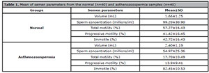
Table 1. Mean of semen parameters from the normal (n=40) and asthenozoospermia samples (n=40)
Conventional sperm parameters in study groups
Motility of sperm cells
Results of motility in the normal and asthenozoospermic groups showed that
the percentage of total motile sperms was increased in the experimental
group (receiving VD) in comparison with the control group. However, there
was a significant difference in the asthenozoospermic group
(p<0.0001). The progressive motility was increased
by VD and the difference was significant in both groups
(p=0.036 in the normal group and p<0.0001 in the asthenozoospermic group). Moreover,
immotile sperms were decreased by VD in both groups, but this difference was
significant (p<0.0001) in the asthenozoospermic group
(Table 2).
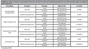
Table 2. Mean of semen parameters from the normal (n=40) and asthenozoospermic (n=40) samples after incubation with vitamin D
Sperm morphology
According to the results (Table 3),
after using VD, no significant difference was observed in the percentage of
sperms with normal morphology in the normal and asthenozoospermic
groups.
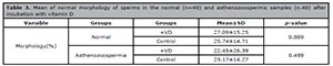
Table 3. Mean of normal morphology of sperms in the normal (n=40) and asthenozoospermic samples (n.40) after incubation with vitamin D
Detection of Apoptosis in the study groups
Apoptosis indicated by PS Expression (Annexin V assay)
Annexin-V assay in normal and asthenozoospermic groups showed that adding VD
causes an increase in the number of normal cells (pale green) in two groups
and the mean apoptosis was decreased in these groups, with a significant
difference (p<0.0001in both groups). The proportions of
necrotic sperm (red color) (PI positive) was significantly different between
the VD and control groups in normal (p=0.007) and
asthenozoospermic individuals (p<0.0001) (Figs. 1 and 2).
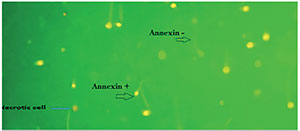
Figure 1. Annexin-V assay by the effect of VD on apoptosis in sperm. Annexin V staining+ (green color), Annexin V staining - (pale color) and necrotic cell (red color).
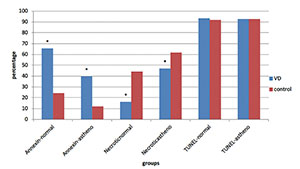
Figure 2. Apoptosis compression in experimental groups, *p-value<0.05
Apoptosis Measured by DNA Double-Strand Breaks (TUNEL assay)
In this study, VD was not able to affect the percentage of DNA fragmentation,
and data analysis showed no significant difference between the VD group and
the control group in normal (p =0.446) and
asthenozoospermic individuals (p=0.998) (Fig. 2).
Detection of chromatin integrity
The chromatin integrity data showed that VD did not have a significant effect on
the protamines of the sperms’ nuclei (aniline blue staining) in the normal and
asthenozoospermic groups (p=0.799 and p=0.071,
respectively); however, this vitamin significantly affected the phosphate groups
(toluidine blue staining) of the sperms’ nuclei and caused an increase of normal
cells in the asthenozoospermic group (p=0.033) (Table 4).
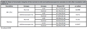
Table 4. Mean of chromatin integrity of the normal (n=40) and asthenozoospermic samples (n=40) after incubation with vitamin D
DISCUSSION
VD has been recognized for maintaining calcium, phosphorus homeostasis, and bone
mineralization. There is some evidence that VD modulates the reproductive processes
in men, and VDR and its metabolizing enzymes are expressed in human spermatozoa
(Blomberg et al., 2011; 2011; Aquila et al., 2008). However, the role of this vitamin
in human male reproduction has not been fully explained to this date (Aquila et al., 2009). In this
study, we assessed the role of VD in the quality and apoptosis of sperms in healthy
and asthenozoospermic men.
Regarding the effects of VD on sperm motility, this study showed that the total sperm
motility was improved, and this improvement was significant in the asthenozoospermic
group; moreover, progressive sperm motility was significantly increased in both
groups. In addition, the immotile sperms were decreased with this vitamin in the
asthenozoospermic samples. Therefore, VD could have an effect on sperm motility,
especially in cases of asthenozoospermia. In agreement with this study, several
studies have shown that VD serum levels are important for semen quality (Aquila et al., 2008; Blomberg et al., 2011). Blomberg Jensen et al. (2011) explained that VD serum levels are positively associated with sperm motility, and
men with VD deficiency (<25 nM) had a lower proportion of total and progressive
motile sperms compared to men with high VD levels (>75 nM). On the other hand,
one study revealed that VDR knockout mice have much less motile sperms (Bouillon et al., 2008). Two
other studies of fertile and infertile men showed that men with vitamin D
sufficiency had more motile spermatozoa than those with vitamin D deficiency (Yang et al., 2012; Blomberg Jensen & Dissing, 2012), but
another study reported that serum vitamin D levels had no relationship with semen
parameter values in a fertile population, while in patients with
oligoasthenozoospermia or teratozoospermia, there was a positive correlation between
vitamin D and sperm quality (Abbasihormozi et
al., 2017). By in vitro studies, Blomberg Jensen et al. (2011) and Aquila et al. (2009) showed
that VD increases intracellular calcium concentration and sperm motility, and it
induces acrosome reaction in mature spermatozoa from healthy men. Other studies
showed that CYP24A1 (VD-inactivating enzyme) is co-expressed with VDR and it
mediates a non-genomic increase in Ca2+ of the human sperm (Blomberg Jensen et al., 2012; Kong et al., 2007). In
addition, one study explained that VD might enhance sperm motility by promoting the
synthesis of ATP, both through the cAMP/PKA pathway and the increase in
intracellular calcium ions (Jueraitetibaike et al., 2019). Although, Ca2+ was not
assessed in our study, its increase may be responsible for the VD-mediated induction
of sperm motility.
Moreover, in this study, normal sperm morphology was evaluated by Diff-Quick staining
in both asthenozoospermic and normal groups, and no improvement in normal morphology
of sperm was seen after adding the vitamin. With regards to the effects of VD on the
morphology of sperms, Ramlau-Hansen et
al. (2011) showed that high serum levels of VD was
associated with low percentage of sperms with normal morphology; however, Blomberg Jensen et al. (2011) explained that men with VD deficiency (<25nM) had a low percentage of
morphologically normal sperm, versus men with high VD levels (>75nM).This
discrepancy with the current study’s data may be due to different methodological
approaches. They evaluated the effects of VD in serum on sperm, but in this study,
vitamin D was used under in vitro conditions, and as a result, VD
was not effective concerning sperm morphology.
Concerning apoptosis in sperm, the fact is that it plays an important role in
regulating spermatogenesis. Several features of apoptosis have been described in
human sperm. These features include PS and DNA fragmentation. These markers are
frequently found in the ejaculations of infertile men (Barroso et al., 2000) and today, researchers
are looking for solutions to reduce apoptosis in the treatment of diseases (Zhang et al., 2007).
In this study, we investigated the effects of VD on apoptosis by PS externalization
(Annexin-V assay) and DNA fragmentation (TUNEL assay) in sperm. The results revealed
that VD caused a decrease in early apoptosis in the asthenozoospermic and normal
groups, and the percentage of normal cells was significantly increased in the two
groups. In explaining these results, one study showed that vitamin D could react
with fatty acid residues in the cell membrane, by its hydrophobic parts and could
protect it from disintegration (Wiseman,
1993). Moreover, in another study, the authors demonstrated that VD could
improve the integrity of sperm membrane during freezing and thawing by decreasing
ROS (Taheri Moghadam et al.,
2019).
In this study as well, the proportion of necrotic sperm was decreased in both groups
that were affected by vitamin D. However, the late apoptosis with TUNEL assay was
not decreased in any of the groups, and this vitamin was not able to improve DNA
fragmentation in sperm within one hour. Like these results, Koppers et al. (2011) induced apoptosis in
spermatozoa by exposure to wortmanina and they showed caspase activation and PS
externalization in sperm, but they did not see any TUNEL positivity. They explained
that the spermatozoa has a mid-tail piece with the mitochondria and cytoplasm in a
distinct subcellular compartment from the nucleus. As a result, even though
endonucleases can be detected in apoptotic human spermatozoa they remain resolutely
locked in the sperm mid-tail piece and never gain access to the nuclear compartment.
In addition, Smith et al.
(2013) showed that spermatozoa does not have APE1 that is needed to
create the 3’-hydroxyl group, targeted by terminal transferases in the TUNEL
reaction. Finally, sperm chromatin is so condensed and it is very difficult for the
reagents used in the TUNEL assay to penetrate the chromatin and register the
presence of a DNA strand break, and 48 hours after the apoptosis starts, the
spermatozoa become TUNEL positive (Smith et
al., 2013). Muratori et al. (2003) assessed the correlation between
sperm’s DNA fragmentation and necrosis, and they stated that the sperms with early
apoptosis may later die by necrosis, and DNA damage occurs further on during
incubation. Another study showed that diets deficient in vitamin D could increase
DNA fragmentation of animal sperms (Merino et al., 2018), and vitamin D has anti-apoptosis
function in some cells (Zhang et
al., 2007); therefore, in the current study, vitamin D may have
improved the late apoptosis at other point-times during sample incubation (that
lasted for more than an hour).
In addition to chromatin integrity detection, only toluidine blue staining was
decreased in the asthenozoospermic group and VD was not able to completely improve
the chromatin integrity of sperms. Sperm DNA is packed with protamines and the sperm
nucleus does not allow for transcription or any other changes by other factors; thus
VD, like late apoptosis, may be able to improve chromatin integrity in longer
incubation periods.
CONCLUSION
The present study revealed that VD could reduce early apoptosis as well as necrosis,
and increase progressive motility in asthenozoospermic and healthy men; in addition,
it improved total motility and immobility in asthenozoospermic men. Further studies
are needed to determine the effects of VD on improving sperm function in
asthenozoospermic men, which may have fundamental therapeutic implication in IVF and
IUI assisting reproductive techniques.
ACKNOWLEDGMENTS
We appreciate the assistance of laboratory technicians of the IVF clinic of Imam
Hospital (Mrs. Arvaneh, R, Lami, F, and Pour shamsa, N.) and those in Cellular and
Molecular Research Center in Ahvaz Jundishapur University of Medical Sciences for
helping us in collecting the samples. This study is the M.Sc. thesis of Ms.
Hosseini.
Financial Support
The Cellular and Molecular Research Center, Ahvaz Jundishapur University of Medical Sciences, Ahvaz, Iran (grant number CMRC-9416), supported this study.
REFERENCES
Abbasihormozi S, Kouhkan A, Alizadeh AR, Shahverdi AH, Nasr-Esfahani
MH, Sadighi Gilani MA, Salman Yazdi R, Matinibehzad A, Zolfaghari Z. Association
of vitamin D status with semen quality and reproductive hormones in Iranian
subfertile men. Andrology. 2017;5:113-8.
Medline Crossref
Aquila S, Guido C, Perrotta I, Tripepi S, Nastro A, Andò S. Human
sperm anatomy: ultrastructural localization of 1α,25-dihydroxyvitamin D3
receptor and its possible role in the human male gamete. J Anat.
2008;213:555-64.
Medline Crossref
Aquila S, Guido C, Middea E, Perrotta I, Bruno R, Pellegrino M, Andò
S. Human male gamete endocrinology: 1alpha, 25-dihydroxyvitamin D3 (1,25(OH)2D3)
regulates different aspects of human sperm biology and metabolism. Reprod Biol
Endocrinol. 2009;7:140.
Medline Crossref
Barroso G, Morshedi M, Oehninger S. Analysis of DNA fragmentation,
plasma membrane translocation of phosphatidylserine andoxidative stress in human
spermatozoa. Hum Reprod. 2000;15:1338-44.
Medline Crossref
Barroso G, Taylor S, Morshedi M, Manzur F, Gaviño F, Oehninger S.
Mitochondrial membrane potential integrity and plasma membrane translocation of
phosphatidylserine as early apoptotic markers: a comparison of two different
sperm subpopulations. Fertil Steril. 2006;85:149-54.
Medline Crossref
Beauchamp PJ, Galle PC, Blasco L. Human sperm velocity and
postinsemination cervical mucus test in the evaluation of the infertile couple.
Arch Androl.1984;13:107-12.
Medline Crossref
Bedu-Addo K, Costello S, Harper C, Machado-Oliveira G, Lefievre L,
Ford C, Barratt C, Publicover S. Mobilisation of stored calcium in the neck
region of human sperm--a mechanism for regulation of flagellar activity. Int J
Dev Biol. 2008;52:615-26.
Medline Crossref
Benyamini Y, Gozlan M, Kokia E. Variability in the difficulties
experienced by women undergoing infertility treatments. Fertil Steril.
2005;83:275-83.
Medline Crossref
Blomberg Jensen M, Nielsen JE, Jørgensen A, Rajpert-De Meyts
E, Kristensen DM, Jørgensen N, Skakkebaek NE, Juul A, Leffers H. Vitamin
D receptor and vitamin D metabolizing enzymes are expressed in the human male
reproductive tract. Hum Reprod . 2010;25:1303-11.
Medline Crossref
Blomberg Jensen M, Bjerrum PJ, Jessen TE, Nielsen JE, Joensen UN,
Olesen IA, Petersen JH, Juul A, Dissing S, Jørgensen N. Vitamin D is
positively associated with sperm motility and increases intracellular calcium in
human spermatozoa. Hum Reprod. 2011;26:1307-17.
Medline Crossref
Blomberg Jensen M, Dissing S. Non-genomic effects of vitamin D in
human spermatozoa. Steroids. 2012;77:903-9.
Medline Crossref
Blomberg Jensen M, Jørgensen A, Nielsen JE, Bjerrum PJ,
Skalkam M, Petersen JH, Egeberg DL, Bangsbøll S, Andersen AN, Skakkebaek
NE, Juul A, Rajpert-De Meyts E, Dissing S, Leffers H, Jørgensen N.
Expression of the vitamin D metabolizing enzyme CYP24A1 at the annulus of human
spermatozoa may serve as a novel marker of semen quality. Int J Androl.
2012;35:499-510.
Medline Crossref
Bouillon R, Carmeliet G, Verlinden L, van Etten E, Verstuyf A,
Luderer HF, Lieben L, Mathieu C, Demay M. Vitamin D and human health: lessons
from vitamin D receptor null mice. Endocr Rev. 2008;29:726-76.
Medline Crossref
Dominguez-Fandos D, Camejo MI, Ballescà JL, Oliva R. Human sperm DNA
fragmentation: correlation of TUNEL results as assessed by flow cytometry and
optical microscopy. Cytometry A. 2007;71:1011-8.
Medline Crossref
Jueraitetibaike K, Ding Z, Wang DD, Peng LP, Jing J, Chen L, Ge X,
Qiu XH, Yao B. The effect of vitamin D on sperm motility and the underlying
mechanism. Asian J Androl. 2019;21:400-7.
Medline Crossref
Kinuta K, Tanaka H, Moriwake T, Aya K, Kato S, Seino Y. Vitamin D is
an important factor in estrogen biosynthesis of both female and male gonads.
Endocrinology. 2000;141:1317-24.
Medline Crossref
Kong LJ, Yang YY, Wang GL. CatSper and sperm hyperactivation.
Zhonghua Nan Ke Xue. 2007;13:164-7.
Medline
Koppers AJ, Mitchell LA, Wang P, Lin M, Aitken RJ. Phosphoinositide
3-kinase signalling pathway involvement in a truncated apoptotic cascade
associated with motility loss and oxidative DNA damage in human spermatozoa.
Biochem J. 2011;436:687-98.
Medline Crossref
Luk J, Torrealday S, Neal Perry G, Pal L. Relevance of vitamin D in
reproduction. Hum Reprod . 2012;27:3015-27.
Medline Crossref
Merino O, Sánchez R, Gregorio BM, Sampaio FJ, Risopatrón J. Effects
of Diet-Induced Obesity and Deficient in Vitamin D on Spermatozoa Function and
DNA Integrity in Sprague-Dawley Rats. Biomed Res Int. 2018;2018:5479057.
Medline Crossref
Moradian Fard Z, Naghdi M, Salehi P, Shahrokhi SZ, Ajam A, Deemeh
MR, Meshkibaf MH. SCSA results correlated with rate of motility reduction after
ejaculation in Asthenozoospermia. Andrologia. 2019;51:e13146.
Medline Crossref
Muratori M, Maggi M, Spinelli S, Filimberti E, Forti G, Baldi E.
Spontaneous DNA fragmentation in swim-up selected human spermatozoa during long
term incubation. J Androl. 2003;24:253-62.
Medline Crossref
Oosterhuis GJ, Mulder AB, Kalsbeek-Batenburg E, Lambalk CB,
Schoemaker J, Vermes I. Measuring apoptosis in human spermatozoa: a biological
assay for semen quality? Fertil Steril . 2000;74:245-50.
Medline Crossref
Ortega C, Verheyen G, Raick D, Camus M, Devroey P, Tournaye H.
Absolute asthenozoospermia and ICSI: what are the options? Hum Reprod Update.
2011;17:648-92.
Medline Crossref
Ramlau-Hansen CH, Moeller UK, Bonde JP, Olsen J, Thulstrup AM. Are
serum levels of vitamin D associated with semen quality? Results from a
cross-sectional study in young healthy men. Fertil Steril . 2011;95:1000-4.
Medline Crossref
Schulte RT, Ohl DA, Sigman M, Smith GD. Sperm DNA damage in male
infertility: etiologies, assays, and outcomes. J Assist Reprod Genet.
2010;27:3-12.
Medline Crossref
Smith TB, Dun MD, Smith ND, Curry BJ, Connaughton HS, Aitken RJ. The
presence of a truncated base excision repair pathway in human spermatozoa that
is mediated by OGG1. J Cell Sci. 2013;126:1488-97.
Medline Crossref
Taheri Moghadam M, Asadifard Y, Saki G, Nikbakht R. Effect of
Vitamin D on apoptotic marker, reactive oxygen species and human sperm
parameters during the process of cryopreservation. Iran J Basic Med Sci.
2019;22:1036-43.
Medline Crossref
Thonneau P, Marchand S, Tallec A, Ferial ML, Ducot B, Lansac J,
Lopes P, Tabaste JM, Spira A. Incidence and main causes of infertility in a
resident population (1,850,000) of three French regions (1988-1989). Hum Reprod.
1991;6:811-6.
Medline Crossref
Wiseman H. Vitamin D is a membrane antioxidant. Ability to inhibit
iron-dependent lipid peroxidation in liposomes compared to cholesterol,
ergosterol and tamoxifen and relevance to anticancer action. FEBS Lett.
1993;326:285-8.
Medline Crossref
Wyllie AH, Kerr JR, Currie A. Cell death: the significance of
apoptosis. Int Rev Cytol. 1980;68:251-306.
Medline Crossref
Yang B, Sun H, Wan Y, Wang H, Qin W, Yang L, Zhao H, Yuan J, Yao B.
Associations between testosterone, bone mineral density, vitamin D and semen
quality in fertile and infertile Chinese men. Int J Androl. 2012;35:783-92.
Medline Crossref
Zhang A, Wang Y, Xie H, Zheng S. Calcitriol inhibits hepatocyte
apoptosis in rat allograft by regulating apoptosis-associated genes. Int
Immunopharmacol. 2007;7:1122-8.
Medline Crossref