JBRA Assist. Reprod. 2021;25(1):90-96
ORIGINAL ARTICLE
doi: 10.5935/1518-0557.20200056
Use of clomiphene citrate protocol for controlled ovarian stimulation impairs endometrial maturity
1Gynecology and Obstetrics Unit, Hospital de Clínicas de Porto Alegre, Porto Alegre, Brazil
2Gynecology and Obstetrics Department, Universidade Federal do Rio Grande do Sul, Porto Alegre, Brazil
3Embryology and Cellular Differentiation Lab, Hospital de Clínicas de Porto Alegre, Porto Alegre, Brazil
4Pathology Unit, Hospital de Clínicas de Porto Alegre, Porto Alegre, Brazil
5Postgraduate Program in Health Sciences: Gynecology and Obstetrics of Universidade Federal do Rio Grande do Sul
CONFLICT OF INTEREST
All authors have completed the ICMJE uniform disclosure and declare that
there was no support from any organization for the submitted paper, no
financial relationships with any organizations that might have an interest
in the submitted study in the previous 3 years and no other relationships or
activities that could appear to have influenced the submitted study.
This study was presented at the:
XXIII Brazilian Congress on Assisted Reproduction 58th Brazilian
Congress on Gynecology and Obstetrics
FUNDING
This research was supported by Fundo de Incentivo à Pesquisa e Eventos (FIPE) of
Hospital de Clínicas de Porto Alegre (number 2017-0180).
ABSTRACT
Introduction: Despite recent advances in assisted reproduction techniques and recent
knowledge regarding embryo and endometrium quality, implantation and birth
rates remain low. The objective of this study was to investigate whether
clomiphene citrate alters endometrial maturation in infertile patients.
Methods: In a prospective self-matched cohort study, we assessed the ovulation of
women in spontaneous and stimulated cycles (with clomiphene citrate). We
determined the ovulation day by ultrasound scanning. In both cycles, we took
four blood samples (BS1 - at early proliferative phase, BS2 - at mid
proliferative phase, BS3 - after ovulation and BS4 - at mid luteal phase) to
determine the serum concentrations of FSH, LH, estradiol and progesterone.
We retrieved an endometrial biopsy five days after ovulation, followed by
blinded analysis and classification according to Noyes criteria, in both
cycles.
Results: Twenty-two participants completed the study. There were significant
differences in FSH BS3 (p=0.001), in LH BS3 and BS4
(p<0.001 and p=0.049,
respectively), in estradiol BS2, BS3 and BS4 (p<0.001, p=0.024 and p<0.001, respectively)
and in progesterone BS3 and BS4 (p=0.028 and p<0.001, respectively). Considering Noyes criteria,
there was a one-day delay when comparing the stimulated cycle with the
spontaneous cycle (p=0.004), and a two-day delay when
comparing the stimulated cycle with the biopsy day.
Conclusion: This study indicates that ovarian stimulation with clomiphene citrate delays
the endometrial maturity, and could possibly impair the implantation process
due to asynchrony.
Keywords: endometrial receptivity, endometrial maturity, Noyes criteria, assisted reproduction techniques, IVF
INTRODUCTION
Despite recent advances in assisted reproduction techniques (ART), implantation rates
remain low after controlled ovarian stimulation (COS) for in vitro fertilization
(IVF), and intracytoplasmic sperm injection (ICSI) treatments (van der Gaast et al., 2008). Embryonic
implantation failure remains the major limitation of ART success (Evans et al., 2018; van der Gaast et al., 2008),
and although embryo quality is the main determinant of implantation success,
maturity and endometrial receptivity are important factors to consider. Embryo
implantation followed by gestation depends on a viable and high quality embryo, a
receptive endometrium and having a molecular "dialogue" between them (Dieamant et al., 2019).
However, what precisely constitutes a receptive endometrium remains uncertain (Bassil et al., 2018; Lessey & Young, 2019; Li & Jin, 2013; Paulson,
2019). The implantation process in humans is complex and depends on
multiple and successive interactions between the embryo and the endometrium, and it
only succeeds when it occurs at a specific time, during the secretory phase of the
menstrual cycle, called "implantation window" (Bassil et al., 2018; Enciso et al., 2018; Lessey
& Young, 2019; Li & Jin,
2013).
Recent research in this area has focused on finding a viable endometrial receptivity
marker that may help to identify the best moment to perform an embryo transfer
(Edgell et al., 2013; Evans et al., 2018; Kliman & Frankfurter, 2019; Miravet-Valenciano et al.,
2015; Siristatidis et
al., 2018). Histologically, endometrial maturity can be
classified using the Noyes criteria (Kliman &
Frankfurter, 2019; Noyes et
al., 1950; Paulson,
2019), which characterizes the endometrium into its phases (Complementary
table).
Clomiphene citrate (CC), commonly used in COS, is a selective estrogen receptor
modulator (SERM) that has been the first-line treatment for patients with
anovulation or oligomenorrhea for more than 40 years with estrogenic and
antiestrogenic effects (Lindheim et
al., 2018). It binds to estrogen receptors (ER), leading to
the misinterpretation of the estradiol feedback mechanism at a hypothalamic level.
The administration of this drug results in increased pituitary gonadotropin release
and enhanced follicular development and ovarian response (Lindheim et al., 2018; Palomino et al., 2005).
An important CC issue is its associated low pregnancy rates, notwithstanding high
ovulation rates. This phenomenon is partially explained by the antiestrogenic effect
on the cervical mucus and the endometrium, causing abnormal endometrial maturation
(Jie et al., 2018; Reed et al., 2018). It is
therefore, tempting to hypothesize that long-lasting ER occupancy by CC might alter
the endometrial cell function, thus affecting the implantation window (Palomino et al., 2005).
To investigate possible changes caused by CC in the pattern of endometrial
receptivity, we obtained endometrial samples during the midsecretory phase from
ovulatory women in a spontaneous cycle and in a CC stimulated cycle.
MATERIALS AND METHODS
Ethical approval
The study was developed in accordance with international laws on procedures for
dealing with human tissue (including the Declaration of Helsinki for Medical
Research involving Human Subjects) and STROBE's publication guidelines (Vandenbroucke et al.,
2014). It was submitted and approved by the ethics committee of the Porto
Alegre University Hospital (HCPA) and all participants signed an informed
consent.
Study population and participants
In a self-matched prospective cohort, we recruited ovulatory women from Rio
Grande do Sul - Brazil, with IVF indication, between the ages of 18 and 35
years, body mass index between 18 and 30 kg/m2, and with tubal
obstruction or male infertility. We excluded those women with anovulation,
endometriosis and uterine abnormalities.
Methodology description
We followed the participants throughout two menstrual cycles. The first one,
spontaneous, and the second, stimulated using a daily dose of 100 mg of
clomiphene citrate (Clomid®, Sanofi, Brazil), for 5 days,
initiated on the third day of the cycle. We followed the follicular growth using
transvaginal ultrasound, from the second day of the cycle, until sonographic
determination of ovulation. During the stimulated cycle, after identifying a
follicle with 17 mm of diameter, the participant received 10.000 UI of chorionic
gonadotrophin (Choriomon M®, Meizler UCB Biopharma S.A.,
Brussels, Belgic), to promote final maturation and ovulation (Figure 1).
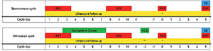
Figure 1.
Schematic representation of the assessments in spontaneous and
stimulated cycles. Following a spontaneous cycle, all 22
participants underwent a stimulated cycle using 100 mg/day of
clomiphene citrate between days 3 and 7 of the cycle. When the
leading follicle had a diameter of 17 mm (represented by **), they
received 10.000 UI of human chorionic gonadotrophin (hCG) for final
oocyte maturation. Endometrial biopsy (EB) was performed 5 days
after ovulation confirmation (represented by *) by ultrasonography.
In both cycles blood samples (BS) were taken at the early
proliferative phase (days 1 -3), in the midproliferative phase (days
8-10), when confirmed ovulation, and five days after
ovulation.
In both cycles, collected blood samples from all participants to analyze the
serum FSH, LH, estradiol and progesterone levels: first blood sample (BS1), at
initial proliferative phase (days 1-3 of the cycle); second blood sample (BS2),
at the mid proliferative phase (days 8-10 of the cycle); third blood sample
(BS3), after determination of ovulation by ultrasound; fourth blood sample
(BS4), five days after determining ovulation by ultrasound. All hormone assays
were performed using electrochemiluminescent immunoassay in the Roche Cobas
e602analyzer, according to correspondent kits for FSH, LH, estradiol and
progesterone (FSH Elecsys and cobas e analyzers, Roche Diagnostics, USA; LH
Elecsys and cobas e analyzers, Roche Diagnostics, USA; Estradiol II Elecsys and
cobas e analyzers, Roche Diagnostics, USA; ProgesteroneIII Elecsys and cobas e
analyzers, Roche Diagnostics, USA).
We submitted the participants to an endometrial biopsy five days after
establishing the ovulation, in both cycles, with a Pipelle curette
(Pipelle® de Cornier for endometrial biopsy, Laboratoire
C.C.D., France). We fixed the biopsy samples in 10% buffered neutral formalin,
and sent them to HCPA´s pathology lab. The specimens were dehydrated with
increasing concentration of ethanol before being embedded in paraffin. A portion
of endometrial tissue was routinely cut, assembled and stained with hematoxylin
for histological dating following the Noyes criteria (Complementary table)
(Noyes et al.,
1950). The same blinded examiner (RCR), an expert in gynecological
pathology at HCPA, evaluated all slides and the result was expressed in whole
numbers, using as a reference, a standard menstrual cycle of 28 days of duration
and ovulation occurring on the 14th day of the cycle.
Statistical analyses
FSH, LH, estradiol and progesterone samples were compared using a Generalized
Estimating Equation (GEE) model concerning values between collections, cycles
and interaction (collection x cycle). We presented the data as mean ±
standard error. For the calculation model, we established the following
parameters: unstructured working correlation matrix, robust estimator covariance
matrix, and normal distributions with identity function. When significant, we
used the Bonferroni post-hoc test to ascertain which blood samples were
different. We analyzed the histopathological results of Noyes criteria using the
Wilcoxon test for related samples, which compares the distribution of the
histopathological data of the endometrial matched samples. We expressed this
data using median values (25-75). For all tests, a p<0.05
was significant.
RESULTS
Demographic data
We recruited thirty-six patients and, of these, 22 completed the two assessment
cycles and were included for analysis of the data collected, description
(presented in the Table 1 and Figure 2) and statistical evaluation.
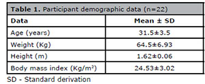
Table 1. Participant demographic data (n=22)
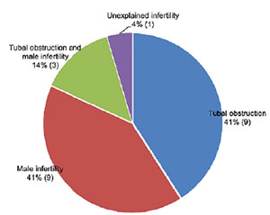
Figure 2.
Participants’ causes of infertility.
Hormonal analysis
Table 2 shows the mean serum levels of
the hormones FSH, LH, estradiol and progesterone. When comparing mean values
between the corresponding samples for each cycle, there were differences in the
FSH dosage comparisons from BS3 (p=0.001); in LH, from BS3
(p<0.001) and BS4 (p=0.049); in
estradiol from BS2 (p<0.001), BS3 (p=0.024)
and BS4 (p<0.001); and in progesterone, from BS3
(p=0.028) and BS4 (p<0.001).
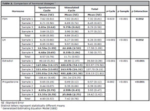
Table 2. Comparison of hormonal dosages †
Endometrial analysis
We evaluated eighteen endometrial matched samples by Noyes criteria tissue
analysis (Figure 3). Four pairs of samples
were withdrawn due to results that showed, at least in one slide, a
stromal-glandular dissociation (2 slides), a basal endometrium (1 slide) or
could not be evaluated (1 slide). The comparison between the median results of
the endometrial biopsy from both cycles expressed a difference
(p<0.004) of one day (Figure 4).
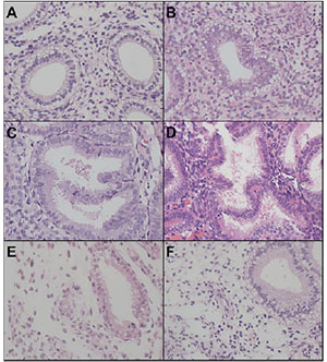
Figure 3.
Endometrial cycle dating based on Noyes et al. (1950) criteria (200x): (A) Day 17: “piano
key” appearance of glandular epithelium with vacuoles at nuclei
level; (B) Day 18: luminal vacuoles with smaller size and the nuclei
approaching the base of the glandular cell; (C) Day 19: presence of
intraluminal secretion with few vacuoles on cellular cytoplasm,
absence of mitosis and pseudo stratification; (D) Day 20: the peak
of intraluminal secretion with stromal edema onset, presence of rare
vacuoles and round nuclei located at the base of the glandular
cell.; (E) Day 23: presence of prominent spiral arterioles with
thickened walls, coiling and endothelial proliferation; (F)
stromal-glandular dissociation - gland on day 17 with decidualized
stroma.
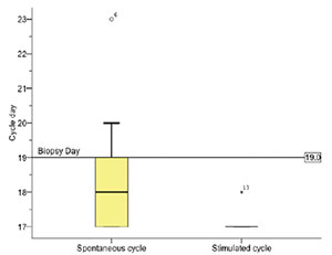
Figure 4. Median comparison of Noyes criteria results.
DISCUSSION
This study found a one-day delay in the CC stimulated cycle, when analyzing the
endometrium following the Noyes criteria. This data supports the idea of asynchrony
between the endometrium in the spontaneous and stimulated cycles. Concerning the day
of blastocyst transfer, in other words, 5 days after ovulation, there was a 2-day
delay - which could explain even lower implantation rates when performing a transfer
on this day in cycles using CC. It was the first to study that analyzed endometrium
matched samples in spontaneous and stimulated cycles from the same infertile
women.
Some investigators report on the lack of correlation between the cycle day and
endometrial dating by Noyes criteria. Murray et al. (2004) concluded that histologic endometrial
dating does not have the accuracy or the precision necessary to provide a valid
method for the diagnosis of luteal phase deficiency and, Coutifaris et al. (2004) stated that the
endometrial biopsy followed by histological dating provides no useful clinical
information as a screening test for infertility. Garrido-Gómez et al. (2013) said that biochemical
markers are ideal as alternatives to classic Noyes criteria and suggest that the
definition of a genomic signature of human endometrial receptivity can be used as a
strategy to overcome subjectivity problems caused by the inter and intracycle
variations in Noyes endometrial receptivity dating. In 2018, Bassil et
al. found no agreement between the endometrial receptivity array (ERA)
and the Noyes histological criteria (Bassil et al., 2018). Despite these relevant findings, they
used different methods to reach this conclusion (none of them using the same
participant in a spontaneous and in a stimulated cycle) and there are no other
validated markers or clinically useful endometrial evaluation methods.
It should be noted that our participants are diagnosed with infertility and with
indication of IVF; however, the ones with anovulation, uterine abnormalities and
endometriosis were carefully excluded to minimize possible selection biases, as it
is considered that these pathologies may modify the endometrial decidualization
process. Yet, we biopsied all the participants in the spontaneous cycle and in a
CC-stimulated cycle, minimizing other types of selection biases.
The significant increases in FSH, LH, estradiol and progesterone dosage confirm that
participants correctly used the medication provided. As known, CC works by
competitive binding to estrogen receptors in the hypothalamus and pituitary,
reducing estrogen signaling via receptors and interfering with the feedback
mechanism of endogenous estrogen, resulting in an increase in FSH and LH secretions
to stimulate ovarian follicular production, and a consequent increase in estradiol
and progesterone (Gadalla et al.,
2018). One of our hypotheses is that the supraphysiological increase in
the estradiol, progesterone, FSH and LH levels in stimulated cycles with clomiphene
citrate, may be one of the factors responsible for the asynchrony of endometrial
histology.
Considering the 22 matched samples of endometrial biopsies, we excluded four pairs
from the statistical calculation, two histological results demonstrated a
stromal-glandular dissociation and another one a basal endometrium. These findings
confirm that CC use for COS causes asynchrony and delayed endometrial development,
although it was not used for analysis.
Bonhoff et al. (1996)
described the same asynchrony effect in patients undergoing COS with CC for
artificial insemination, comparing them with fertile controls. Gonzalez et al. (2001) also reported an
endometrial asynchrony and integrins alteration in CC. stimulated cycles and
intrauterine device users, when compared to fertile controls.
Looking at these results, we hypothesize that the antiestrogenic effects of CC on the
endometrium impairs endometrial proliferation in the proliferative phase of the
cycle, which reflects a delayed endometrial maturation in the luteal phase. This
finding is confirmed by several studies which concluded that ovulation induction
with CC might result in lower endometrial thickness (Gadalla et al., 2018; Reed et al., 2018) and, despite the high rate of
ovulation, pregnancy rates are low when using CC (Jie et al., 2018).
There are few limitations to our study. We did not compare these findings to
implantation, pregnancy and birth rates, as we consider these outcomes the best
predictors for any developed endometrial receptivity test. Despite the small sample
of this study, our results show a significant difference in endometrial maturity
comparing the spontaneous and stimulated cycle with CC. Regardless of the Noyes
criteria limits, there are no other validated test to assess endometrial maturity,
so that we use the same test, performed by the same blinded observer, in samples of
the same participant in order to minimize this bias.
In conclusion, our study found statistically significant changes in the endometrial
compartment in patients who used clomiphene citrate for ovarian stimulation,
represented by an asynchrony demonstrated by a one-day delay in histological
endometrial maturity when comparing spontaneous and stimulated cycles; and a
two-days delay when compared the stimulated cycle with the probable day of
blastocyst transfer. The freeze-all strategy is a possible approach to be adopted in
IVF cycles, to avoid this asynchrony with subsequent transfer in a spontaneous cycle
or associated with endometrial preparation. In order to confirm this finding, we
need larger prospective studies.
ACKNOWLEDGEMENTS
We acknowledge the Fundo de Incentivo à Pesquisa e Eventos (FIPE) of Hospital de
Clínicas de Porto Alegre for funding this research. We would like to thank the staff
from the Gynecology and Obstetrics Unit, Embryology and Cellular Differentiation Lab
and Pathology Unit of Hospital de Clínicas de Porto Alegre and staff of Gynecology
and Obstetrics Department of Universidade Federal do Rio Grande do Sul involved in
this research.

Complementary table. Noyes criteria
REFERENCES
Bassil R, Casper R, Samara N, Hsieh TB, Barzilay E, Orvieto R, Haas
J. Does the endometrial receptivity array really provide personalized embryo
transfer? J Assist Reprod Genet. 2018;35:1301-5.
Medline Crossref
Bonhoff AJ, Naether OG, Johannisson E. Effects of clomiphene citrate
stimulation on endometrial structure in infertile women. Hum Reprod.
1996;11:844-9.
Medline Crossref
Coutifaris C, Myers ER, Guzick DS, Diamond MP, Carson SA, Legro RS,
McGovern PG, Schlaff WD, Carr BR, Steinkampf MP, Silva S, Vogel DL, Leppert PC ;
NICHD National Cooperative Reproductive Medicine Network . Histological dating
of timed endometrial biopsy tissue is not related to fertility status. Fertil
Steril. 2004;82:1264-72.
Medline Crossref
Dieamant F, Vagnini LD, Petersen CG, Mauri AL, Renzi A, Petersen B,
Mattila MC, Nicoletti A, Oliveira JBA, Baruffi R, Franco JG Jr. New therapeutic
protocol for improvement of endometrial receptivity (PRIMER) for patients with
recurrent implantation failure (RIF) - A pilot study. JBRA Assist Reprod.
2019;23:250-4.
Medline Crossref
Edgell TA, Rombauts LJ, Salamonsen LA. Assessing receptivity in the
endometrium: the need for a rapid, non-invasive test. Reprod Biomed Online.
2013;27:486-96.
Medline Crossref
Enciso M, Carrascosa JP, Sarasa J, Martínez-Ortiz PA, Munné S,
Horcajadas JA, Aizpurua J. Development of a new comprehensive and reliable
endometrial receptivity map (ER Map/ER Grade) based on RT-qPCR gene expression
analysis. Hum Reprod. 2018;33:220-8.
Medline Crossref
Evans GE, Phillipson GTM, Sykes PH, McNoe LA, Print CG, Evans JJ.
Does the endometrial gene expression of fertile women vary within and between
cycles? Hum Reprod. 2018;33:452-63.
Medline Crossref
Gadalla MA, Huang S, Wang R, Norman RJ, Abdullah SA, El Saman AM,
Ismail AM, van Wely M, Mol BWJ. Effect of clomiphene citrate on endometrial
thickness, ovulation, pregnancy and live birth in anovulatory women: systematic
review and meta-analysis. Ultrasound Obstet Gynecol. 2018;51:64-76.
Medline Crossref
Garrido-Gómez T, Ruiz-Alonso M, Blesa D, Diaz-Gimeno P, Vilella F,
Simón C. Profiling the gene signature of endometrial receptivity: clinical
results. Fertil Steril. 2013;99:1078-85.
Medline Crossref
Gonzalez RR, Palomino A, Vantman D, Gabler F, Devoto L. Abnormal
pattern of integrin expression at the implantation window in endometrium from
fertile women treated with clomiphene citrate and users of intrauterine device.
Early Pregnancy. 2001;5:132-43.
Medline
Jie L, Li D, Yang C, Haiying Z. Tamoxifen versus clomiphene citrate
for ovulation induction in infertile women. Eur J Obstet Gynecol Reprod Biol.
2018;228:57-64.
Medline Crossref
Kliman HJ, Frankfurter D. Clinical approach to recurrent
implantation failure: evidence-based evaluation of the endometrium. Fertil
Steril. 2019;111:618-28.
Medline Crossref
Lessey BA, Young SL. What exactly is endometrial receptivity? Fertil
Steril. 2019;111:611-7.
Medline Crossref
Li MQ, Jin LP. Ovarian stimulation for in vitro fertilization alters
the protein profile expression in endometrial secretion. Int J Clin Exp Pathol.
2013;6:1964-71.
Medline
Lindheim SR, Glenn TL, Smith MC, Gagneux P. Ovulation Induction for
the General Gynecologist. J Obstet Gynaecol India. 2018;68:242-52.
Medline Crossref
Miravet-Valenciano JA, Rincon-Bertolin A, Vilella F, Simon C.
Understanding and improving endometrial receptivity. Curr Opin Obstet Gynecol.
2015;27:187-92.
Medline Crossref
Murray MJ, Meyer WR, Zaino RJ, Lessey BA, Novotny DB, Ireland K,
Zeng D, Fritz MA. A critical analysis of the accuracy, reproducibility, and
clinical utility of histologic endometrial dating in fertile women. Fertil
Steril. 2004;81:1333-43.
Medline Crossref
Noyes RW, Hertig AT, Rock J. Dating the endometrial biopsy. Obstet Gynecol Surv. 1950;5:561-4.
Palomino WA, Fuentes A, González RR, Gabler F, Boric MA, Vega M,
Devoto L. Differential expression of endometrial integrins and progesterone
receptor during the window of implantation in normo-ovulatory women treated with
clomiphene citrate. Fertil Steril. 2005;83:587-93.
Medline Crossref
Paulson RJ. Introduction: Endometrial receptivity: evaluation,
induction and inhibition. Fertil Steril. 2019;111:609-10.
Medline Crossref
Reed BG, Wu JL, Nemer LB, Carr BR, Bukulmez O. Use of Clomiphene
Citrate in minimal stimulation in vitro fertilization negatively impacts
endometrial thickness: an argument for a freeze-all approach. JBRA Assist
Reprod. 2018;22:355-62.
Medline Crossref
Siristatidis CS, Sertedaki E, Vaidakis D, Varounis C, Trivella M.
Metabolomics for improving pregnancy outcomes in women undergoing assisted
reproductive technologies. Cochrane Database Syst Rev. 2018;3:CD011872.
Medline Crossref
van der Gaast MH, Classen-Linke I, Krusche CA, Beier-Hellwig K,
Fauser BC, Beier HM, Macklon NS. Impact of ovarian stimulation on mid-luteal
endometrial tissue and secretion markers of receptivity. Reprod Biomed Online.
2008;17:553-63.
Medline Crossref
Vandenbroucke JP, von Elm E, Altman DG, Gøtzsche PC, Mulrow
CD, Pocock SJ, Poole C, Schlesselman JJ, Egger M, STROBE initiative.
Strengthening the Reporting of Observational Studies in Epidemiology (STROBE):
explanation and elaboration. Int J Surg. 2014;12:1500-24.
Medline Crossref