JBRA Assist. Reprod. 2022;26(1):105-122
REVIEW
doi: 10.5935/1518-0557.20210026
Oocyte quality and aging
1Cellular & Molecular Research Center, Medical Basic Sciences Research Institute, Ahvaz Jundishapur University of Medical Sciences, Ahvaz, Iran
2Department of Anatomical Science, Faculty of Medicine, Ahvaz Jundishapur University of Medical Sciences, Ahvaz, Iran
The authors declare that there is no conflict of interests.
ABSTRACT
It is well known that female reproduction ability decreases during the forth decade of life due to age-related changes in oocyte quality and quantity; although the number of women trying to conceive has today increased remarkably between the ages of 36 to 44. The causes of reproductive aging and physiological aspects of this phenomenon are still elusive. With increase in the women’s age, during Assisted Reproductive Technologies (ART) we have perceived a significant decline in the number and quality of retrieved oocytes, as well as in ovarian follicle reserves. This is because of increased aneuploidy due to factors such as spindle apparatus disruption; oxidative stress and mitochondrial damage. The aim of this review paper is to study data on the potential role of the aging process impacting oocyte quality and female reproductive ability. We present the current evidence that show the decreased oocyte quality with age, related to reductions in female reproductive outcome. The aging process is complicated and it is caused by many factors that control cellular and organism life span. Although the factors responsible for reduced oocyte quality remain unknown, the present review focuses on the potential role of ovarian follicle environment, oocyte structure and its organelles. To find a way to optimize oocyte quality and ameliorate clinical outcomes for women with aging-related causes of infertility.
Keywords: oocyte quality, aging, ovary, female infertility
INTRODUCTION
The human female reproductive system has a faster rate in aging than any other system of the body. According to population studies and reports the biological ability of a woman to fertilize decreases with age, similar to other species (Wood, 1989). The number of women delivering their first child after the age of 35 increased from 1/100 to 1/12 from 1970 to 2006 (Cil et al., 2015). The fecundity capacity of females peaks is their 20s, it fails in late 30s, in defiance of regular menstrual cycles, ending in menopause at the mean age of 50 - 51 years (te Velde & Pearson, 2002). The in vitro fertilization (IVF) study shows that the mother’s age is an essential cause that leads to impressing clinical results (Tan et al., 2014). While, between 1970 and 2002, the percentage of first births in women over the age of 30 years increased 6-fold (Crawford & Steiner, 2015). According to an American report based on >120.000 assisted reproduction technologies (ART), successful delivery rate per embryo transfer dropped from 43.2% in women <35 years old to 15.1% in women aged 41- 42 years and to 5.9% in women > 42 years old (Wright et al., 2005). Today, in developed countries, because of the women’s decision to delay their conception, families faced female aging-related infertility (Balasch, 2010). Despite neuroendocrine and uterine factors, it is well defined that ovaries and ovarian follicles are prominent regulators of reproductive aging. In humans, changes to the ovarian follicle pool and diminishing of ovarian reserve are the chief causes of decline in female reproduction ability (Faddy, 2000). This claim is confirmed by oocyte donation from younger women, in overcoming age-related infertility (Sauer et al., 1990; te Velde & Pearson, 2002). By the fifth month of prenatal development, the size of ovarian follicle pool is estimate at 7 million oogonia (Sadler, 2011). At this time large numbers of oogonia, as well as primary oocytes become atretic, except for a few near the ovarian surface. The surviving oogonia enter the prophase of first meiotic division and transform into primary oocytes, and are surrounded by flat epithelial cells to forming the pool of primary follicles (Pepling, 2006). Primary oocytes stay arrested in prophase in the so-called diplotene stage and do not complete their first meiotic division before puberty is reached. At birth, one million of primordial follicles are present, a number which will disappear by atresia. During childhood, this number reaches the amount of approximately 300,000 at the time of menarche (Faddy et al., 1992). Over the past decades there has been controversy about the origin of ovarian germ cells and oogenesis during one’s life span, which indicates the neo-oogenesis in mice ovaries and adult humans’ (Bukovsky, 2011; de Souza et al., 2017). During the female reproductive life span, under the influence of intra and extra ovarian factors, such as cytokines, growth factors and gonadal steroid follicles leave the resting pool to enter the growing pool on a regular manner (Pangas, 2007). Therefore, follicles behave differently in response to such stimulatory and regulatory signals (Craig et al., 2007). As a result of the ovarian follicular pool diminishing exponentially with age, decline is more prominent at the age of 38-39 and above (Faddy et al., 1992).
Therefore, according to the observations mentioned above, assessment of cellular and molecular aspects of follicles/oocytes need precise attention to some factors. First, some oocytes and their surrounding cells remain in quiescent phase (diplotene stage) for ~ 40 years or more, it is unknown how this stage can retain the cells against environmental influences (Absalan et al., 2017; Sadler, 2011). The fact that the increase in maternal age is directly related to chromosomal abnormalities in the fetus confirms that primary oocytes are susceptible to damage as they age (Sadler, 2011). Second, the growth of a mature oocyte depends on fine cross-talk signalling between all ovarian compartments. Age-related infertility is a multifactorial process and understanding factors affecting follicle/oocyte aging requires the comparison of the theories on aging mechanisms based on studies on tissues and organs other than the ovary (Hamet & Tremblay, 2003). Ultrastructural changes in oocytes, such as meiotic spindle disruption and zona pellucida hardening are some of the factors on age-related declined fecundity (Eftekhari Moghadam et al., 2018). In addition, higher tendency for acquired conditions such as uterine problems, hormonal changes, endometriosis, fibroids and pelvic infections occur in aged women (Schwartz & Mayaux, 1982). The most relevant theory for ovarian aging, conveys a reduction in oocytes and its surrounding cells’ capacity to nullify reactive oxygen species (Sousa et al., 2015), which is the most essential cellular damage inducer with aging (Harman, 2006). Loss of active DNA repair apparatus, bioenergetics status and mitochondrial damage of the oocytes are possibilities for follicle/oocyte aging phenomenon (Titus et al., 2013). Other feasible mechanisms, like molecular pathways that could lead to loss of germ cells in aged ovaries, are poorly defined (Ben-Meir et al., 2015).
In this paper, we review studies on human and mice models about the basic age-related aspects of follicles/oocytes, ovarian microenvironment and involvement of oxidative stress in progressive reproductive aging.
OVARY AGING
The ovarian tissue, like every organ of the body undergoes aging with no exclusion. Every day, in an ovary, a certain number of primordial follicles are engaged from resting follicular pool (ovarian reserve) for more growth, a process called folliculogenesis. This action, takes approximately 16 - 24 weeks, during which time the oocyte and its encompassing somatic cells endure a string of changes that finally result in an antral follicle to release a mature ovum (Baird et al., 2005). Primordial follicle recruitment ratio raises with age and it is reciprocally related to ovarian reserve (Orisaka et al., 2009). Ovarian follicles are essential for keeping endocrine function of female gonads, as well as fertility accordingly, the number of follicles present in the tissue control ovarian lifespan (Tilly & Sinclair, 2013). In older women, it appears that follicles enter precociously the growing phase in contrast with their younger counterparts. This condition may illustrate the quantitative and qualitative features of follicle aging (Westergaard et al., 2007). Based on research, the increased FSH serum level, reported to occur in the early follicular phase during reproductive aging, increases the final reduction in ovarian follicular reserve (Richardson & Nelson, 1990; te Velde & Pearson, 2002). However, studies on transgenic mice models with rising FSH serum levels show that elevation of this hormone decreases the proportion of good fertilized oocytes without significant alteration in ovarian follicle pool (McTavish et al., 2007), therewith indicating the intricate role of this hormone in ovarian aging-related changes. Anti-Müllerian hormone (AMH) is a 140kDa glycoprotein that does not belong to the pituitary-gonadal axis, secreted by granulosa cells of growing non-selected follicles. The serum levels accurately reflect the size of antral follicles’ pool, representing the quantity of the remaining primordial follicles. AMH is the best currently available and effective assay for ovarian reserve in several clinical cases. In addition, AMH has a role in predicting reproductive lifespan, ovarian dysfunction and the impact of gonadotoxic cancer treatment, or ovarian surgery (Broer et al., 2014). There is a positive relation between follicular fluid AMH concentrations and embryo implantation rates, but its correlation with the qualitative aspects of follicle aging remains to be elucidated (Fanchin et al., 2007).
Based on a central dogma, irreversible age-alteration in ovarian reserve in mammals depends on the lack of proliferative germ cells in adult ovarian tissues. However, there are new studies in rodents and humans offering the evidence of germline stem cells (GSCs), and consequently generating oocytes to form new follicles in adults -a process called neo-oogenesis (White et al., 2012). Therefore, these paradigm shifting evidences support GSCs as arguments on how female fertility and ovarian lifespan might be regulated in mammals.
OOCYTE CHARACTERISTICS IN AGING
Here, we provide a comprehensive discussion on morphological criteria related to maturation competence of oocytes for understanding their role in the age-related pathophysiology of human oocytes, including the oocytes and their extracellular compartments and cells.
Cumulus Cells
The cells immediately encompassing oocyte are called Cumulus Cells. These cells accompany the oocyte throughout development from an immature to a fully mature ovulated oocyte, and play a central role in supporting the oocyte, whether in vivo or in vitro. There is a bilateral dependency between the two-cell compartments of the cumulus-oocyte complex (COC). Indeed, the cumulus cells rely on the oocyte for their normal differentiation, regulation, and functions through intercellular interaction gap junctions and paracrine signaling (Gilchrist et al., 2004; 2008). In addition, the direct interaction with COCs appears to depend on members of transforming growth factor-β (TGF-β) - a family of signaling molecules (Knight & Glister, 2006). The cumulus-oocyte complex is embedded in a hyaluronic acid-rich extracellular matrix (ECM) which are secreted by them. This matrix is essential not only in ovulation but also for the fertilization process (Knight & Glister, 2006). In the pre-ovulatory follicle, the cumulus cells are firmly packed around the oocyte, because the follicle is stimulated by luteinizing hormone (Hemadi et al., 2009) before ovulation, the cumulus cells producing ECM become much less tightly packed by the time of ovulation. The ovulated oocyte is still restricted by a number of cumulus cells called corona radiata. In the ampullary portion of the uterine tube ala, the place where the spermatozoa first encounter the oocyte, they are confronted by the corona radiate, and some of cumulus cells. Recent studies suggest that mammalian spermatozoa have odorant receptors as similar to olfactory receptors in the nose, so they can respond to chemically specific odorants (Spehr et al., 2006).
The COCs collected from mature ovarian follicles are categorized based on compactness of the cumulus and appearance of oocyte cytoplasm (de Wit et al., 2000). For instance, the quality of bovine COCs are classified in three groups:
1. Class A COCs. Five layers of dense and compact cumulus cells enclose the oocyte and the cytoplasm of oocytes, it is homogeneous, transparent with a dark ring encircling the cytoplasm.
2. Class B COCs. Less compact cumulus cells along with an oocyte that has a dark and moderately granular cytoplasm.
3. Class C COCs. Expanded cumulus cells and oocyte with a dark and granular cytoplasm (Mayes & Sirard, 2001) (Figure 1). The cumulus cells’ layers are a remarkable factor in determining oocyte quality (Hemadi et al., 2009), and the oocytes which belong to the group B of COCs classification showed a better developmental ability when compared to other groups (B > A > C) (Boni et al., 2002).
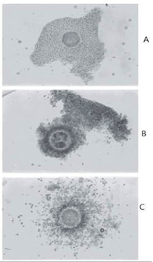
Figure 1. Different classes of COCs (de Wit et al., 2000). Class A. Oocyte is surrounded by a compact cumulus with a homogeneous cytoplasm. Class B. Oocyte is surrounded by a less compact and darker cumulus. The cytoplasm is dark and moderately granular. Class C. Oocyte is surrounded by dispersed cumulus cells. Oocyte has a granular cytoplasm. Reproduced from Lasiene et al. (2009) with permission.
An aged follicular microenvironment could highly affect oocytes and put a specific transcriptional footprint in the surrounding CCs (Al-Edani et al., 2014). New evidence shows that female age may have influence on the gene expression profiles of CCs that are crucial for oocyte quality and competence (McReynolds et al., 2012). For the first time as Al-Edani et al. (2014) showed that the angiogenic genes were notably overexpressed compared to patients of the younger groups. In contrast, the genes involved in TGF-ß signaling pathway such as AMH, TGF-ß1, inhibin, and activin receptor were under-expressed (Al-Edani et al., 2014). Miao et al. (2005), hypothesis suggests that CCs would accelerate the progression of in vitro aging of mice oocytes. Also, they assessed the effects of CCs on the susceptibility to activating stimuli, maturation promoting factor (MPF) activity, exocytosis of cortical granules (CGs), and anaphase onset during aging process of both in vivo and in vitro matured mice oocytes. They concluded that CCs accelerated the aging progression of both in vivo and in vitro-matured mice oocytes (Miao et al., 2005).
Zona Pellucida
Zona pellucida (ZP) is a glycoprotein interface among oocytes and their surrounding cells that coat and protect the mammalian ovum. In humans, it compromises four glycoproteins-hZP1 to hZP4 (Lefièvre et al., 2004). ZP2 and ZP3 combine to form basic units that polymerize into long filaments. These filaments are periodically linked by cross-bridges of ZP1 and ZP4 molecules. But, the mice ZP is composed of three major glycoproteins: mZP1, mZP2, and mZP3 (Wassarman et al., 2005). In the mice, the acrosome reaction inductor and the sperm receptor is the glycoprotein ZP3, a component of all mammalian oocyte ZPs (Wassarman, 1990). Because of difficulties with direct observation of the human oocyte, there has been a large number of investigations exploring the molecular mechanism of fertilization in mice. It is estimated that the ZP of unfertilized mice oocyte include more than 1 billion copies of the ZP3 protein (Carlson, 2018). In mammals, there is less species alteration in ZP3composition; this may illustrate why penetration of the zona pellucida by spermatozoa of nearly related mammalian species is sometimes feasible, whilst it is rare amongst lower beasts (Carlson, 2018). The normal maturation of follicles and oocytes rely on presence of the ZP (Rawe & Combelles, 2009). The zona pellucida plays an essential role in the sperm-oocyte interaction, acrosomal reaction, sufficient block to polyspermy, preimplantation embryos and following troubles in developmental progression (Ebner et al., 2008). After penetrating the corona radiata, the spermatozoa attach firmly to the ZP via plasma membrane of the sperm head. Spermatozoa binds in a specific manner to a sialic acid molecule, which is the terminal part of a sequence of four sugars at the end of an O-linked oligosaccharides that are attached to the polypeptide core of the ZP3 molecule (Carlson, 2018). Ultrastructural study of mice ZP by conventional scanning electron microscopy (SEM) (Figures 2 and 3) shows various patterns in its outer and inner surfaces. The external surface is defined by the presence of abundant fenestrations, giving it a somewhat spongy configuration, whilst inner surface is smooth and condensed (Phillips et al., 1985). Two main forms of zona pellucida are identified as a mesh-like (spongy structure) ZP in mature oocytes and a more compact (smooth surface) one in atretic or immature oocytes (Familiari et al., 2006). Studies have revealed a direct association between the ZP surface structure and the grade of oocyte development and (Sousa et al., 2015), hence it can be a valid predictor of oocyte quality (Calafell et al., 1992). Familiari et al. (2006) postulated that the condensation of the outer side of the ZP causes sperm-binding site confusion, which would cause decreased sperm binding and reduced penetration ability, so it goes to in vitro oocyte fertilization disability. Calafell et al. (1992) reported four types of ZP in mice oocytes, aged in vivo or in vitro from immature, young, and elderly females, termed A, B, C, and D. Type-A oocytes have a structure observed in prepuberal females that may be related to immature oocytes. Type B was the usual type of the ZP between the groups reported by these authors and its rate decreases with advancing age. The A/B type ZP was specified by abundant large pores with fully cytoplasmic and matured nuclear and freshly ovulated oocytes. Type C was observed before degeneration and raise conspicuously with increasing female age (both in vivo and in vitro). This concept is suggesting that there is a direct relation among the properties of the ZP fertilization ability of the oocyte. Type D with amorphous appearance, seemingly corresponded to degenerated oocytes, because the surface was similar to that seen in fragmented eggs (Familiari et al., 2006). In routine clinical intracytoplasmic sperm injection (ICSI) trials, it is possible to recognize the ZP alterations. In spite of IVF, removal of cumulus cells enables clinicians to assess the ZP variants such as thickness, appearance, irregularities and composition (Kilani et al., 2006). We know that the thicker ZPs are related to fertilization, implantation and fecundity rate (Figure 4) (Bertrand et al., 1995; Rama Raju et al., 2007). The zona pellucida of fresh oocytes consists of granulofibrillar, interrelated reticulum with pores; whereas the zona pellucida of aged oocytes have ‘cobblestone-like’ cover, which is composed of narrow clusters of granulofibrillar material, apart from one another by an obvious space (0.3mm in width). Zona pellucida hardening (explained increased time to enzymatically digest the ZP and decreased sperm binding and penetration) evaluations have shown that the time for chymotrypsin-mediated dissolution of the zona pellucida increases notably in aged oocytes, compared with fresh oocytes (Miao et al., 2005). In the clinic, assisted hatching (AH) of the ZP is sometimes applied to help the embryo escape its ZP. Some environmental agents such as non-physiological and prolonged culture conditions can lead to irreversible chemical changes or Zona hardening. Despite such knowledge, we need to increase our understanding about age-related abnormalities in zona morphology and their possible clinical importance (De Vos & Van Steirteghem, 2000).
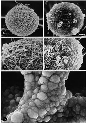
Figure 2. Conventional SEM. (a): Unfertilized human oocyte. Porousnet appearance of ZP (X 2,000). (b): Unfertilized human oocyte. Compact and smooth surfaced ZP (X 2,000). (c): Higher magnification of (a). The spongy ZP structure is evident (X 4,000). (d): Higher magnification of (b). It shows a dense and compact ZP structure (X 4,000). (e): Unfertilized mouse oocyte with very high magnification showing a branch of the spongy structure of the ZP (X 350,000). Familiari et al. (2006) with permission.

Figure 3. (a): The outer surface of the ZP of a human mature oocyte. Many fenestrations are present in which the filaments form a large meshed network (X 9,000). (b): The outer surface of the ZP of a human atretic oocyte. The filaments form a tight meshed network (X 9,000). Familiari et al. (2006) with permission.

Figure 4. An ideal oocyte which displaying good spindle retardance and length, trilaminar structure of zona with good retardance of inner layer. Magnification ×200. Reproduced from Rama Raju et al. (2007) with permission.

Figure 5. Calculation method for measuring the size of each part of the oocyte. ZP: Zona pellucida. PS: perivitelline space. PB: 1st polar body. Diameter of cytoplasm (A) (A1+A2) / 2. Inner diameter of zona pellucida (B) = (B1+B2) / 2. Outer diameter of zona pellucida (C) = (C1+C2) / 2. Thickness of zona pellucida = (C B) / 2. Size of perivitelline space = (B A) / 2. Yoshida & Niimura (2010) with permission.
Perivitelline Space
The perivitelline space (PVS) demonstrates that the acellular portion between the plasma membrane of the oocyte (oolema) and the ZP PVS contains a hyaluronan-rich extracellular matrix that is not visible under light microscopy. In a mature human oocyte, it is fluctuating in size, and evident with the expelled polar body located in its most eminent part (Figure 5). PVS is one of the most significant parts of extracytoplasmic maturation. There are three kinds of PVS classifications that are possible: immediately opposed to the plasma membrane, apart and recognizable, exaggerated (Rawe & Combelles, 2009; Talbot & Dandekar, 2003). Despite these differences, there is a clear disagreement that still remains over PVS arrangement regarding what may be typical or deviant from healthy. PVS-size is important as it affects the prevalence of the polyspermy, fertilization rate and pronuclear morphology (Yoshida & Niimura, 2010). A retrospective study shows a direct relationship between PVS width and reduction in fertilization and embryo quality, while others reported no similar relevance (Plachot et al., 2002). Ten et al. showed no PVS effect on fertilization, but oocytes with large PVS were associated with higher likelihoods of yielding good-quality embryos (Ten et al., 2007). Anyway, there is no agreement as to whether PVS evaluation alone may influence oocyte quality. In another study on hamsters by Li et al. (2017), they reported that old hamsters produced a meaningfully lower percentage of healthy oocytes with minimal PVS, compared with those in younger hamsters (Figure 6). They also gathered that a large PVS has been related to lower fertilization rates in humans (Rienzi et al., 2008). The reason for the high occurrence of polyspermy after insemination in oocytes with small perivitelline space is vague. The precise source of the PVS chemicals remain unknown, although the cumulus cells and oocytes maybe candidates. Hyaluronic acid (HA) has been shown to have an inhibitory impact on oocyte plasma membrane fusion. Therefore, it is believed that large amounts of HA in the expanded PVS may hinder the membrane fusion of sperm and oocytes, resulting in a lower rate of polyspermy in oocytes with larger PVS (Yoshida & Niimura, 2010). Finally, Yoshida & Niimura (2010), in rodents, reported that, oocytes with smaller perivitelline space show higher incidence of polyspermy after insemination, and suggested the size of PVS, whereas, evidence confirmed the presence of cortical granule material in the PVS, in addition to creation of cortical granule envelope inside the PVS. In this manner PVS can impact sperm penetration and appropriate block to polyspermy. However, Yoshida & Niimura (2010) showed that the diffusion of cortical granules in some rodents with smaller PVS is similar to those with larger PVS. They discussed the higher frequency of polyspermy in oocytes with small PVS, which is not caused by the lack of cortical granules in those oocytes. Because of the lack of knowledge, we can understand that a large PVS may break the connection between the cumulus cells and the oocyte, specifically by means of transzonal projections and gap junctions. On the other hand, it may be useful to disconnect some ways of cell communication at the suitable time, and there may be a mechanism by which PVS controls oocyte competence (Rawe & Combelles, 2009). Overall, morphological assessments of the three extracellular parts of the oocyte yields a definite measure of oocyte evaluation while not freely rendering predictors of oocyte quality. According to the intricacy in morphological diversity, we still require much research to detect and recognize these aspects, and then in determining their clinical importance. Randomized controlled experiments may then test the impact of aging on extracytoplasmic evaluation on fecundity yields.
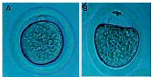
Figure 6. Young oocytes (A) expanded against the zona pellucida with a minimal perivitelline space (PVS) and a clear indentation at the polar body, whereas old oocytes (B) had wide PVS with no indentation from the polar body. Reproduced from Li et al. (2017) with permission.
INTRACYTOPLASMIC COMPONENTS
Polar Body
Oocytes from mammalian species is arrested in the prophase meiosis I (MI). At puberty, a surge in luteinizing hormone [LH] impels a preovulatory growth phase and MI is complete, resulting in the formation of two unequal size cells with 23 chromosomes. One cell receives most of the cytoplasm (secondary oocyte), and the other cell, that extruded between the ZP and the plasma membrane of the secondary oocyte in the form of a first polar body (PBI), has no function. Then the oocyte enters the metaphase meiosis II (MII) stage, almost 3 hours before ovulation (Rawe & Combelles, 2009). Eichenlaub-Ritter et al. (1995) reported that the PBI morphology revealed the postovulatory age of the oocyte. Several studies have revealed that the PB shape, size and contour can help anticipate oocyte quality, but other documents show that there is no association between PB properties and oocyte developmental ability or genetic form for the embryo (Ciotti et al., 2004; De Santis et al., 2005). In a study by De Santis et al. (2005), they suggested that PBI morphology cannot give provide embryos with high developmental competency, and they proposed alternative parameters for oocyte selection. Halvaei et al. (2011) analyzed the role of PBI morphology on rates of fertilization and embryo development in ICSI cycles. They concluded that PBI morphology does not seem to be a prognostic factor for rates of fertilization and embryo development in ICSI cycles (Figure 7) (Halvaei et al., 2011). Another study conducted by Zhou et al. (2016), on the relationship of polar body-morphology to embryo quality and pregnancy outcome at 16-18 hours after insemination. They reported that no remarkable differences in pregnancy rate (PR) or implantation rate (IR) were seen between the intact and fragmented groups of PBI. Although they reported little effects of PR or IR in fresh embryo transfer cycles, and concluded that better embryo quality can be attained in the PB-intact group, which is important for embryo selection (Zhou et al., 2016). Other studies show that oocytes with intact and good shaped PBI yield higher fertilization rates and higher embryo quality (Ebner et al., 2000). Evidence on the relationship between PBI and mother’s age show that the PBI from young individuals is intact and adjacent to the MII spindle, while aged oocytes contain degenerated and deviated PBI from the MII spindle, and space between PBI and the spindle raises with time throughout the oocyte aging (Miao et al., 2004). PBI displacement can be due to increased PVS with time and facilitated moving from its usual place (Miao et al., 2004). Polar bodies due to their amount of cytoplasm, organelles and chromatids, can yield applicable data (by cytogenetic studies like fluorescent in situ hybridization) about the genetic condition of oocytes, without possibly endangering it. Does aging-related changes cause molecular alterations in the PBI in spite of apparent morphological modification? This question was the aim of a study by Jiao et al. (2012). In a prospective mice model survey, they assessed the oocyte-specific mRNAs detection in the PB of MII oocytes and define the effects of age on oocyte-specific transcript levels. They showed that transcript levels were lower in aged PBs compared with young PBs. Finally, they concluded that there is a remarkable difference in the transcript levels of oocyte-specific genes in aged vs. young PB, that correlates with age-related reduction in oocyte capability (Jiao et al., 2012). This gene expression alteration in PB may be a possible biomarker of MII oocyte competence (Jiao et al., 2012). Therefore, the assessment of PBI could work as a strong genetic diagnostic device during pre-fertilization screening without the requirement of embryo biopsy (Gitlin et al., 2003), but some limitations should also be understood when PB analysis is taken into account (Rawe & Combelles, 2009). These restrictions include: first, the maternal genetic portion, which can be studied alone. In second, possible nondisjunction of sister chromatids during MII can happen, which requires second polar body analysis. In third, PB chromosomes are shorter, which makes the technique more difficult (Rawe & Combelles, 2009).

Figure 7. MII oocytes after denudation of cumulus cells before microinjection. (A) normal PBI and (B) fragmented PBI. Halvaei et al. (2011) with permission.
Ooplasm
Cytoplasmic and nuclear factors are important criteria in oocyte quality, which directly influence the developmental competence of embryos in ART. For this, nuclear maturity indicates meiosis continuation and the progression to metaphase II, the arrested stage at the time of ovulation. MPF can be created by cyclin B and p34cdc2, which supports the transition from G2 to M phase. Active MPF starts nuclear maturation (e.g., germinal vesicle breakdown) and condensation of metaphase I (MI) chromosomes, then a reduction in MPF causes entry into anaphase I, and a second peak in MPF activity drives the oocyte to metaphase II (Eppig, 1996). Even if oocytes get nuclear competence, they still may be lacking cytoplasmic maturation, which indicates all processes construct the oocytes for activation, fertilization, and embryo development. Completion of both kinds of maturation are highly sensitive to changes in follicular hormonal environment and/or in vitro culture status (e.g., pH, temperature, oxygen) that may lead to alterations in oocyte morphology. Some of these oocyte cytoplasmic aberrations are visible at the light-microscopic level (Hu et al., 2001). Elimination of cumulus cells with the ICSI method enables the clinician to directly evaluate the morphological attributes of oocytes under light microscopy. Ooplasmic morphological characteristics can be categorized by the presence of vacuoles, inclusion, debris, fragmented corpuscles, dark cytoplasm, and organelle clustering (Kahraman et al., 2000; Moussa et al., 2015). In fact, more than 50% of human oocytes show at least one of these morphological abnormal features in the center or in the periphery of the Ooplasm (Ebner et al., 2003). Nagano et al. (2006) in an animal model study revealed that the dark cytoplasm has an accumulation of lipids and good developmental potential of oocytes after in vitro fertilization. They found that a light-coloured cytoplasm indicated a low density of organelles and poor developmental potential. An oocyte with a dark ooplasm declines by 83% in its likelihood of yielding good-quality embryos (Ten et al., 2007). The most evident cytoplasmic characteristics that hinders developmental ability are aggregations of smooth endoplasmic reticulum (sER) and vacuoles (Figure 8). Vacuoles originate spontaneously by fusion of pre-existing vesicles derived from the sER or Golgi apparatus (Shafie et al., 2000; Van Blerkom, 1990). Few studies have been carried out on the effects of oocyte vacuoles on human fertility, but a large number of reports noted a negative influence on fertilization outcomes (Loutradis et al., 1999). Otsuki et al. (2004) for the first time evaluated the origin of smooth Endoplasmic Reticulum Clusters (ERCs) and defined their effect on embryonic development and clinical fecundity rates. They reported that pregnancy rates remain low with sERC-negative cohort oocytes in sER C-positive cycles (Otsuki et al., 2004). Another physiological consequence of women’s aging is FSH rise, that can be correlated with morphometric parameters. In a study by Santos et al. (2015) they concluded that patients with high FSH levels displayed low-quality oocytes when compared with controls. This can be suggestive of ovarian aging, which negatively affects oocyte development into viable embryos. These findings were confirmed by other studies carried out by Kdous et al. (2016). Although, Valeri et al. (2011) showed that there is no significant relationship between FSH levels and morphometric oocyte parameters, they concluded that it maybe because other factors have an effect on this relationship. Despite several studies on the relationship between oocyte morphology and embryo quality, it is clear that microscopic observation alone cannot determine fecundity pathology or its relative effect in ART, and we need more research and studies in this field.

Figure 8. (A) Mature oocyte. Tubules (SER) and vesicles (V) of smooth endoplasmic reticulum associated with mitochondria (M) to form M-SER aggregates and MV complexes. TEM, bar is 0,5 μm. (B) Early degenerating mature oocyte. Numerous vacuoles (Va) associated with lysosomes (Ly) are found in the ooplasm. TEM bar is 1,2 μm. Nottola et al. (2007) with permission.
Meiotic Spindle
The spindle is an important barrel-shaped cellular structure, primarily composed of microtubules and centrosomes, that are responsible for the precise segregation of homologous chromosomes (meiosis I) or two sets of chromatids (meiosis II) within germ cell division (Karsenti & Vernos, 2001; Wang & Sun, 2006). Failure of equal separation will cause aneuploidy, which is thought to be a source of a lot of genetic problems and aneuploid embryos (Hassold & Hunt, 2001; Wang & Sun, 2006). Some aneuploid embryos implant in the uterus undergo spontaneous abortion, while others develop to full term and bear genetic disorders (Bruyère et al., 2000; Robinson et al., 2001). Aneuploidy is indeed a main problem in getting reproductive success. It is estimated that 20% of all human oocytes are aneuploid, although this can fluctuate from 10% to as high as 40-60% (Kuliev et al., 2005; Pacchierotti et al., 2007). The meiotic spindle organization and morphology is essential for ensuring accurate chromosome separation during MI and MII. The structural features of the meiotic spindle, such as length and location, are usually used for assessing oocyte quality. In somatic cells, there is a spindle check point mechanism that controls chromosome segregation (Eichenlaub-Ritter, 2012), while in mammalian oocytes these monitoring apparatus are not exactly engaged during the meiotic procedure, which may cause chromosome mis-segregation (Jones, 2008). An intact meiotic spindle is critically significant for precise segregation of chromosomes to the dividing blastomeres, thus ensuring accurate embryo development (Miao et al., 2009). Spindle size is approximately 11.2±3.4 mm. Fresh human oocytes carry compact anastral spindles (spindles without centrosomes) placed orthogonally to the oolemma, with the pole close to the oolemma being smaller than that related to the center of the oocyte (Miao et al., 2009). The human meiotic spindle size in the first day is shorter (8.08±0.84 mm) than the spindle in fresh oocytes. In 2-day oocytes, the spindles are smaller and can be bi- or multipolar, which will have serious aftermath for chromosome segregation and, therefore, result in aneuploidies (Figure 9) (Miao et al., 2009). Microtubules of meiotic spindles in 2-day-aged oocytes radiate to cell periphery and make additional microtubule asters in the cytoplasm. Immunocytochemical of tubulin staining in aged oocyte assessments shows increased staining through the meiotic spindle, compared with that of fresh oocytes (Wang et al., 2001). This staining shape exhibits a loss of centrosome structure in the meiotic poles, which accompanies the loss of microtubule integrity (Schatten, 2008). Studies show that different patterns of spindle disruption can be seen in aged, failed-to-fertilize oocytes. This spindle disorganization includes: tetrapolar spindles, abnormal expression of the nuclear mitotic apparatus (NuMA) protein, which gives spindle stability to fresh oocytes, and alters the microtubule kinesin motor protein EG5 (Hall et al., 2007). Because of ethical issues and limitations of human oocytes for research, mice and pigs have been used for many studies with the purpose to infer data to humans (Prather, 2007). These studies indicate that microtubules in fresh mice oocytes are obviously detected in the meiotic spindle, additional small microtubule asters are detected in the cytoplasm, which are arranged by cytoplasmic centrosomes. Microtubules in aged mice oocytes become slowly misplaced from the spindle, with preferential loss in the central spindle area, close to the chromosomes. Astral fibers radiate out from the polar centrosomes into the cytoplasm, whereas the mean pole-to-pole distance becomes remarkably decreased (Miao et al., 2009). Concurrently, astral microtubules in the cytoplasm become steadily depolymerized (Segers et al., 2008). Studies revealed that the expression of the spindle checkpoint protein MAD2 (mitotic arrest deficient protein) is gradually decreased in pig and mice oocyte aging (Steuerwald et al., 2005). Increases in premature chromosome segregation is an oocyte aging-related change that causes embryo aneuploidy (Steuerwald et al., 2005). Oocyte from older women grown in suboptimal environments are prone to a poorly-controlled mechanism of chromosome segregation. Rama Raju et al. (2007) investigated the meiotic spindle and other oocyte structure characteristics using the PolScope microscope, and analyzed their relationship to the embryonic development potential. They showed that delays and the meiotic spindle length have a positive predictive value in relation to progression to blastocysts. In addition, they reported a significant change in retardance and spindle length in aged patients, and deduced that retardance reduction and spindle length is correlated with advanced age (Rama Raju et al., 2007). Eichenlaub-Ritter et al. (2002) found that human oocytes without birefringent spindles may still be at the telophase or prometaphase I stage, and as a result, have worse prognosis after IVF/ICSI. It has been indicated, that’s why meiotic spindle assessment is an important structure for evaluating oocyte quality (Moon et al., 2003; Rienzi et al., 2003).

Figure 9. Confocal micrographs of fresh and aged mice oocytes. Configuration of meiotic spindle and chromosomes in (A) Normal spindle morphology of fresh oocytes, microtubules traversing between both poles and chromosomes aligned in a compact group on the equatorial plane (B) and (c) abnormal spindle morphologies of aged oocytes. Miao et al. (2009) with permission.
Mitochondria
Mitochondrias are double membrane-bound organelles present in cytoplasm of eukaryotic cells, with a highly specialized function and morphology that generate necessary energy for different cellular purposes (Kelly & Scarpulla, 2004). They are vital for metabolism, signalling, and apoptosis (Danial & Korsmeyer, 2004). Oocyte mitochondria produce ATP via oxidative phosphorylation (OXPHOS), which renders the energy demanded from fertilization through the blastocyst stage (Dumollard et al., 2007). In response to extra and intra cellular requirements, mitochondrial function is improved by complexed mechanisms that regulate various morphologies and distributions. In most types of cells, mitochondrial injury and oxidative stress represent the principle of cellular aging (Liochev, 2013). Each mitochondrion consists of 1 to 15 mitochondrial DNA (mtDNA) molecules and developing oocyte competence is associated with a copy of mtDNA per oocyte (Wai et al., 2010). The mitochondrial genome, which contains approximately 16k base pairs (bp) and encodes core complexes for cellular respiration like ATP synthase, cytochrome c-oxidase, cytochrome b and so on (Kogo et al., 2011). Mitochondrial DNA has no histone safeguarding, antioxidant system and impression correction apparatus (Zhang et al., 2017). Thus, oxidative factors such Reactive Oxygen Species (ROS) can simply lead to mtDNA and cellular damage, when there is oxidative/antioxidant imbalance (Zhang et al., 2017). In accordance to oocyte ATP demands for energy consumption in cytoplasmic and nuclear maturation, meiotic spindle formation, the mitochondria are able to spread and concentrate in ooplasmic regions (Figure 10). In addition to this, it appears that the arrangement of mitochondrial distribution is different in the mature oocyte of various species (Yu et al., 2010). Mitochondria, which are the main factors of oocyte quality, may be directly affected throughout ovarian aging. Studies revealed that there is a relationship between the mtDNA content, copy number, maternal age and ovarian reserve indexes (Duran et al., 2011). Studies have also indicated that mtDNA copy number in human oocytes reduces with advancing maternal age (Chan et al., 2005). It is possible that mitochondrial abnormalities may also play an essential role in age-related oocyte competence and aneuploidy rates (Barritt et al., 2000). Large numbers of evidence have revealed that mtDNA levels are remarkably higher in aneuploid embryos, compared with those in chromosomally-normal embryos (Fragouli et al., 2015). In humans and rodents, ultrastructural studies of aged mitochondria (Figure 11) show morphological and functional abnormalities such as mitochondrial swelling, vacuolization and cristae alterations in comparison with young individuals (Kushnir et al., 2012; Simsek-Duran et al., 2013). Mitochondrial membrane potential (Delta ψ), as is ATP production, which indicates mitochondrial activity is another age-related change in human and mice (Ben-Meir et al., 2015). Mitochondrial ATP production requires the operation of the electron transport chain, situated on the inner mitochondrial membrane. Complexes I and II oxidize the tricarboxylic acid (TCA) cycle products and transfers the electrons to ubiquinone, also the well-known coenzyme Q (CoQ). CoQ has an important antioxidant characteristic, controls cellular redox and impacts transcriptional activity, necessary to the electron transport chain (Crane, 2001). CoQ proteins make a big mitochondrial complex and the existence of all protein elements is essential for steadiness (Wang et al., 2009). Evidence has shown that coenzyme Q10 supplementation in aged animal models (but not in younger animals), can be used to postpone the reduction of ovarian reserve, fortify the expression of mitochondrial genes in oocytes and ameliorate the mitochondrial activity. Ben-Meir et al. (2015) concluded that the reduction of mitochondrial activity produced by suboptimal CoQ10 availability plays a key role in age-related oocytes, and its lack causes infertility. Different studies on animal models have shown that injection of mitochondria or cytoplasm from young donor oocytes into an aged oocyte enhances oocyte competence, embryo quality, decreased fragmentation, and finally enabling successful implantation (May-Panloup et al., 2016). With this definition, cytoplasmic mtDNA, ATP content and mitochondrial distribution may be considered as prognostic factors of oocyte developmental competence. However, these procedures are invasive and therefore have no value as prediction implements in the clinical IVF laboratory.

Figure 10. Confocal microscopy images demonstrating mitochondria distribution during maturation of human oocytes. (A) GV oocyte displays the distribution of mitochondria in the peripheral zone. (B) GV oocyte with mitochondria diffused fairly in the cytoplasm. (C) MI oocyte with mitochondria displaying a peripheral type of distribution. (D) In vitro matured oocyte indicating the semiperipheral distribution of mitochondria in the oocyte. (E) In vitro matured oocyte showing the distribution of mitochondria throughout the cytoplasm. (F) In vivo matured oocyte: mitochondria are disseminated throughout the cytoplasm, and there are mitochondria in the central region. Scale bar represents 50 μm. Liu et al. (2010) with permission.
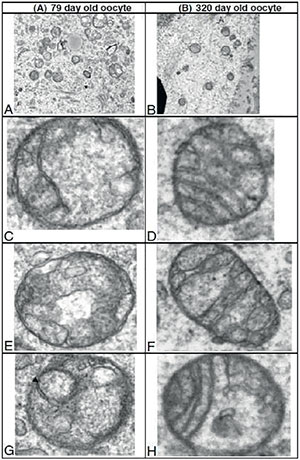
Figure 11. TEM images of mitochondria from young and old mice oocytes. MII oocytes from hyperstimulated young and old mice were evaluated and the mitochondrial structures were compared. Representative electron micrographs of ooplasm at 11,000x magnification are shown; higher magnification views of individual mitochondria are also presented. (A) abundant mitochondria per field, also notice different size mitochondria compared to B. (B) relatively few mitochondria of uniform size per field in ooplasm of an aged animal. (C & E) undifferentiated round mitochondria with an electron dense matrix vs D, F & H. More differentiated mitochondria with an elongated shape and distinct cristae G. arrow indicating vacuoles within mitochondria, Kushnir et al. (2012) with permission.
EPIGENETICS
Waddington (1942) was the first who presented the word of epigenetics in the early 1940s. He explained epigenetics as ‘‘the branch of biology which studies the causal interactions among genes and their products, which cause the phenotype into being (Waddington, 1968). In recent years, with the rapid growth of genetics, the meaning of this term has gradually changed. Epigenetics defined the process that regulates gene function and does not affect DNA sequence, and is heritable through cell division (Baird et al., 2005). Gene activation in the zygote and early embryonic development are controlled by both genetic and epigenetic mechanisms. Epigenetic mechanisms are not DNA sequence-based, while the genetic mechanism relies on DNA sequence and codes. Epigenetic mechanisms establish inheritable alterations that play an important role in regulating gene expression (Lucifero et al., 2004). The main epigenetic alterations include DNA methylation, modification of histones and chromatin remodelling; they are intimately related and operated at the transcriptional level. Epigenetic reprogramming takes place at gametogenesis and the accurate establishment of epigenetic modifications are crucial for normal embryo growth and viability (Baird et al., 2005). The DNA methylation process is mostly catalysed by DNA methyltransferase 3s (DMT3s) (Tomizawa et al., 2012). Studies imply that the epigenetic modifications in oocytes may be influenced by advanced maternal age, owing to the expression of DNMTs and histone acetyltransferases that varies with aging (Hamatani et al., 2004). In women older than 38 years, the expression pattern of TAP73 in oocytes is lower, and down regulated when compared with oocytes from younger women (Guglielmino et al., 2011). However, there is still no clear evidence that the DNA methylation in human oocytes is influenced by aging. Immunocytochemical studies report histone deacetylation at the MI and MII stages by histone deacetylase (HDAC) activity in mammalian oocytes. Akiyama et al. (2004) revealed that if meiotic histone deacetylation is restrained, aneuploidy raised in fertilized mice oocytes, and this culminated in embryonic death in the uterus at an early stage of development. Hamatani et al. (2004) showed that HDAC is down-regulated at transcript level in aging mice oocytes older than 42 weeks, while histone remains acetylated in young female mice. Evidence revealed the relationship between histone acetylation and maternal age. The results show advanced maternal age negatively affects the deacetylation of some histone proteins (H4K12) in human MII oocytes (Steuerwald et al., 2007). Studies indicate that alterations in enzymes comprising methyltransferases (DNMTs) and demethylases (TETs), are possibly the direct cause of epigenetic changes in aged oocytes. However, whether/how aging impacts the modifications in their expression requires more explanation (Ge et al., 2015). Investigations recommend that age-related epigenetic changes on oocyte might be inhibited by diets, medicine or other methods.
NEW INSIGHTS IN OVARIAN STEM CELLS
Recent evidence indicate that components of adult mammalian ovaries in primary follicles; that is, primitive granulosa and germ cells may differentiate from mesenchymal progenitor cells (MPC) resting in the ovarian tunica albuginea (Bukovsky, 2015). Jonathan Tilly’s team for the first time explained stem cells in adult mice ovary (Johnson et al., 2004). Their results challenged old central dogmas that ovaries have a finite number of follicles that get reduced with age, resulting in menopause. Ovarian stem cells have opened a new landscape for a further understanding of human oogenesis, infertility preservation, and therapy. Recent findings reported Ovarian Stem cells (OSCs) isolated from rabbits, marmosets, sheep, and human ovaries. Researchers characterized two distinct populations of stem cells based on their size, which comprises quiescent Very Small Embryonic-Like (VSEL) stem cells that expresses nuclear OCT-4 and slightly bigger cells expressing cytoplasmic OCT-4 (Bhartiya et al., 2019; Kumar et al., 2009; Sriraman et al., 2015). Evidence shows that ovarian stem cells on culture medium developed spontaneously to oocyte-like cells (OLCs) and expresses oocyte and germ cell specific markers (Kumar et al., 2009). Our recent findings showed that ovarian derived SCs could differentiate to OLCs in BMP15 conditioned medium, and expresses ZP genes and proteins, which confirm earlier reports (Taheri Moghadam et al., 2021). Increasing our knowledge about ovarian stem cells and their differentiation into OLCs can help us better understand this phenomenon and retard the aging process.
CONCLUSION(S) AND PERSPECTIVES
The main role of oocyte competence and aging in relation to an embryo’s development has encouraged wide research for reliable predictors of oocyte quality. A series of age-related molecular, cellular and morphological alterations take place during the process of oocyte aging and fertilization. These changes not only influence pre- and post-implantation of embryo development quality but also the later life of the offspring. Essays to describe morphological features related to oocyte quality and aging have attained little results. A number of chemicals and physical facilities have been used to characterize a good oocyte quality and postpone the oocyte aging process, to provide hopeful chances for intervention in the aging phenomenon. These specific steps are essential for ART procedures to elevate the rate of successful delivery, which leads to optimal production of healthy babies. With modern ART, the successful rate of this technology is affected by oocyte aging; therefore, control of oocyte aging would provide an important advantage in allowing adequate manipulation and selection of high quality oocytes. Hence, creating methods to control aging might increase progress in assisted reproduction technologies. Because in the lack of a good oocyte grading plan, the ability of morphological monitoring to aid oocyte/embryo selection is reduced.
In this review, we summarized the oocyte quality, and mechanisms of oocyte aging, and delay or reversibility of the aging process. Then, we showed briefly the importance of procedures to control oocyte quality and age-related changes in oocyte competence. There are still many unanswered questions, but several paths have now been paved unto much awaited advances and the detection of oocytes with compromised quality.
ACKNOWLEDGMENTS
The authors thank Mrs. Elina Asl Sharhani for English language revision.
REFERENCES
Absalan F, Saremy S, Mansori E, Taheri Moghadam M, Eftekhari Moghadam AR, Ghanavati R. Effects of Mono-(2-Ethylhexyl) Phthalate and Di-(2-Ethylhexyl) Phthalate Administrations on Oocyte Meiotic Maturation, Apoptosis and Gene Quantification in Mouse Model. Cell J. 2017;18:503-13. PMID: 28042535 DOI: 10.22074/cellj.2016.4717
Medline Crossref
Akiyama T, Kim JM, Nagata M, Aoki F. Regulation of histone acetylation during meiotic maturation in mouse oocytes. Mol Reprod Dev. 2004;69:222-7. PMID: 15293224 DOI: 10.1002/mrd.20121
Medline Crossref
Al-Edani T, Assou S, Ferrières A, Bringer Deutsch S, Gala A, Lecellier CH, Aït-Ahmed O, Hamamah S. Female aging alters expression of human cumulus cells genes that are essential for oocyte quality. Biomed Res Int. 2014;2014:964614. PMID: 25276836 DOI: 10.1155/2014/964614
Medline Crossref
Baird DT, Collins J, Egozcue J, Evers LH, Gianaroli L, Leridon H, Sunde A, Templeton A, Van Steirteghem A, Cohen J, Crosignani PG, Devroey P, Diedrich K, Fauser BC, Fraser L, Glasier A, Liebaers I, Mautone G, Penney G, Tarlatzis B; ESHRE Capri Workshop Group. Fertility and ageing. Hum Reprod Update. 2005;11:261-76. PMID: 15831503 DOI: 10.1093/humupd/dmi006
Medline Crossref
Balasch J. Ageing and infertility: an overview. Gynecol Endocrinol. 2010;26:855-60. PMID: 20642380 DOI: 10.3109/09513590.2010.501889
Medline Crossref
Barritt JA, Cohen J, Brenner CA. Mitochondrial DNA point mutation in human oocytes is associated with maternal age. Reprod Biomed Online. 2000;1:96-100. PMID: 12804188 DOI: 10.1016/S1472-6483(10)61946-3
Medline Crossref
Ben-Meir A, Burstein E, Borrego-Alvarez A, Chong J, Wong E, Yavorska T, Naranian T, Chi M, Wang Y, Bentov Y, Alexis J, Meriano J, Sung HK, Gasser DL, Moley KH, Hekimi S, Casper RF, Jurisicova A. Coenzyme Q10 restores oocyte mitochondrial function and fertility during reproductive aging. Aging Cell. 2015;14:887-95. PMID: 26111777 DOI: 10.1111/acel.12368
Medline Crossref
Bertrand E, Van den Bergh M, Englert Y. Does zona pellucida thickness influence the fertilization rate? Hum Reprod. 1995;10:1189-93. PMID: 7657763 DOI: 10.1093/oxfordjournals.humrep.a136116
Medline Crossref
Boni R, Cuomo A, Tosti E. Developmental potential in bovine oocytes is related to cumulus-oocyte complex grade, calcium current activity, and calcium stores. Biol Reprod. 2002;66:836-42. PMID: 11870093 DOI: 10.1095/biolreprod66.3.836
Medline Crossref
Broer SL, Broekmans FJ, Laven JS, Fauser BC. Anti-Müllerian hormone: ovarian reserve testing and its potential clinical implications. Hum Reprod Update. 2014;20:688-701. PMID: 24821925 DOI: 10.1093/humupd/dmu020
Medline Crossref
Bruyère H, Rupps R, Kuchinka BD, Friedman JM, Robinson WP. Recurrent trisomy 21 in a couple with a child presenting trisomy 21 mosaicism and maternal uniparental disomy for chromosome 21 in the euploid cell line. Am J Med Genet. 2000;94:35-41. PMID: 10982480 DOI:10.1002/1096-8628(20000904)94:1<35::AID-AJMG8>3.0.CO;2-9
Medline Crossref
Bukovsky A. Ovarian stem cell niche and follicular renewal in mammals. Anat Rec (Hoboken). 2011;294:1284-306. PMID: 21714105 DOI: 10.1002/ar.21422
Medline Crossref
Bukovsky A. Novel methods of treating ovarian infertility in older and POF women, testicular infertility, and other human functional diseases. Reprod Biol Endocrinol. 2015;13:10. PMID: 25889983 DOI: 10.1186/s12958-015-0001-8
Medline Crossref
Calafell JM, Nogués C, Ponsà M, Santaló J, Egozcue J. Zona pellucida surface of immature and in vitro matured mouse oocytes: analysis by scanning electron microscopy. J Assist Reprod Genet. 1992;9:365-72. PMID: 1472816 DOI: 10.1007/BF01203961
Medline Crossref
Carlson BM. Human Embryology and Developmental Biology. E-book. Elsevier Health Sciences; 2018.
Chan CC, Liu VW, Lau EY, Yeung WS, Ng EH, Ho PC. Mitochondrial DNA content and 4977 bp deletion in unfertilized oocytes. Mol Hum Reprod. 2005;11:843-6. PMID: 16421213 DOI: 10.1093/molehr/gah243
Medline Crossref
Cil AP, Turkgeldi L, Seli E. Oocyte Cryopreservation as a Preventive Measure for Age-Related Fertility Loss. Semin Reprod Med. 2015;33:429-35. PMID: 26562287 DOI: 10.1055/s-0035-1567819
Medline Crossref
Ciotti PM, Notarangelo L, Morselli-Labate AM, Felletti V, Porcu E, Venturoli S. First polar body morphology before ICSI is not related to embryo quality or pregnancy rate. Hum Reprod. 2004;19:2334-9. PMID: 15347596 DOI: 10.1093/humrep/deh433
Medline Crossref
Craig J, Orisaka M, Wang H, Orisaka S, Thompson W, Zhu C, Kotsuji F, Tsang BK. Gonadotropin and intra-ovarian signals regulating follicle development and atresia: the delicate balance between life and death. Front Biosci. 2007;12:3628-39. PMID: 17485326 DOI: 10.2741/2339
Medline Crossref
Crane FL. Biochemical functions of coenzyme Q10. J Am Coll Nutr. 2001;20:591-8. PMID: 11771674 DOI: 10.1080/07315724.2001.10719063
Medline Crossref
Crawford NM, Steiner AZ. Age-related infertility. Obstet Gynecol Clin North Am. 2015;42:15-25. PMID: 25681837 DOI: 10.1016/j.ogc.2014.09.005
Medline Crossref
Danial NN, Korsmeyer SJ. Cell death: critical control points. Cell. 2004;116:205-19. PMID: 14744432 DOI: 10.1016/S0092-8674(04)00046-7
Medline Crossref
De Santis L, Cino I, Rabellotti E, Calzi F, Persico P, Borini A, Coticchio G. Polar body morphology and spindle imaging as predictors of oocyte quality. Reprod Biomed Online. 2005;11:36-42. PMID: 16102284 DOI: 10.1016/S1472-6483(10)61296-5
Medline Crossref
de Souza GB, Costa J, da Cunha EV, Passos J, Ribeiro RP, Saraiva M, van den Hurk R, Silva J. Bovine ovarian stem cells differentiate into germ cells and oocyte-like structures after culture in vitro. Reprod Domest Anim. 2017;52:243-50. PMID: 27925309 DOI: 10.1111/rda.12886
Medline Crossref
De Vos A, Van Steirteghem A. Zona hardening, zona drilling and assisted hatching: new achievements in assisted reproduction. Cells Tissues Organs. 2000;166:220-7. PMID: 10729729 DOI: 10.1159/000016734
Medline Crossref
de Wit AA, Wurth YA, Kruip TA. Effect of ovarian phase and follicle quality on morphology and developmental capacity of the bovine cumulus-oocyte complex. J Anim Sci. 2000;78:1277-83. PMID: 10834583 DOI: 10.2527/2000.7851277x
Medline Crossref
Dumollard R, Duchen M, Carroll J. The role of mitochondrial function in the oocyte and embryo. Curr Top Dev Biol. 2007;77:21-49. PMID: 17222699 DOI: 10.1016/S0070-2153(06)77002-8
Medline Crossref
Duran HE, Simsek-Duran F, Oehninger SC, Jones HW Jr, Castora FJ. The association of reproductive senescence with mitochondrial quantity, function, and DNA integrity in human oocytes at different stages of maturation. Fertil Steril. 2011;96:384-8. PMID: 21683351 DOI: 10.1016/j.fertnstert.2011.05.056
Medline Crossref
Ebner T, Yaman C, Moser M, Sommergruber M, Feichtinger O, Tews G. Prognostic value of first polar body morphology on fertilization rate and embryo quality in intracytoplasmic sperm injection. Hum Reprod . 2000;15:427-30. PMID: 10655316 DOI: 10.1093/humrep/15.2.427
Medline Crossref
Ebner T, Moser M, Sommergruber M, Tews G. Selection based on morphological assessment of oocytes and embryos at different stages of preimplantation development: a review. Hum Reprod Update. 2003;9:251-62. PMID: 12859046 DOI: 10.1093/humupd/dmg021
Medline Crossref
Ebner T, Shebl O, Moser M, Sommergruber M, Tews G. Developmental fate of ovoid oocytes. Hum Reprod. 2008;23:62-6. PMID: 17977865 DOI: 10.1093/humrep/dem280
Medline Crossref
Eftekhari Moghadam AR, Saki G, Hemadi M, Mazaheri Z, Khodadadi A. Effect of dehydroepiandrosterone on meiotic spindle structure and oocyte quality in mice. Iran J Basic Med Sci. 2018;21:1020-5. PMID: 30524675 DOI: 10.22038/IJBMS.2018.27111.6629
Medline Crossref
Eichenlaub-Ritter U. Oocyte ageing and its cellular basis. Int J Dev Biol. 2012;56:841-52. PMID: 23417406 DOI: 10.1387/ijdb.120141ue
Medline Crossref
Eichenlaub-Ritter U, Schmiady H, Kentenich H, Soewarto D. Recurrent failure in polar body formation and premature chromosome condensation in oocytes from a human patient: indicators of asynchrony in nuclear and cytoplasmic maturation. Hum Reprod. 1995;10:2343-9. PMID: 8530664 DOI: 10.1093/oxfordjournals.humrep.a136297
Medline Crossref
Eichenlaub-Ritter U, Shen Y, Tinneberg HR. Manipulation of the oocyte: possible damage to the spindle apparatus. Reprod Biomed Online. 2002;5:117-24. PMID: 12419035 DOI: 10.1016/S1472-6483(10)61613-6
Medline Crossref
Eppig JJ. Coordination of nuclear and cytoplasmic oocyte maturation in eutherian mammals. Reprod Fertil Dev. 1996;8:485-9. PMID: 8870074 DOI: 10.1071/RD9960485
Medline Crossref
Faddy MJ. Follicle dynamics during ovarian ageing. Mol Cell Endocrinol. 2000;163:43-8. PMID: 10963872 DOI: 10.1016/S0303-7207(99)00238-5
Medline Crossref
Faddy MJ, Gosden RG, Gougeon A, Richardson SJ, Nelson JF. Accelerated disappearance of ovarian follicles in mid-life: implications for forecasting menopause. Hum Reprod. 1992;7:1342-6. PMID: 1291557 DOI: 10.1093/oxfordjournals.humrep.a137570
Medline Crossref
Familiari G, Relucenti M, Heyn R, Micara G, Correr S. Three-dimensional structure of the zona pellucida at ovulation. Microsc Res Tech. 2006;69:415-26. PMID: 16703610 DOI: 10.1002/jemt.20301
Medline Crossref
Fanchin R, Mendez Lozano DH, Frydman N, Gougeon A, di Clemente N, Frydman R, Taieb J. Anti-Müllerian hormone concentrations in the follicular fluid of the preovulatory follicle are predictive of the implantation potential of the ensuing embryo obtained by in vitro fertilization. J Clin Endocrinol Metab. 2007;92:1796-802. PMID: 17327387 DOI: 10.1210/jc.2006-1053
Medline Crossref
Fragouli E, Spath K, Alfarawati S, Kaper F, Craig A, Michel CE, Kokocinski F, Cohen J, Munne S, Wells D. Altered levels of mitochondrial DNA are associated with female age, aneuploidy, and provide an independent measure of embryonic implantation potential. PLoS Genet. 2015;11:e1005241. PMID: 26039092 DOI: 10.1371/journal.pgen.1005241
Medline Crossref
Ge ZJ, Schatten H, Zhang CL, Sun QY. Oocyte ageing and epigenetics. Reproduction. 2015;149:R103-14. PMID: 25391845 DOI: 10.1530/REP-14-0242
Medline Crossref
Gilchrist RB, Ritter LJ, Armstrong DT. Oocyte-somatic cell interactions during follicle development in mammals. Anim Reprod Sci. 2004;82-83:431-46. PMID: 15271471 DOI: 10.1016/j.anireprosci.2004.05.017
Medline Crossref
Gilchrist RB, Lane M, Thompson JG. Oocyte-secreted factors: regulators of cumulus cell function and oocyte quality. Hum Reprod Update. 2008;14:159-77. PMID: 18175787 DOI: 10.1093/humupd/dmm040
Medline Crossref
Gitlin SA, Gibbons WE, Gosden RG. Oocyte biology and genetics revelations from polar bodies. Reprod Biomed Online. 2003;6:403-9. PMID: 12831582 DOI: 10.1016/S1472-6483(10)62158-X
Medline Crossref
Guglielmino MR, Santonocito M, Vento M, Ragusa M, Barbagallo D, Borzì P, Casciano I, Banelli B, Barbieri O, Astigiano S, Scollo P, Romani M, Purrello M, Di Pietro C. TAp73 is downregulated in oocytes from women of advanced reproductive age. Cell Cycle. 2011;10:3253-6. PMID: 21946516 DOI: 10.4161/cc.10.19.17585
Medline Crossref
Hall VJ, Compton D, Stojkovic P, Nesbitt M, Herbert M, Murdoch A, Stojkovic M. Developmental competence of human in vitro aged oocytes as host cells for nuclear transfer. Hum Reprod. 2007;22:52-62. PMID: 16957049 DOI: 10.1093/humrep/del345
Medline Crossref
Halvaei I, Khalili MA, Soleimani M, Razi MH. Evaluating the Role of First Polar Body Morphology on Rates of Fertilization and Embryo Development in ICSI Cycles. Int J Fertil Steril. 2011;5:110-5. PMID: 24963368
Medline
Hamatani T, Falco G, Carter MG, Akutsu H, Stagg CA, Sharov AA, Dudekula DB, VanBuren V, Ko MS. Age-associated alteration of gene expression patterns in mouse oocytes. Hum Mol Genet. 2004;13:2263-78. PMID: 15317747 DOI: 10.1093/hmg/ddh241
Medline Crossref
Hamet P, Tremblay J. Genes of aging. Metabolism. 2003;52:5-9. PMID: 14577056 DOI: 10.1016/S0026-0495(03)00294-4
Medline Crossref
Harman D. Free radical theory of aging: an update: increasing the functional life span. Ann N Y Acad Sci. 2006;1067:10-21. PMID: 16803965 DOI: 10.1196/annals.1354.003
Medline Crossref
Hassold T, Hunt P. To err (meiotically) is human: the genesis of human aneuploidy. Nat Rev Genet. 2001;2:280-91. PMID: 11283700 DOI: 10.1038/35066065
Medline Crossref
Hemadi M, Abolhassani F, Akbari M, Sobhani A, Pasbakhsh P, Ahrlund-Richter L, Modaresi MH, Salehnia M. Melatonin promotes the cumulus-oocyte complexes quality of vitrified-thawed murine ovaries; with increased mean number of follicles survival and ovary size following heterotopic transplantation. Eur J Pharmacol. 2009;618:84-90. PMID: 19622351 DOI: 10.1016/j.ejphar.2009.07.018
Medline Crossref
Hu Y, Betzendahl I, Cortvrindt R, Smitz J, Eichenlaub-Ritter U. Effects of low O2 and ageing on spindles and chromosomes in mouse oocytes from pre-antral follicle culture. Hum Reprod. 2001;16:737-48. PMID: 11278227 DOI: 10.1093/humrep/16.4.737
Medline Crossref
Jiao ZX, Xu M, Woodruff TK. Age-associated alteration of oocyte-specific gene expression in polar bodies: potential markers of oocyte competence. Fertil Steril. 2012;98:480-6. PMID: 22633262 DOI: 10.1016/j.fertnstert.2012.04.035
Medline Crossref
Johnson J, Canning J, Kaneko T, Pru JK, Tilly JL. Germline stem cells and follicular renewal in the postnatal mammalian ovary. Nature. 2004;428:145-50. PMID: 15014492 DOI: 10.1038/nature02316
Medline Crossref
Jones KT. Meiosis in oocytes: predisposition to aneuploidy and its increased incidence with age. Hum Reprod Update. 2008;14:143-58. PMID: 18084010 DOI: 10.1093/humupd/dmm043
Medline Crossref
Kahraman S, Yakin K, Dönmez E, Samli H, Bahçe M, Cengiz G, Sertyel S, Samli M, Imirzalioğlu N. Relationship between granular cytoplasm of oocytes and pregnancy outcome following intracytoplasmic sperm injection. Hum Reprod. 2000;15:2390-3. PMID: 11056140 DOI: 10.1093/humrep/15.11.2390
Medline Crossref
Karsenti E, Vernos I. The mitotic spindle: a self-made machine. Science. 2001;294:543-7. PMID: 11641489 DOI: 10.1126/science.1063488
Medline Crossref
Kdous M, Merdassi G, Zhioua F, Elloumi H, Kacem K, Zhioua A. Basal follicle stimulating hormone level correlated to age is a good prognostic criterion for the outcome of intracytoplasmic sperm microinjection. Tunis Med. 2016;94:181-5. PMID: 27575500
Medline
Kelly DP, Scarpulla RC. Transcriptional regulatory circuits controlling mitochondrial biogenesis and function. Genes Dev. 2004;18:357-68. PMID: 15004004 DOI: 10.1101/gad.1177604
Medline Crossref
Kilani SS, Cooke S, Kan AK, Chapman MG. Do age and extended culture affect the architecture of the zona pellucida of human oocytes and embryos? Zygote. 2006;14:39-44. PMID: 16700974 DOI: 10.1017/S0967199406003625
Medline Crossref
Knight PG, Glister C. TGF-beta superfamily members and ovarian follicle development. Reproduction. 2006;132:191-206. PMID: 16885529 DOI: 10.1530/rep.1.01074
Medline Crossref
Kogo N, Tazaki A, Kashino Y, Morichika K, Orii H, Mochii M, Watanabe K. Germ-line mitochondria exhibit suppressed respiratory activity to support their accurate transmission to the next generation. Dev Biol. 2011;349:462-9. PMID: 21112323 DOI: 10.1016/j.ydbio.2010.11.021
Medline Crossref
Kuliev A, Cieslak J, Verlinsky Y. Frequency and distribution of chromosome abnormalities in human oocytes. Cytogenet Genome Res. 2005;111:193-8. PMID: 16192694 DOI: 10.1159/000086889
Medline Crossref
Kumar N, Hinduja I, Nagvenkar P, Pillai L, Zaveri K, Mukadam L, Telang J, Desai S, Mangoli V, Mangoli R, Padgaonkar S, Kaur G, Puri C, Bhartiya D. Derivation and characterization of two genetically unique human embryonic stem cell lines on in-house-derived human feeders. Stem Cells Dev. 2009;18:435-45. PMID: 18699724 DOI: 10.1089/scd.2008.0234
Medline Crossref
Kushnir VA, Ludaway T, Russ RB, Fields EJ, Koczor C, Lewis W. Reproductive aging is associated with decreased mitochondrial abundance and altered structure in murine oocytes. J Assist Reprod Genet. 2012;29:637-42. PMID: 22527902 DOI: 10.1007/s10815-012-9771-5
Medline Crossref
Lasiene K, Vitkus A, Valanciūte A, Lasys V. Morphological criteria of oocyte quality. Medicina (Kaunas). 2009;45:509-15. PMID: 19667744 DOI: 10.3390/medicina45070067
Medline Crossref
Lefièvre L, Conner SJ, Salpekar A, Olufowobi O, Ashton P, Pavlovic B, Lenton W, Afnan M, Brewis IA, Monk M, Hughes DC, Barratt CL. Four zona pellucida glycoproteins are expressed in the human. Hum Reprod. 2004;19:1580-6. PMID: 15142998 DOI: 10.1093/humrep/deh301
Medline Crossref
Li F, Castora FJ, Ford W, Alarid K, Jones HW, Swanson RJ. Reproductive competency and mitochondrial variation in aged Syrian hamster oocytes. Reprod Fertil Dev. 2017;29:1384-91. PMID: 27327865 DOI: 10.1071/RD15404
Medline Crossref
Liochev SI. Reactive oxygen species and the free radical theory of aging. Free Radic Biol Med. 2013;60:1-4. PMID: 23434764 DOI: 10.1016/j.freeradbiomed.2013.02.011
Medline Crossref
Liu S, Li Y, Gao X, Yan JH, Chen ZJ. Changes in the distribution of mitochondria before and after in vitro maturation of human oocytes and the effect of in vitro maturation on mitochondria distribution. Fertil Steril. 2010;93:1550-5. PMID: 19423101 DOI: 10.1016/j.fertnstert.2009.03.050
Medline Crossref
Loutradis D, Drakakis P, Kallianidis K, Milingos S, Dendrinos S, Michalas S. Oocyte morphology correlates with embryo quality and pregnancy rate after intracytoplasmic sperm injection. Fertil Steril. 1999;72:240-4. PMID: 10438988 DOI: 10.1016/S0015-0282(99)00233-2
Medline Crossref
Lucifero D, Chaillet JR, Trasler JM. Potential significance of genomic imprinting defects for reproduction and assisted reproductive technology. Hum Reprod Update. 2004;10:3-18. PMID: 15005460 DOI: 10.1093/humupd/dmh002
Medline Crossref
May-Panloup P, Boucret L, Chao de la Barca JM, Desquiret-Dumas V, Ferré-L’Hotellier V, Morinière C, Descamps P, Procaccio V, Reynier P. Ovarian ageing: the role of mitochondria in oocytes and follicles. Hum Reprod Update. 2016;22:725-43. PMID: 27562289 DOI: 10.1093/humupd/dmw028
Medline Crossref
Mayes MA, Sirard MA. The influence of cumulus-oocyte complex morphology and meiotic inhibitors on the kinetics of nuclear maturation in cattle. Theriogenology. 2001;55:911-22. PMID: 11291914 DOI: 10.1016/S0093-691X(01)00453-8
Medline Crossref
McReynolds S, Dzieciatkowska M, McCallie BR, Mitchell SD, Stevens J, Hansen K, Schoolcraft WB, Katz-Jaffe MG. Impact of maternal aging on the molecular signature of human cumulus cells. Fertil Steril. 2012;98:1574-80.e5. PMID: 22968048 DOI: 10.1016/j.fertnstert.2012.08.012
Medline Crossref
McTavish KJ, Jimenez M, Walters KA, Spaliviero J, Groome NP, Themmen AP, Visser JA, Handelsman DJ, Allan CM. Rising follicle-stimulating hormone levels with age accelerate female reproductive failure. Endocrinology. 2007;148:4432-9. PMID: 17540727 DOI: 10.1210/en.2007-0046
Medline Crossref
Miao Y, Ma S, Liu X, Miao D, Chang Z, Luo M, Tan J. Fate of the first polar bodies in mouse oocytes. Mol Reprod Dev. 2004;69:66-76. PMID: 15278906 DOI: 10.1002/mrd.20148
Medline Crossref
Miao YL, Liu XY, Qiao TW, Miao DQ, Luo MJ, Tan JH. Cumulus cells accelerate aging of mouse oocytes. Biol Reprod. 2005;73:1025-31. PMID: 15987819 DOI: 10.1095/biolreprod.105.043703
Medline Crossref
Miao YL, Kikuchi K, Sun QY, Schatten H. Oocyte aging: cellular and molecular changes, developmental potential and reversal possibility. Hum Reprod Update. 2009;15:573-85. PMID: 19429634 DOI: 10.1093/humupd/dmp014
Medline Crossref
Moon JH, Hyun CS, Lee SW, Son WY, Yoon SH, Lim JH. Visualization of the metaphase II meiotic spindle in living human oocytes using the Polscope enables the prediction of embryonic developmental competence after ICSI. Hum Reprod. 2003;18:817-20. PMID: 12660277 DOI: 10.1093/humrep/deg165
Medline Crossref
Moussa M, Shu J, Zhang XH, Zeng F. Maternal control of oocyte quality in cattle “a review”. Anim Reprod Sci. 2015;155:11-27. PMID: 25726438 DOI: 10.1016/j.anireprosci.2015.01.011
Medline Crossref
Nagano M, Katagiri S, Takahashi Y. Relationship between bovine oocyte morphology and in vitro developmental potential. Zygote. 2006;14:53-61. PMID: 16700976 DOI: 10.1017/S0967199406003510
Medline Crossref
Nottola SA, Macchiarelli G, Coticchio G, Bianchi S, Cecconi S, De Santis L, Scaravelli G, Flamigni C, Borini A. Ultrastructure of human mature oocytes after slow cooling cryopreservation using different sucrose concentrations. Hum Reprod. 2007;22:1123-33. PMID: 17158818 DOI: 10.1093/humrep/del463
Medline Crossref
Orisaka M, Tajima K, Tsang BK, Kotsuji F. Oocyte-granulosa-theca cell interactions during preantral follicular development. J Ovarian Res. 2009;2:9. PMID: 19589134 DOI: 10.1186/1757-2215-2-9
Medline Crossref
Otsuki J, Okada A, Morimoto K, Nagai Y, Kubo H. The relationship between pregnancy outcome and smooth endoplasmic reticulum clusters in MII human oocytes. Hum Reprod. 2004;19:1591-7. PMID: 15180981 DOI: 10.1093/humrep/deh258
Medline Crossref
Pacchierotti F, Adler ID, Eichenlaub-Ritter U, Mailhes JB. Gender effects on the incidence of aneuploidy in mammalian germ cells. Environ Res. 2007;104:46-69. PMID: 17292877 DOI: 10.1016/j.envres.2006.12.001
Medline Crossref
Pangas SA. Growth factors in ovarian development. Semin Reprod Med. 2007;25:225-34. PMID: 17594603 DOI: 10.1055/s-2007-980216
Medline Crossref
Pepling ME. From primordial germ cell to primordial follicle: mammalian female germ cell development. Genesis. 2006;44:622-32. PMID: 17146778 DOI: 10.1002/dvg.20258
Medline Crossref
Phillips DM, Shalgi R, Dekel N. Mammalian fertilization as seen with the scanning electron microscope. Am J Anat. 1985;174:357-72. PMID: 4072947 DOI: 10.1002/aja.1001740314
Medline Crossref
Plachot M, Selva J, Wolf JP, Bastit P, de Mouzon J. Consequences of oocyte dysmorphy on the fertilization rate and embryo development after intracytoplasmic sperm injection. A prospective multicenter study. Gynecol Obstet Fertil. 2002;30:772-9. PMID: 12484343 DOI: 10.1016/S1297-9589(02)00437-X
Medline Crossref
Prather RS. Targeted genetic modification: xenotransplantation and beyond. Cloning Stem Cells. 2007;9:17-20. PMID: 17386008 DOI: 10.1089/clo.2006.0085
Medline Crossref
Rama Raju GA, Prakash GJ, Krishna KM, Madan K. Meiotic spindle and zona pellucida characteristics as predictors of embryonic development: a preliminary study using PolScope imaging. Reprod Biomed Online. 2007;14:166-74. PMID: 17298718 DOI: 10.1016/S1472-6483(10)60784-5
Medline Crossref
Richardson SJ, Nelson JF. Follicular depletion during the menopausal transition. Ann N Y Acad Sci. 1990;592:13-20; discussion 44-51. PMID: 2197939 DOI: 10.1111/j.1749-6632.1990.tb30312.x
Medline Crossref
Rienzi L, Ubaldi F, Martinez F, Iacobelli M, Minasi MG, Ferrero S, Tesarik J, Greco E. Relationship between meiotic spindle location with regard to the polar body position and oocyte developmental potential after ICSI. Hum Reprod. 2003;18:1289-93. PMID: 12773461 DOI: 10.1093/humrep/deg274
Medline Crossref
Rienzi L, Ubaldi FM, Iacobelli M, Minasi MG, Romano S, Ferrero S, Sapienza F, Baroni E, Litwicka K, Greco E. Significance of metaphase II human oocyte morphology on ICSI outcome. Fertil Steril. 2008;90:1692-700. PMID: 18249393 DOI: 10.1016/j.fertnstert.2007.09.024
Medline Crossref
Robinson WP, McFadden DE, Stephenson MD. The origin of abnormalities in recurrent aneuploidy/polyploidy. Am J Hum Genet. 2001;69:1245-54. PMID: 11675616 DOI: 10.1086/324468
Medline Crossref
Sadler TW, ed. Langman’s medical embryology. Philadelphia: Lippincott Williams & Wilkins; 2011.
Santos M, Cordts EB, Bianco B, Barbosa CP, Christofolini DM. Oocyte Quality in Patients with Increased FSH Levels. JBRA Assist Reprod. 2015;19:227-9. PMID: 27203197 DOI: 10.5935/1518-0557.20150044
Medline Crossref
Sauer MV, Paulson RJ, Lobo RA. A preliminary report on oocyte donation extending reproductive potential to women over 40. N Engl J Med. 1990;323:1157-60. PMID: 2136370 DOI: 10.1056/NEJM199010253231702
Medline Crossref
Schatten H. The mammalian centrosome and its functional significance. Histochem Cell Biol. 2008;129:667-86. PMID: 18437411 DOI: 10.1007/s00418-008-0427-6
Medline Crossref
Schwartz D, Mayaux MJ. Female fecundity as a function of age: results of artificial insemination in 2193 nulliparous women with azoospermic husbands. Federation CECOS. N Engl J Med. 1982;306:404-6. PMID: 7057832 DOI: 10.1056/NEJM198202183060706
Medline Crossref
Segers I, Adriaenssens T, Coucke W, Cortvrindt R, Smitz J. Timing of nuclear maturation and postovulatory aging in oocytes of in vitro-grown mouse follicles with or without oil overlay. Biol Reprod. 2008;78:859-68. PMID: 18184922 DOI: 10.1095/biolreprod.107.062539
Medline Crossref
Simsek-Duran F, Li F, Ford W, Swanson RJ, Jones HW Jr, Castora FJ. Age-associated metabolic and morphologic changes in mitochondria of individual mouse and hamster oocytes. PLoS One. 2013;8:e64955. PMID: 23741435 DOI: 10.1371/journal.pone.0064955
Medline Crossref
Sousa M, Teixeira da Silva J, Silva J, Cunha M, Viana P, Oliveira E, Sá R, Soares C, Oliveira C, Barros A. Embryological, clinical and ultrastructural study of human oocytes presenting indented zona pellucida. Zygote. 2015;23:145-57. PMID: 23992046 DOI: 10.1017/S0967199413000403
Medline Crossref
Spehr M, Schwane K, Riffell JA, Zimmer RK, Hatt H. Odorant receptors and olfactory-like signaling mechanisms in mammalian sperm. Mol Cell Endocrinol. 2006;250:128-36. PMID: 16413109 DOI: 10.1016/j.mce.2005.12.035
Medline Crossref
Sriraman K, Bhartiya D, Anand S, Bhutda S. Mouse Ovarian Very Small Embryonic-Like Stem Cells Resist Chemotherapy and Retain Ability to Initiate Oocyte-Specific Differentiation. Reprod Sci. 2015;22:884-903. PMID: 25779995 DOI: 10.1177/1933719115576727
Medline Crossref
Steuerwald NM, Steuerwald MD, Mailhes JB. Post-ovulatory aging of mouse oocytes leads to decreased MAD2 transcripts and increased frequencies of premature centromere separation and anaphase. Mol Hum Reprod. 2005;11:623-30. PMID: 16207798 DOI: 10.1093/molehr/gah231
Medline Crossref
Steuerwald NM, Bermúdez MG, Wells D , Munné S, Cohen J. Maternal age-related differential global expression profiles observed in human oocytes. Reprod Biomed Online. 2007;14:700-8. PMID: 17579982 DOI: 10.1016/S1472-6483(10)60671-2
Medline Crossref
Taheri Moghadam M, Saki G, Nikbakht R, Eftekhari AR Moghadam. Bone morphogenetic protein 15 induces differentiation of mesenchymal stem cells derived from human follicular fluid to oocyte-like cell. Cell Biol Int. 2021;45:127-39. PMID: 32997425 DOI: 10.1002/cbin.11475
Medline Crossref
Talbot P, Dandekar P. Perivitelline space: does it play a role in blocking polyspermy in mammals? Microsc Res Tech. 2003;61:349-57. PMID: 12811740 DOI: 10.1002/jemt.10348
Medline Crossref
Tan TY, Lau SK, Loh SF, Tan HH. Female ageing and reproductive outcome in assisted reproduction cycles. Singapore Med J. 2014;55:305-9. PMID: 25017405 DOI: 10.11622/smedj.2014081
Medline Crossref
te Velde ER, Pearson PL. The variability of female reproductive ageing. Hum Reprod Update. 2002;8:141-54. PMID: 12099629 DOI: 10.1093/humupd/8.2.141
Medline Crossref
Ten J, Mendiola J, Vioque J, de Juan J, Bernabeu R. Donor oocyte dysmorphisms and their influence on fertilization and embryo quality. Reprod Biomed Online. 2007;14:40-8. PMID: 17207330 DOI: 10.1016/S1472-6483(10)60762-6
Medline Crossref
Tilly JL, Sinclair DA. Germline energetics, aging, and female infertility. Cell Metab. 2013;17:838-50. PMID: 23747243 DOI: 10.1016/j.cmet.2013.05.007
Medline Crossref
Titus S, Li F, Stobezki R, Akula K, Unsal E, Jeong K, Dickler M, Robson M, Moy F, Goswami S, Oktay K. Impairment of BRCA1-related DNA double-strand break repair leads to ovarian aging in mice and humans. Sci Transl Med. 2013;5:172ra21. PMID: 23408054 DOI: 10.1126/scitranslmed.3004925
Medline Crossref
Tomizawa S, Nowacka-Woszuk J, Kelsey G. DNA methylation establishment during oocyte growth: mechanisms and significance. Int J Dev Biol. 2012;56:867-75. PMID: 23417409 DOI: 10.1387/ijdb.120152gk
Medline Crossref
Valeri C, Pappalardo S, De Felici M, Manna C. Correlation of oocyte morphometry parameters with woman’s age. J Assist Reprod Genet. 2011;28:545-52. PMID: 21468654 DOI: 10.1007/s10815-011-9555-3
Medline Crossref
Van Blerkom J. Occurrence and developmental consequences of aberrant cellular organization in meiotically mature human oocytes after exogenous ovarian hyperstimulation. J Electron Microsc Tech. 1990;16:324-46. PMID: 2250186 DOI: 10.1002/jemt.1060160405
Medline Crossref
Waddington CH. The Epigenotype. Endeavour. 1942;1:18-20.
Waddington CH. Towards a theoretical biology. Nature. 1968;218:525-7. PMID: 5650959 DOI: 10.1038/218525a0
Medline Crossref
Wai T, Ao A, Zhang X, Cyr D, Dufort D, Shoubridge EA. The role of mitochondrial DNA copy number in mammalian fertility. Biol Reprod. 2010;83:52-62. PMID: 20130269 DOI: 10.1095/biolreprod.109.080887
Medline Crossref
Wang Q, Ratchford AM, Chi MM, Schoeller E, Frolova A, Schedl T, Moley KH. Maternal diabetes causes mitochondrial dysfunction and meiotic defects in murine oocytes. Mol Endocrinol. 2009;23:1603-12. PMID: 19574447 DOI: 10.1210/me.2009-0033
Medline Crossref
Wang WH, Meng L, Hackett RJ, Odenbourg R, Keefe DL. The spindle observation and its relationship with fertilization after intracytoplasmic sperm injection in living human oocytes. Fertil Steril. 2001;75:348-53. PMID: 11172838 DOI: 10.1016/S0015-0282(00)01692-7
Medline Crossref
Wang WH, Sun QY. Meiotic spindle, spindle checkpoint and embryonic aneuploidy. Front Biosci. 2006;11:620-36. PMID: 16146756 DOI: 10.2741/1822
Medline Crossref
Wassarman PM. Profile of a mammalian sperm receptor. Development. 1990;108:1-17. PMID: 2161729 DOI: 10.1242/dev.108.1.1
Medline Crossref
Wassarman PM, Jovine L, Qi H, Williams Z, Darie C, Litscher ES. Recent aspects of mammalian fertilization research. Mol Cell Endocrinol. 2005;234:95-103. PMID: 15836958 DOI: 10.1016/j.mce.2004.08.017
Medline Crossref
Westergaard CG, Byskov AG, Andersen CY. Morphometric characteristics of the primordial to primary follicle transition in the human ovary in relation to age. Hum Reprod. 2007;22:2225-31. PMID: 17580300 DOI: 10.1093/humrep/dem135
Medline Crossref
White YA, Woods DC, Takai Y, Ishihara O, Seki H, Tilly JL. Oocyte formation by mitotically active germ cells purified from ovaries of reproductive-age women. Nat Med. 2012;18:413-21. PMID: 22366948 DOI: 10.1038/nm.2669
Medline Crossref
Wood JW. Fecundity and natural fertility in humans. Oxf Rev Reprod Biol. 1989;11:61-109. PMID: 2697833
Medline
Wright VC, Schieve LA, Reynolds MA, Jeng G; Division of Reproductive Health, National Center for Chronic Disease Prevention and Health Promotion, Centers for Disease Control and Prevention (CDC). Assisted reproductive technology surveillance--United States, 2002. MMWR Surveill Summ. 2005;54:1-24. PMID: 15931153
Medline
Yoshida N, Niimura S. Size of the perivitelline space and incidence of polyspermy in rabbit and hamster oocytes. Reprod Med Biol. 2010;10:31-41. PMID: 29662352 DOI: 10.1007/s12522-010-0067-0
Medline Crossref
Yu Y, Dumollard R, Rossbach A, Lai FA, Swann K. Redistribution of mitochondria leads to bursts of ATP production during spontaneous mouse oocyte maturation. J Cell Physiol. 2010;224:672-80. PMID: 20578238 DOI: 10.1002/jcp.22171
Medline Crossref
Zhang D, Keilty D, Zhang ZF, Chian RC. Mitochondria in oocyte aging: current understanding. Facts Views Vis Obgyn. 2017;9:29-38. PMID: 28721182
Medline
Zhou W, Fu L, Sha W, Chu D, Li Y. Relationship of polar bodies morphology to embryo quality and pregnancy outcome. Zygote . 2016;24:401-7. PMID: 26198980 DOI: 10.1017/S0967199415000325
Medline Crossref