JBRA Assist. Reprod. 2022;26(3):508-521
REVIEW
doi: 10.5935/1518-0557.20210076
In vitro growth of the ovarian follicle: taking stock of advances in research
1Department of Anatomical Sciences, School of Medicine, Shiraz University of Medical Sciences, Shiraz, Iran
2Department of Anatomical Sciences, Faculty of Medicine, Fasa University of Medical Sciences, Fasa, Iran
3Abadan School of Medical sciences, Abadan, Iran
4Men's health and reproductive health research center, Shahid Beheshti University of Medical Sciences, Tehran, Iran
The authors have no conflict of interest to declare.
ABSTRACT
Several factors are necessary for the growth and survival of healthy follicles in the folliculogenesis process, including endocrine and paracrine glands, and a regulated ratio of granulosa cells to oocytes. One of the most powerful methods for studying folliculogenesis is the culture of ovarian follicles and oogenesis within a completely controlled environment. Follicle culture systems are highly developed and are rapidly evolving. However, the methods for separating the follicles, the cultivation techniques, the culture medium, and the dietary and hormonal supplements vary depending on the species studied. This study made a literature review of follicular culture techniques, and we investigated the heterogeneity among these key variables in follicular culture.
Keywords: follicle culture, follicle, folliculogenesis
INTRODUCTION
The ovaries produce steroid hormones as well as fertilized eggs. The ovarian function unit is the follicle. Each follicle contains one egg surrounded by granulosa and Theca cells (Edson et al., 2009). Folliculogenesis starts with the transformation of primordial follicles into primary follicles and the transformation of granulosa cells into cube cells. Granulosa cells proliferate, the oocyte grows, and a secondary follicle takes shape. Theca cells produce androgens. They differentiate outside the basal membrane, and the follicles are dependent on gonadotropins. When a cavity filled with follicular fluid forms, this is called the antral follicle. Depending on the species, folliculogenesis completes one or more follicles and ovulation occurs, but the remaining follicles are involved in the growth process, and suffer from atresia (Mesbah et al., 2018; Green & Shikanov, 2016; West et al., 2007; Bahmanpour et al., 2020). Folliculogenesis and oogenesis are controlled by complicated paracrine, autocrine and juxtacrine genetic factors, and are vital to sustainable fertility (Dehghani et al., 2018; Matzuk & Burns, 2012; Richards & Ascoli, 2018) (Figure 1).
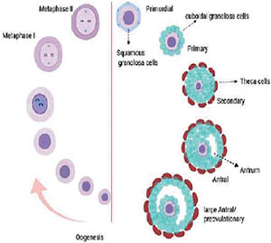
Figure 1.
Key steps in developing follicles and oocytes.
A number of in vitro follicular culture systems have been developed to preserve the reproductive ability of threatened species or iatrogenic infertility in women (Marin et al., 2018). In addition, it is used as a method to identify the toxicity of medications and undesirable fertility chemicals in vitro (Xu et al., 2015a). There is now a broad spectrum of culture techniques. Here we investigated the follicular culture variables in detail. Including species differences, age, isolation techniques, two-dimensional (2D) vs. three-dimensional (3D) systems, cultivation medium and hormonal supplementation.
Follicle culture systems in different species
Follicle cultures occur in a variety of species. Oocyte growth rate and follicle size (Griffin et al., 2006) vary between species (Pepling et al., 2010). Follicles are usually classified according to diameter. The term "preantral follicles" is used to describe their different phases (Mehrabianfar et al., 2020). Follicles produced in vitro are small compared to follicles produced in vivo (Xiao et al., 2015; Rodrigues et al., 2015). Rodents and mammals are the most prevalent models, approximately one-fifth of the studies use human follicles (Xiao et al., 2015; Telfer et al., 2008), and other mammalian follicles, like the Rhesus monkey (Rodrigues et al., 2015; Xu et al., 2009a; Peluffo et al., 2010; Xu et al., 2011a; Hornick et al., 2012; Xu et al., 2013; Xu et al., 2015b; Xu et al., 2018; Baba et al., 2017); baboon (Xu et al., 2011b); bovine (Yamamoto et al., 1999; Rossetto et al., 2013a;b; Araújo et al., 2015); ovine (Arunakumari et al., 2010; Muruvi et al., 2005); caprine (Rossetto et al., 2013a; Ferreira et al., 2018; Silva et al., 2015; Magalhães et al., 2011); swine (Hirao et al., 1994; Wu et al., 2001); cats (Songsasen et al., 2017; Thongkittidilok et al., 2018); dogs (Songsasen et al., 2011); horses (Haag et al., 2013); wildcats (Wiedemann et al., 2013).
The main reason for the differences between species is the difference in follicular culture outcomes. For instance, the diameter of the follicles in large mammal species in the preantral stage is much larger than in rodents. In the pre-ovulation stage, adult mouse follicles have a diameter of 420 µm; ovine 600 µm; 750 microns in goats; swine 800 µm and 20 000-23 000 µm in cattle and humans; respectively (Simon et al., 2020).
Folliculogenesis and ovulation stages are also different from one species to another. For example, mice follicles reach their maximum diameter within 19 days (Hoage & Cameron, 1976); but large mammals require months (Scaramuzzi et al., 2011). Because the growth stages of the follicles in large mammals are long, the presence of nutrients, gas exchange, and hormonal needs are the main challenges of cultivation (Telfer & Zelinski, 2013; West et al., 2007). Follicle structures vary from species to species as well. For instance, in large mammals, the theca cellular layer is thicker and affects the exchange of food and gas. Follicle culture and live birth have occurred in mice (Xu et al., 2006a;b). However, the follicle culture of rats (Daniel et al., 1989), pigs (Wu et al., 2001), buffaloes (Manjunatha et al., 2007), sheep (Arunakumari et al., 2010), goats (Magalhães et al., 2011), and Rhesus monkeys (Peluffo et al., 2012) were successful in pre-implantation after fertilization. In vitro oocyte maturation (IVM) have also been observed in rhesus monkey (Peluffo et al., 2010) and Baboon (Xu et al., 2011b) follicles. In general, for different reasons, particular species have been used in different follicular culture studies.
Age and growth stages for follicle culture in different species
In most rodent follicle culture studies, prepubertal follicles have been used, and in less than 30% of adult follicles (Diaz et al., 2007; Simon et al., 2020). Young animals of reproductive age have been used in studies of mammalian follicles such as sheep (Thomas et al., 2003; Arunakumari et al., 2010), goats (Ferreira et al., 2018; Magalhães et al., 2011), and cattle (Gutierrez et al., 2000; Itoh et al., 2002; Araújo et al., 2014a;b; 2015). Prepubertal follicles and smaller follicles have been used to evaluate the use of FSH supplementation in cattle and sheep (Wandji et al., 1996; Cecconi et al., 1999; Muruvi et al., 2005). Prepubertal follicles were used in comparison to the follicles of young and adult goats in 2D or 3D culture system (Leal et al., 2018). Prepubertal follicles were used to assess whether smaller preantral follicles could develop into antral follicles in vitro (Wu et al., 2001). In dogs, different stages of the estrus (Songsasen et al., 2011), and in marsupials (Nation & Selwood, 2009) were used in follicular cultures. In the rhesus monkey, the follicles used were primarily of young animals of reproductive age (Rodrigues et al., 2015; Baba et al., 2017; Xu et al., 2018). Small adult follicles were cultivated in adult baboons and were capable of producing live embryos (Xu et al., 2011b). Follicular culture studies have been conducted on different species at different ages and cycle stages; and demonstrate that these factors are chosen based on study objectives and ease of access to ovarian tissue.
Procedures for isolating the follicle
The separation of the follicle from the ovary tissue is the first step in follicle cultivation. Isolated follicles should have a similar morphology (Demeestere et al., 2002). Generally, the techniques of separating the follicles from the ovarian tissue include enzymatic, mechanical, or both. In the enzymatic separation of the follicles, proteolytic extracellular matrix (ECM) digestion such as collagenase, deoxyribonucleic, or liberase is utilized. The number of follicles obtained is typically higher in the enzyme digestion method and in compared to mechanical separation methods, they require less time, particularly for fibrous tissues in house mammals (Araújo et al., 2014a;b). However, in the enzyme digestion method, the follicles are more likely to be damaged. In mice, for example, collagenase leads to the production of preantral granulosa cell-oocyte complexes (PGOCs) and cell-oocyte complexes (COCs) from ovarian tissues, rather than whole follicles. In the mechanical separation method, special needles are used to separate the follicles of the ovarian stroma or tissue grinders, homogenizers, and cell strainers (Songsasen et al., 2017; Mahalingam et al., 2016a;b; Craig et al., 2010). The mechanical separation method results in less damage to the follicle than the enzyme method, and provides improved protection to the theca layer and follicular morphology (Araújo et al., 2014a;b), but the worst problem is that this method takes a lot of time (Demeestere et al., 2002). Usually, the selection of the isolating method depends on the follicular stage and the species used in the study. Generally, a short enzymatic digestion step and mechanical separation are used to maintain the structure of the follicle and obtain the maximum number of follicles (Table 1).
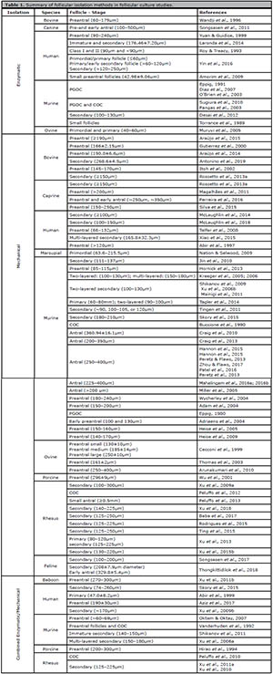
Table 1. Summary of follicular isolation methods in follicular culture studies.
Culture systems
Follicular culture systems are known as two-dimensional (2D) or three-dimensional (3D) (Figure 2). In 2D cultures, the follicles are static, but in 3D cultures, the follicles float in biomaterial matter (West et al., 2007). 2D-systems include the droplet method, substrate method (ECM coating), and membrane insert systems. In general, the 2D-method is used for small culturing follicles, hormonal studies, and gene expression studies. It is difficult to evaluate folliculogenesis and oocyte maturation in the 2D-method, because during oocyte proliferation, granulosa cells migrate to the surface of the culture medium (Kreeger et al., 2006). Logout of the communication between follicular cells stops follicular growth, inhibits ovulation, and meiosis in the egg (Green & Shikanov, 2016; West et al., 2007). In general, the follicles may be maintained for a short period of time in the 2D-culture.
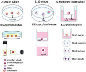
Figure 2. Schematic overview of 2D, 3D and multi-stage culture systems.
A - 2D-culture systems
1. Droplet culture
Within the droplet system, each follicle is implanted into a drop of culture medium, and each drop is covered with oil. There are drop methods for different stages and different species including mice (Adam et al., 2004; Wycherley et al., 2004; Adriaens et al., 2004), Rhesus monkey (Peluffo et al., 2012), sheep (Arunakumari et al., 2010), marsupial (Nation & Selwood, 2009), goat (Rossetto et al., 2013a; Ferreira et al., 2018) and cows (Araújo et al., 2014a;b) have been used. It typically takes about 6-18 days for the droplet method (Nation & Selwood, 2009; Arunakumari et al., 2010), A18 (Ferreira et al., 2018; Magalhães et al., 2011) and 32 (Araújo et al., 2014a;b) (Figure 2-A).
2 - 2D-culture
In the two-dimensional method, the follicles are grown directly on a surface covered by ECM compounds, such as collagen, laminin, or Matrigel. ECM plays an important role in folliculogenesis and affects cellular behavior, differentiation, and secretory activity (Desai et al., 2010). Collagen compounds have elasticity properties and contribute to intercellular communication, while Matrigel promotes cell proliferation and differentiation (Belli et al., 2012). Larger follicles such as preantral and antral follicles, PGOCs, and COCs have been used more in systems with 2D plastic substrates (Zhou & Flaws, 2017; Xu et al., 2018; Araújo et al., 2015; Patel et al., 2016; Mahalingam et al., 2016a;b; Peluffo et al., 2010). In large mammals, the follicles are larger and require more time in the culture environment to grow. Thus, the growing time of larger follicles may be reduced (Araújo et al., 2014a;b), and the duration of the culture varies in hours and days. For example, some studies have used the method to grow mammalian follicles such as those of Rhesus and cattle (Xu et al., 2018; Gutierrez et al., 2000). A fibronectin-coated plate was also used to culture the primordial and primary follicles of sheep. The growth of follicles was not much different from the follicles cultured in fibronectin-free plates (Muruvi et al., 2005) (Figure 2B).
3 - Membrane insert culture
Membrane insertion systems function in the same way as 2D-systems, and may contain ECM protein coatings, but in this method, the follicles are in an insert within a well of a culture plate and immersed in the environment. The mice follicles were cultured using a membrane inserting system, which improved the growth and ovulation of the follicles (Adam et al., 2004). For the first time, human follicles were cultured with a membrane insert system for 4 weeks. COC culture studies using membranes coated with ECM proteins (Sugiura et al., 2010) were also reported. Other 2D methods of follicle culture, include the use of glass coverslips coated with various ECM components. Although the earliest methods for cultivating ovarian follicles are 2D-systems, the 2D-methods damages the structure of the follicles, so that it is better suited for short-term cultures and small follicles (Figure 2C).
B - 3D-culture systems
3D-culture acts as in vivo and is adapted to long-term follicle culture. A major disadvantage of two-dimensional systems is that it damages the structure of the follicle surrounded by granulosa cells. This system is problematic for the culture of large mammalian follicles, which require culture and long-term communication among cells. In a 3D culture system, the structure of the intact follicles retains, in which the follicles are surrounded by biomaterials or have little access to a substrate. There are different types of 3D-systems, some using different scaffolding and encapsulation follicles, others using floating culture, or using in situ culture. To encapsulate the follicles, several matrices are used, which, in vivo, creates a very restricted environment, similar to that of the ovary and maintains the follicular structure and intercellular communication (Belli et al., 2012). Matrix compounds include natural substances such as collagen, alginate, or matrigel, or synthetic compounds such as polyethylene glycol (PEG) hydrogels that bind to protein-sensitive peptides (Figure 2).
1. Suspension culture
In this 3D-system, there is no scaffold and the structure of the follicles is protected by a system of rolls, inversion, or magnetic grains (Nation & Selwood, 2009; Wycherley et al., 2004). In marsupials, using inverted droplets, mature oocytes were obtained, which were more effective than follicles cultivated in different systems such as vertical droplets and roller systems. In tubes containing polypropylene, rat follicles produced eggs capable of performing meiosis, and were fertilized with intra cytoplasmic sperm injection (ICSI). Using a 3D magnetic system, cattle follicles produced live eggs that resumed meiosis after in vitro maturation (IVM) (Antonino et al., 2019) and follicle survival was higher than in the 2D-system (Figure 2D).
2. Encapsulated culture
In these culture systems, a biocompatible substance such as agar and collagen surround the follicle and protects its 3D structure. These materials are placed in layers on culture sheets to insert the follicles between these layers. In the first report of using the collagen gel matrix in the three-dimensional method, due to the stiffness of the matrix, no antrum was formed. Other studies have used collagen and agar matrices to grow follicles in mice (Vanderhyden et al., 1992) and pigs (Hirao et al., 1994), which, in comparison to 2D-systems, has maintained follicle structure and extended culture. In human studies, the use of collagen and agar in the 3D system made it possible to maintain the structure of the follicle and the egg for only 24 to 120 hours.
Brown algae are capable of producing a hydrogel called alginate that is biocompatible and can be used as a matrix in follicle culture (Belli et al., 2012). Alginate was first used in the culture of mice COCs. The results showed that alginate maintains intercellular communication, the proliferation of granulosa cells, and increases egg volume. Usually, ovarian cortex follicles move from the hard medulla to softer layers as they develop. Results of studies have demonstrated that concentrated alginate contributes to the growth of mice primary follicles, but it is not suitable for the development of larger follicles and the formation of antrum (Xu et al., 2006a;b; Skory et al., 2015). Also, studies of follicular culture in a 3D-system containing alginate have shown that low levels of alginate contribute to follicular growth, but, concentrated alginate is appropriate for hormone production (Songsasen et al., 2011).
Alginate encapsulation was used in other mammals, such as the Rhesus monkey, which could produce embryos at the cleavage stage (Xu et al., 2011a). By culturing the follicles in the combination system, the first mature human metaphase II (MII) oocytes were produced. First, the preantral follicles were cultured in 0.5% alginate for 10-15 days, and then the antral follicles were placed in low attachment plates for up to 40 days (Xiao et al., 2015). Supplements can impact 2D and 3D-culture systems. For example, one study found that vascular endothelium growth factor (VEGF) contributes to the growth of bovine secondary follicles in the 2D-system, and the growth hormone (GH) induces estradiol production in the 3D-alginate system (Araújo et al., 2014a;b). In a study using the caprine model, the encapsulation of 3D alginate was compared to the 2D substrate system that increased follicular survival and increased the number of eggs appropriate for IVM and IVF in the 3D-system. But in the 2D-culture, the follicles produced higher levels of progesterone.
Using the combination of alginate and fibrin, a dynamic permeable fibrin-alginate (IFN) network was developed (Shikanov et al., 2009). Within this matrix, follicular proteases degrade fibrin, reducing alginate concentration and matrix rigidity. This matrix mimics the internal environment of the ovary, as in ovarian tissue, follicles smaller than the hard cortex move into the soft marrow (Shikanov et al., 2011). With IFN in rodents, high meiotic follicles were developed (Jin et al., 2010) but in monkeys, it did not increase secondary follicle production. Embryonic stromal cells and fibroblasts (MEF) in mice were also grown with alginate-encapsulated follicles (Tagler et al., 2014). Ovarian stromal cells are involved in the growth, survival, and production of androgens in primary and secondary mice follicles. Culture of MEF cells with primary follicles containing alginate enhanced growth but decreased cell survival. Matrigel matrix is also used in three-dimensional culture, which in addition to maintaining the structure of the follicle, creates a protein-rich environment for folliculogenesis.
In matrigel, with fibrin and alginate, baboon follicles were enclosed, grew, and were able to produce mature eggs. The hyaluronan matrix was also used to grow follicles (Belli et al., 2012). The hyaluronan-ECM (no alginate) matrix on rat follicles increased follicular survival and increased the steroid hormone (Desai et al., 2012). The synthetic matrix of polyethylene glycol (PEG) acts like fibrin and is degraded by follicular proteases. Using the PEG matrix increased follicle growth in mouse models by 17 times (Shikanov et al., 2011) (Figure 2E).
3. Multi-step culture
Multi-step methods have been developed for follicle growth and the creation of a more similar physiological environment that primordial, primary, and early-secondary stage follicles can be cultured. First, the small follicles are grown in situ in the ovarian natural environment, and then the cultured follicles are separated from this tissue (McLaughlin et al., 2014; Jin et al., 2010; McLaughlin et al., 2018; Telfer et al., 2008). This method helps grow human follicles until they become mature gametes. For example, in one study using human ovarian tissue, secondary follicles were isolated and encapsulated in alginate. As the follicles grew and the antrum formed, they were released from the alginate matrix and transferred to low attachment plates for 40 days. Which turned human follicles into mature eggs (Xiao et al., 2015). In the next study, an alternative multistage method was used. In the first stage, cortical strips were cultured for 8 days. Secondly, the follicles were cultured for 8 days, and the COC cells were cultured on the membranes for 4 more days (Step 3). In the fourth stage, eggs larger than 100 µm were selected for IVM (McLaughlin et al., 2018). Also, a multi-step method was used for follicle growth in rodents. Generally, these systems have been very useful for long-term cultures of large mammal follicles. Therefore, the introduction of microfluidic systems or other natural scaffolding can be very useful in healthy in vitro follicles (Gargus et al., 2020) (Figure 2F).
Media composition and supplements
To grow the follicles, it is necessary to enrich the growing medium with nutrients, growth supplements, and hormone compounds. The selected culture medium should protect the growth of follicles and the maturation of eggs. As a result, the main media used in follicular culture typically include minimal essential medium (MEM), Dulbecco's modified eagle medium (DMEM), Waymouth's medium, McCoy's 5a medium), balanced salt solutions (Earle's balanced salt solution) (EBSS)), or mixed media (DMEM + F12, α-MEM + Glutamax).
Also, supplements are added to the follicular culture medium. For example, glucose as a source of carbon energy (Nation & Selwood, 2009), L-glutamine or fetuin as a source of amino acids (Asadi et al., 2017); ascorbic acid for reducing apoptosis and maintaining follicular structure, penicillin, streptomycin, and kanamycin as antibiotics (Demeestere et al., 2005) is used. Additionally, for the growth of follicles in vitro from the combination of ITS (insulin, transferrin, selenium) to increase the absorption of amino acids (Abedelahi et al., 2008). Protein supplements such as fetal calf serum (FCS), fetal bovine serum (FBS), and bovine serum albumin (BSA) are used in culture medium. Results from a mice model study showed that over a 10-day period, α-MEM, DMEM, and DMEM + F12 media had a better effect on antrum formation, follicle growth than Waymouth, M199, IMDM, and RPM1640. Also resulted in an increase in the number of MII oocytes (Simon et al., 2020). For culturing the human ovarian cortical tissue over a 10-day period, the MEM medium enriched with 10% human serum and 300 mIU/mL FSH may have a greater effect on follicular growth than the Waymouth and EBSS media (Wright et al., 1999). In another study, TCM-199 enriched with 10 ng/ml EGF was used over a 7-day period and had a better effect on the growth of goat and sheep follicles than α-MEM with the EMF (Andrade et al., 2014).
The TCM199 medium also increased the rate of antrum formation from bovine preantral follicles, relative to a-MEM or McCoy 5a medium (Rossetto et al., 2013a;b). Another factor affecting folliculogenesis is oxygen stress. Oxygen 5% is near the physiological oxygen levels. High oxygen stresses may produce reactive oxygen radicals (ROS) with cytotoxic effects (Rajabi et al., 2018). In one study, oxygen stress was induced in the follicle culture environment in rats. Which resulted in the production of mature eggs with higher performance in terms of static control. The rate of antrum formation in culture with 5% oxygen from caprine, ovine, and bovine (Gigli et al., 2006) follicles had more than 20% oxygen. Also, the culture of dog COCs in 5% oxygen decreased cell apoptosis compared to that in 20% oxygen (Silva et al., 2009). Low-oxygen stress along with high FSH and high fetuin in rhesus monkey, increased follicle growth, and antrum formation (Xu et al., 2011a). In general, these studies show that the selection of a suitable culture medium for follicle growth depends on the species. Furthermore, the protective effect of oxygen is much more important at the physiological level (Table 2).
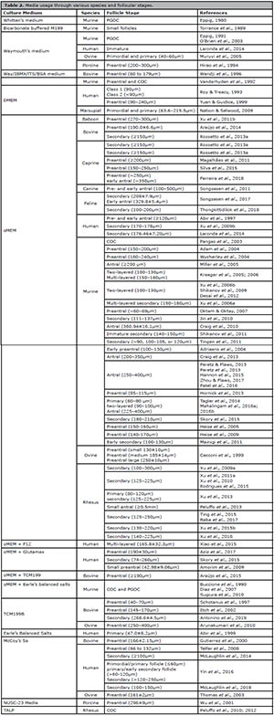
Table 2. Media usage through various species and follicular stages.
CONCLUSIONS
The general process of follicular culture has changed a lot from the past until now, and the main purpose of these changes has been to imitate the natural ovarian environment. By identifying the structure of the ovarian scaffold, information about 3D printing of the ovary was obtained. Ovarian function was thoroughly investigated by making 3D-printed scaffolds (Laronda et al., 2017). In addition, depending on physiological needs of the cell, other technologies such as microfluidics can be used to grow follicles. In static models, the use of a microfluidic system can be very effective. Because in addition to oxygenation, nutrient exchange and cellular communication, it provides a three-dimensional environment for the follicles (Desai et al., 2010). In order to reconstruct the human ovary environment in vitro, factoring plays a major role in the menstrual cycle. Therefore, in the context of a microfluidic chip (Scaramuzzi et al., 2011), alginate encapsulation (Gomes et al., 2015) was used to mimic the hormonal changes of the menstrual cycle in follicle culture. Microfluidics have made possible the successfully recombine the 28-day human menstrual cycle by fusion of tissues, such as mice ovaries and human fallopian tubes, ectopic uterus, and liver (Xiao et al., 2017). Microfluidic operating systems should be readily available and promote follicular culture among different species. Follicle culture methods vary depending on the species, the age of the animals, and the stage of the follicle.
ACKNOWLEDGEMENT
We thank our colleagues from the Shiraz University of Medical Sciences, who provided insight and expertise that greatly assisted the study.
REFERENCES
Abedelahi A, Salehnia M, Allameh AA. The effects of different concentrations of sodium selenite on the in vitro maturation of preantral follicles in serum-free and serum supplemented media. J Assist Reprod Genet. 2008;25:483-8. PMID: 18814023 DOI: 10.1007/s10815-008-9252-z
Medline Crossref
Abir R, Franks S, Mobberley MA, Moore PA, Margara RA, Winston RM. Mechanical isolation and in vitro growth of preantral and small antral human follicles. Fertil Steril. 1997;68:682-8. PMID: 9341611 DOI: 10.1016/S0015-0282(97)00264-1
Medline Crossref
Abir R, Roizman P, Fisch B, Nitke S, Okon E, Orvieto R, Ben Rafael Z. Pilot study of isolated early human follicles cultured in collagen gels for 24 hours. Hum Reprod. 1999;14:1299-301. PMID: 10325281 DOI: 10.1093/humrep/14.5.1299
Medline Crossref
Adam AA, Takahashi Y, Katagiri S, Nagano M. In vitro culture of mouse preantral follicles using membrane inserts and developmental competence of in vitro ovulated oocytes. J Reprod Dev. 2004;50:579-86. PMID: 15514465 DOI: 10.1262/jrd.50.579
Medline Crossref
Adriaens I, Cortvrindt R, Smitz J. Differential FSH exposure in preantral follicle culture has marked effects on folliculogenesis and oocyte developmental competence. Hum Reprod. 2004;19:398-408. PMID: 14747188 DOI: 10.1093/humrep/deh074
Medline Crossref
Amorim CA, Van Langendonckt A, David A, Dolmans MM, Donnez J. Survival of human pre-antral follicles after cryopreservation of ovarian tissue, follicular isolation and in vitro culture in a calcium alginate matrix. Hum Reprod. 2009;24:92-9. PMID: 18815120 DOI: 10.1093/humrep/den343
Medline Crossref
Antonino DC, Soares MM, Júnior JM, de Alvarenga PB, Mohallem RFF, Rocha CD, Vieira LA, de Souza AG, Beletti ME, Alves BG, Jacomini JO, Goulart LR, Alves KA. Three-dimensional levitation culture improves in-vitro growth of secondary follicles in bovine model. Reprod Biomed Online. 2019;38:300-11. PMID: 30639159 DOI: 10.1016/j.rbmo.2018.11.013
Medline Crossref
Araújo VR, Gastal MO, Wischral A, Figueiredo JR, Gastal EL. In vitro development of bovine secondary follicles in two- and three-dimensional culture systems using vascular endothelial growth factor, insulin-like growth factor-1, and growth hormone. Theriogenology. 2014a;82:1246-53. PMID: 25219848 DOI: 10.1016/j.theriogenology.2014.08.004
Medline Crossref
Araújo VR, Gastal MO, Figueiredo JR, Gastal EL. In vitro culture of bovine preantral follicles: a review. Reprod Biol Endocrinol. 2014b;12:78. PMID: 25117631 DOI: 10.1186/1477-7827-12-78
Medline Crossref
Araújo VR, Gastal MO, Wischral A, Figueiredo JR, Gastal EL. Long-term in vitro culture of bovine preantral follicles: Effect of base medium and medium replacement methods. Anim Reprod Sci. 2015;161:23-31. PMID: 26304751 DOI: 10.1016/j.anireprosci.2015.07.006
Medline Crossref
Arunakumari G, Shanmugasundaram N, Rao VH. Development of morulae from the oocytes of cultured sheep preantral follicles. Theriogenology. 2010;74:884-94. PMID: 20615540 DOI: 10.1016/j.theriogenology.2010.04.013
Medline Crossref
Asadi E, Najafi A, Moeini A, Pirjani R, Hassanzadeh G, Mikaeili S, Salehi E, Adutwum E, Soleimani M, Khosravi F, Barati M, Abolhassani F. Ovarian tissue culture in the presence of VEGF and fetuin stimulates follicle growth and steroidogenesis. J Endocrinol. 2017;232:205-19. PMID: 27852727 DOI: 10.1530/JOE-16-0368
Medline Crossref
Aziz AUR, Fu M, Deng J, Geng C, Luo Y, Lin B, Yu X, Liu B. A Microfluidic Device for Culturing an Encapsulated Ovarian Follicle. Micromachines. 2017;8:335. PMID: 30400524 DOI: 10.3390/mi8110335
Medline Crossref
Baba T, Ting AY, Tkachenko O, Xu J, Stouffer RL. Direct actions of androgen, estrogen and anti-Müllerian hormone on primate secondary follicle development in the absence of FSH in vitro. Hum Reprod. 2017;32:2456-64. PMID: 29077845 DOI: 10.1093/humrep/dex322
Medline Crossref
Bahmanpour S, Moradiyan E, Dehghani F, Zarei-Fard N. Chemoprotective effects of plasma derived from mice of different ages and genders on ovarian failure after cyclophosphamide treatment. J Ovarian Res. 2020;13:138. PMID: 33239062 DOI: 10.1186/s13048-020-00735-3
Medline Crossref
Belli M, Vigone G, Merico V, Redi CA, Zuccotti M, Garagna S. Towards a 3D culture of mouse ovarian follicles. Int J Dev Biol. 2012;56:931-7. PMID: 23417415 DOI: 10.1387/ijdb.120175mz
Medline Crossref
Buccione R, Vanderhyden BC, Caron PJ, Eppig JJ. FSH-induced expansion of the mouse cumulus oophorus in vitro is dependent upon a specific factor(s) secreted by the oocyte. Dev Biol. 1990;138:16-25. PMID: 2155145 DOI: 10.1016/0012-1606(90)90172-F
Medline Crossref
Cecconi S, Barboni B, Coccia M, Mattioli M. In vitro development of sheep preantral follicles. Biol Reprod. 1999;60:594-601. PMID: 10026104 DOI: 10.1095/biolreprod60.3.594
Medline Crossref
Craig ZR, Leslie TC, Hatfield KP, Gupta RK, Flaws JA. Mono-hydroxy methoxychlor alters levels of key sex steroids and steroidogenic enzymes in cultured mouse antral follicles. Toxicol Appl Pharmacol. 2010;249:107-13. PMID: 20840852 DOI: 10.1016/j.taap.2010.09.001
Medline Crossref
Craig ZR, Hannon PR, Wang W, Ziv-Gal A, Flaws JA. Di-n-butyl phthalate disrupts the expression of genes involved in cell cycle and apoptotic pathways in mouse ovarian antral follicles. Biol Reprod. 2013;88:23. PMID: 23242528 DOI: 10.1095/biolreprod.112.105122
Medline Crossref
Daniel SA, Armstrong DT, Gore-Langton RE. Growth and development of rat oocytes in vitro. Gamete Res. 1989;24:109-21. PMID: 2591848 DOI: 10.1002/mrd.1120240113
Medline Crossref
Dehghani F, Aboutalebi H, Esmaeilpour T, Panjehshahin MR, Bordbar H. Effect of platelet-rich plasma (PRP) on ovarian structures in cyclophosphamide-induced ovarian failure in female rats: a stereological study. Toxicol Mech Methods. 2018;28:653-9. PMID: 29968488 DOI: 10.1080/15376516.2018.1491662
Medline Crossref
Demeestere I, Delbaere A, Gervy C, Van Den Bergh M, Devreker F, Englert Y. Effect of preantral follicle isolation technique on in-vitro follicular growth, oocyte maturation and embryo development in mice. Hum Reprod. 2002;17:2152-9. PMID: 12151451 DOI: 10.1093/humrep/17.8.2152
Medline Crossref
Demeestere I, Centner J, Gervy C, Englert Y, Delbaere A. Impact of various endocrine and paracrine factors on in vitro culture of preantral follicles in rodents. Reproduction. 2005;130:147-56. PMID: 16049152 DOI: 10.1530/rep.1.00648
Medline Crossref
Desai N, AbdelHafez F, Drazba J, Goldfarb J, Falcone T. A simple and efficient method for preparation of isolated ovarian follicles for transmission electron microscopy. J Assist Reprod Genet. 2010;27:97-101. PMID: 20140639 DOI: 10.1007/s10815-010-9389-4
Medline Crossref
Desai N, Abdelhafez F, Calabro A, Falcone T. Three dimensional culture of fresh and vitrified mouse pre-antral follicles in a hyaluronan-based hydrogel: a preliminary investigation of a novel biomaterial for in vitro follicle maturation. Reprod Biol Endocrinol. 2012;10:29. PMID: 22513305 DOI: 10.1186/1477-7827-10-29
Medline Crossref
Diaz FJ, Wigglesworth K, Eppig JJ. Oocytes are required for the preantral granulosa cell to cumulus cell transition in mice. Dev Biol. 2007;305:300-11. PMID: 17368609 DOI: 10.1016/j.ydbio.2007.02.019
Medline Crossref
Edson MA, Nagaraja AK, Matzuk MM. The mammalian ovary from genesis to revelation. Endocr Rev. 2009;30:624-712. PMID: 19776209 DOI: 10.1210/er.2009-0012
Medline Crossref
Eppig JJ. Regulation of cumulus oophorus expansion by gonadotropins in vivo and in vitro. Biol Reprod. 1980;23:545-52. PMID: 6778513 DOI: 10.1095/biolreprod23.3.545
Medline Crossref
Eppig JJ. Maintenance of meiotic arrest and the induction of oocyte maturation in mouse oocyte-granulosa cell complexes developed in vitro from preantral follicles. Biol Reprod. 1991;45:824-30. PMID: 1666849 DOI: 10.1095/biolreprod45.6.824
Medline Crossref
Ferreira ACA, Maside C, Sá NAR, Guerreiro DD, Correia HHV, Leiva-Revilla J, Lobo CH, Araújo VR, Apgar GA, Brandão FZ, Figueiredo JR, Campello CC. Balance of insulin and FSH concentrations improves the in vitro development of isolated goat preantral follicles in medium containing GH. Anim Reprod Sci. 2016;165:1-10. PMID: 26723481 DOI: 10.1016/j.anireprosci.2015.10.010
Medline Crossref
Ferreira ACA, Cadenas J, Sá NAR, Correia HHV, Guerreiro DD, Lobo CH, Alves BG, Maside C, Gastal EL, Rodrigues APR, Figueiredo JR. In vitro culture of isolated preantral and antral follicles of goats using human recombinant FSH: Concentration-dependent and stage-specific effect. Anim Reprod Sci. 2018;196:120-9. PMID: 30049427 DOI: 10.1016/j.anireprosci.2018.07.004
Medline Crossref
Gargus ES, Rogers HB, McKinnon KE, Edmonds ME, Woodruff TK. Engineered reproductive tissues. Nat Biomed Eng. 2020;4:381-93. PMID: 32251392 DOI: 10.1038/s41551-020-0525-x
Medline Crossref
Gigli I, Byrd DD, Fortune JE. Effects of oxygen tension and supplements to the culture medium on activation and development of bovine follicles in vitro. Theriogenology. 2006;66:344-53. PMID: 16442155 DOI: 10.1016/j.theriogenology.2005.11.021
Medline Crossref
Gomes RG, Lisboa LA, Silva CB, Max MC, Marino PC, Oliveira RL, González SM, Barreiros TR, Marinho LS, Seneda MM. Improvement of development of equine preantral follicles after 6 days of in vitro culture with ascorbic acid supplementation. Theriogenology. 2015;84:750-5. PMID: 26074067 DOI: 10.1016/j.theriogenology.2015.05.006
Medline Crossref
Green LJ, Shikanov A. In vitro culture methods of preantral follicles. Theriogenology. 2016;86:229-38. PMID: 27173961 DOI: 10.1016/j.theriogenology.2016.04.036
Medline Crossref
Griffin J, Emery BR, Huang I, Peterson CM, Carrell DT. Comparative analysis of follicle morphology and oocyte diameter in four mammalian species (mouse, hamster, pig, and human). J Exp Clin Assist Reprod. 2006;3:2. PMID: 16509981 DOI: 10.1186/1743-1050-3-2
Medline Crossref
Gutierrez CG, Ralph JH, Telfer EE, Wilmut I, Webb R. Growth and antrum formation of bovine preantral follicles in long-term culture in vitro. Biol Reprod. 2000;62:1322-8. PMID: 10775183 DOI: 10.1095/biolreprod62.5.1322
Medline Crossref
Haag KT, Magalhães-Padilha DM, Fonseca GR, Wischral A, Gastal MO, King SS, Jones KL, Figueiredo JR, Gastal EL. In vitro culture of equine preantral follicles obtained via the Biopsy Pick-Up method. Theriogenology. 2013;79:911-7. PMID: 23434205 DOI: 10.1016/j.theriogenology.2013.01.001
Medline Crossref
Hannon PR, Brannick KE, Wang W, Gupta RK, Flaws JA. Di(2-ethylhexyl) phthalate inhibits antral follicle growth, induces atresia, and inhibits steroid hormone production in cultured mouse antral follicles. Toxicol Appl Pharmacol. 2015;284:42-53. PMID: 25701202 DOI: 10.1016/j.taap.2015.02.010
Medline Crossref
Heise M, Koepsel R, Russell AJ, McGee EA. Calcium alginate microencapsulation of ovarian follicles impacts FSH delivery and follicle morphology. Reprod Biol Endocrinol. 2005;3:47. PMID: 16162282 DOI: 10.1186/1477-7827-3-47
Medline Crossref
Heise MK, Koepsel R, McGee EA, Russell AJ. Dynamic oxygen enhances oocyte maturation in long-term follicle culture. Tissue Eng Part C Methods. 2009;15:323-32. PMID: 19552585 DOI: 10.1089/ten.tec.2007.0418
Medline Crossref
Hirao Y, Nagai T, Kubo M, Miyano T, Miyake M, Kato S. In vitro growth and maturation of pig oocytes. J Reprod Fertil. 1994;100:333-9. PMID: 8021848 DOI: 10.1530/jrf.0.1000333
Medline Crossref
Hoage TR, Cameron IL. Folliculogenesis in the ovary of the mature mouse: a radioautographic study. Anat Rec. 1976;184:699-709. PMID: 1259183 DOI: 10.1002/ar.1091840409
Medline Crossref
Hornick JE, Duncan FE, Shea LD, Woodruff TK. Isolated primate primordial follicles require a rigid physical environment to survive and grow in vitro. Hum Reprod. 2012;27:1801-10. PMID: 22456922 DOI: 10.1093/humrep/der468
Medline Crossref
Hornick JE, Duncan FE, Shea LD, Woodruff TK. Multiple follicle culture supports primary follicle growth through paracrine-acting signals. Reproduction. 2013;145:19-32. PMID: 23108112 DOI: 10.1530/REP-12-0233
Medline Crossref
Itoh T, Kacchi M, Abe H, Sendai Y, Hoshi H. Growth, antrum formation, and estradiol production of bovine preantral follicles cultured in a serum-free medium. Biol Reprod. 2002;67:1099-105. PMID: 12297524 DOI: 10.1095/biolreprod67.4.1099
Medline Crossref
Jin SY, Lei L, Shikanov A, Shea LD, Woodruff TK. A novel two-step strategy for in vitro culture of early-stage ovarian follicles in the mouse. Fertil Steril. 2010;93:2633-9. PMID: 20004373 DOI: 10.1016/j.fertnstert.2009.10.027
Medline Crossref
Kreeger PK, Fernandes NN, Woodruff TK, Shea LD. Regulation of mouse follicle development by follicle-stimulating hormone in a three-dimensional in vitro culture system is dependent on follicle stage and dose. Biol Reprod. 2005;73:942-50. PMID: 15987824 DOI: 10.1095/biolreprod.105.042390
Medline Crossref
Kreeger PK, Deck JW, Woodruff TK, Shea LD. The in vitro regulation of ovarian follicle development using alginate-extracellular matrix gels. Biomaterials. 2006;27:714-23. PMID: 16076485 DOI: 10.1016/j.biomaterials.2005.06.016
Medline Crossref
Laronda MM, Duncan FE, Hornick JE, Xu M, Pahnke JE, Whelan KA, Shea LD, Woodruff TK. Alginate encapsulation supports the growth and differentiation of human primordial follicles within ovarian cortical tissue. J Assist Reprod Genet. 2014;31:1013-28. PMID: 24845158 DOI: 10.1007/s10815-014-0252-x.
Medline Crossref
Laronda MM, Rutz AL, Xiao S, Whelan KA, Duncan FE, Roth EW, Woodruff TK, Shah RN. A bioprosthetic ovary created using 3D printed microporous scaffolds restores ovarian function in sterilized mice. Nat Commun. 2017;8:15261. PMID: 28509899 DOI: 10.1038/ncomms15261
Medline Crossref
Leal ÉSS, Vieira LA, Sá NAR, Silva GM, Lunardi FO, Ferreira ACA, Campello CC, Alves BG, Cibin FWS, Smitz J, Figueiredo JR, Rodrigues APR. In vitro growth and development of isolated secondary follicles from vitrified caprine ovarian cortex. Reprod Fertil Dev. 2018;30:359-70. PMID: 28768567 DOI: 10.1071/RD16487
Medline Crossref
Magalhães DM, Duarte AB, Araújo VR, Brito IR, Soares TG, Lima IM, Lopes CA, Campello CC, Rodrigues AP, Figueiredo JR. In vitro production of a caprine embryo from a preantral follicle cultured in media supplemented with growth hormone. Theriogenology. 2011;75:182-8. PMID: 20875671 DOI: 10.1016/j.theriogenology.2010.08.004
Medline Crossref
Mahalingam S, Gao L, Gonnering M, Helferich W, Flaws JA. Equol inhibits growth, induces atresia, and inhibits steroidogenesis of mouse antral follicles in vitro. Toxicol Appl Pharmacol. 2016a;295:47-55. PMID: 26876617 DOI: 10.1016/j.taap.2016.02.009
Medline Crossref
Mahalingam S, Gao L, Eisner J, Helferich W, Flaws JA. Effects of isoliquiritigenin on ovarian antral follicle growth and steroidogenesis. Reprod Toxicol. 2016b;66:107-14. PMID: 27773742 DOI: 10.1016/j.reprotox.2016.10.004
Medline Crossref
Mainigi MA, Ord T, Schultz RM. Meiotic and developmental competence in mice are compromised following follicle development in vitro using an alginate-based culture system. Biol Reprod. 2011;85:269-76. PMID: 21490243 DOI: 10.1095/biolreprod.111.091124
Medline Crossref
Manjunatha BM, Gupta PS, Ravindra JP, Devaraj M, Ramesh HS, Nandi S. In vitro developmental competence of buffalo oocytes collected at various stages of the estrous cycle. Theriogenology. 2007;68:882-8. PMID: 17706758 DOI: 10.1016/j.theriogenology.2007.07.001
Medline Crossref
Marin D, Yang M, Wang T. In Vitro growth of human ovarian follicles for fertility preservation. Reprod Dev Med. 2018;2:230-6. DOI: 10.4103/2096-2924.249892
Crossref
Matzuk MM, Burns KH. Genetics of mammalian reproduction: modeling the end of the germline. Annu Rev Physiol. 2012;74:503-28. PMID: 22335799 DOI: 10.1146/annurev-physiol-020911-153248
Medline Crossref
McLaughlin M, Kinnell HL, Anderson RA, Telfer EE. Inhibition of phosphatase and tensin homologue (PTEN) in human ovary in vitro results in increased activation of primordial follicles but compromises development of growing follicles. Mol Hum Reprod. 2014;20:736-44. PMID: 24830779 DOI: 10.1093/molehr/gau037
Medline Crossref
McLaughlin M, Albertini DF, Wallace WHB, Anderson RA, Telfer EE. Metaphase II oocytes from human unilaminar follicles grown in a multi-step culture system. Mol Hum Reprod. 2018;24:135-42. PMID: 29390119 DOI: 10.1093/molehr/gay002
Medline Crossref
Mehrabianfar P, Dehghani F, Karbalaei N, Mesbah F. The effects of metformin on stereological and ultrastructural features of the ovary in streptozotocin -induced diabetes adult rats: An experimental study. Int J Reprod Biomed. 2020;18:651-66. PMID: 32923931 DOI: 10.18502/ijrm.v13i8.7506
Medline Crossref
Mesbah F, Bordbar H, Talaei Khozani T, Dehghani F, Mirkhani H. The non-preventive effects of human menopausal gonadotropins on ovarian tissues in Nandrolone decanoate-treated female rats: A histochemical and ultra-structural study. Int J Reprod Biomed. 2018;16:159-74. PMID: 29766147 DOI: 10.29252/ijrm.16.3.159
Medline Crossref
Miller DW, Harrison JL, Brown YA, Doyle U, Lindsay A, Adam CL, Lea RG. Immunohistochemical evidence for an endocrine/paracrine role for ghrelin in the reproductive tissues of sheep. Reprod Biol Endocrinol. 2005;3:60. PMID: 16259638 DOI: 10.1186/1477-7827-3-60
Medline Crossref
Muruvi W, Picton HM, Rodway RG, Joyce IM. In vitro growth of oocytes from primordial follicles isolated from frozen-thawed lamb ovaries. Theriogenology. 2005;64:1357-70. PMID: 16139612 DOI: 10.1016/j.theriogenology.2005.02.010
Medline Crossref
Nation A, Selwood L. The production of mature oocytes from adult ovaries following primary follicle culture in a marsupial. Reproduction. 2009;138:247-55. PMID: 19494049 DOI: 10.1530/REP-09-0028
Medline Crossref
O'Brien MJ, Pendola JK, Eppig JJ. A revised protocol for in vitro development of mouse oocytes from primordial follicles dramatically improves their developmental competence. Biol Reprod. 2003;68:1682-6. PMID: 12606400 DOI: 10.1095/biolreprod.102.013029
Medline Crossref
Oktem O, Oktay K. The role of extracellular matrix and activin-A in in vitro growth and survival of murine preantral follicles. Reprod Sci. 2007;14:358-66. PMID: 17644808 DOI: 10.1177/1933719107303397
Medline Crossref
Pangas SA, Saudye H, Shea LD, Woodruff TK. Novel approach for the three-dimensional culture of granulosa cell-oocyte complexes. Tissue Eng. 2003;9:1013-21. PMID: 14633385 DOI: 10.1089/107632703322495655
Medline Crossref
Patel S, Peretz J, Pan YX, Helferich WG, Flaws JA. Genistein exposure inhibits growth and alters steroidogenesis in adult mouse antral follicles. Toxicol Appl Pharmacol. 2016;293:53-62. PMID: 26792615 DOI: 10.1016/j.taap.2015.12.026
Medline Crossref
Peluffo MC, Barrett SL, Stouffer RL, Hennebold JD, Zelinski MB. Cumulus-oocyte complexes from small antral follicles during the early follicular phase of menstrual cycles in rhesus monkeys yield oocytes that reinitiate meiosis and fertilize in vitro. Biol Reprod. 2010;83:525-32. PMID: 20519694 DOI: 10.1095/biolreprod.110.084418
Medline Crossref
Peluffo MC, Ting AY, Zamah AM, Conti M, Stouffer RL, Zelinski MB, Hennebold JD. Amphiregulin promotes the maturation of oocytes isolated from the small antral follicles of the rhesus macaque. Hum Reprod. 2012;27:2430-7. PMID: 22593432 DOI: 10.1093/humrep/des158
Medline Crossref
Peluffo MC, Hennebold JD, Stouffer RL, Zelinski MB. Oocyte maturation and in vitro hormone production in small antral follicles (SAFs) isolated from rhesus monkeys. J Assist Reprod Genet. 2013;30:353-9. PMID: 23423613 DOI: 10.1007/s10815-013-9937-9
Medline Crossref
Pepling ME, Sundman EA, Patterson NL, Gephardt GW, Medico L Jr, Wilson KI. Differences in oocyte development and estradiol sensitivity among mouse strains. Reproduction. 2010;139:349-57. PMID: 19846484 DOI: 10.1530/REP-09-0392
Medline Crossref
Peretz J, Flaws JA. Bisphenol A down-regulates rate-limiting Cyp11a1 to acutely inhibit steroidogenesis in cultured mouse antral follicles. Toxicol Appl Pharmacol. 2013;271:249-56. PMID: 23707772 DOI: 10.1016/j.taap.2013.04.028
Medline Crossref
Peretz J, Neese SL, Flaws JA. Mouse strain does not influence the overall effects of bisphenol a-induced toxicity in adult antral follicles. Biol Reprod. 2013;89:108. PMID: 24025742 DOI: 10.1095/biolreprod.113.111864
Medline Crossref
Rajabi Z, Khokhar Z, Yazdekhasti H. The Growth of Preantral Follicles and the Impact of Different Supplementations and Circumstances: A Review Study with Focus on Bovine and Human Preantral Follicles. Cell Reprogram. 2018;20:164-77. PMID: 29782184 DOI: 10.1089/cell.2017.0068
Medline Crossref
Richards JS, Ascoli M. Endocrine, Paracrine, and Autocrine Signaling Pathways That Regulate Ovulation. Trends Endocrinol Metab. 2018;29:313-25. PMID: 29602523 DOI: 10.1016/j.tem.2018.02.012
Medline Crossref
Rodrigues JK, Navarro PA, Zelinski MB, Stouffer RL, Xu J. Direct actions of androgens on the survival, growth and secretion of steroids and anti-Müllerian hormone by individual macaque follicles during three-dimensional culture. Hum Reprod. 2015;30:664-74. PMID: 25567619 DOI: 10.1093/humrep/deu335
Medline Crossref
Rossetto R, Santos RR, Silva GM, Duarte ABG, Silva CMG, Campello CC, Figueiredo JR. Comparative study on the in vitro development of caprine and bovine preantral follicles. Small Rumin Res. 2013a;113:167-70. DOI: 10.1016/j.smallrumres.2013.03.004
Crossref
Rossetto R, Saraiva MV, Santos RR, Silva CM, Faustino LR, Chaves RN, Brito IR, Rodrigues GQ, Lima IM, Donato MA, Peixoto CA, Figueiredo JR. Effect of medium composition on the in vitro culture of bovine pre-antral follicles: morphology and viability do not guarantee functionality. Zygote. 2013b;21:125-8. PMID: 22717039 DOI: 10.1017/S0967199412000044
Medline Crossref
Roy SK, Treacy BJ. Isolation and long-term culture of human preantral follicles. Fertil Steril. 1993;59:783-90. PMID: 8458497 DOI: 10.1016/S0015-0282(16)55860-9
Medline Crossref
Scaramuzzi RJ, Baird DT, Campbell BK, Driancourt MA, Dupont J, Fortune JE, Gilchrist RB, Martin GB, McNatty KP, McNeilly AS, Monget P, Monniaux D, Viñoles C, Webb R. Regulation of folliculogenesis and the determination of ovulation rate in ruminants. Reprod Fertil Dev. 2011;23:444-67. PMID: 21426863 DOI: 10.1071/RD09161
Medline Crossref
Schotanus K, Hage WJ, Vanderstichele H, van den Hurk R. Effects of conditioned media from murine granulosa cell lines on the growth of isolated bovine preantral follicles. Theriogenology. 1997;48:471-83. PMID: 16728143 DOI: 10.1016/S0093-691X(97)00256-2
Medline Crossref
Shikanov A, Xu M, Woodruff TK, Shea LD. Interpenetrating fibrin-alginate matrices for in vitro ovarian follicle development. Biomaterials. 2009;30:5476-85. PMID: 19616843 DOI: 10.1016/j.biomaterials.2009.06.054
Medline Crossref
Shikanov A, Xu M, Woodruff TK, Shea LD. A method for ovarian follicle encapsulation and culture in a proteolytically degradable 3 dimensional system. J Vis Exp. 2011;2695. PMID: 21445043 DOI: 10.3791/2695
Medline Crossref
Silva AE, Rodriguez P, Cavalcante LF, Rodrigues BA, Rodrigues JL. The influence of oxygen tension on cumulus cell viability of canine COCs matured in high-glucose medium. Reprod Domest Anim. 2009;44:259-62. PMID: 19754582 DOI: 10.1111/j.1439-0531.2009.01406.x
Medline Crossref
Silva GM, Rossetto R, Chaves RN, Duarte AB, Araújo VR, Feltrin C, Bernuci MP, Anselmo-Franci JA, Xu M, Woodruff TK, Campello CC, Figueiredo JR. In vitro development of secondary follicles from pre-pubertal and adult goats cultured in two-dimensional or three-dimensional systems. Zygote. 2015;23:475-84. PMID: 24666604 DOI: 10.1017/S0967199414000070
Medline Crossref
Simon LE, Kumar TR, Duncan FE. In vitro ovarian follicle growth: a comprehensive analysis of key protocol variables. Biol Reprod. 2020;103:455-70. PMID: 32406908 DOI: 10.1093/biolre/ioaa073
Medline Crossref
Skory RM, Xu Y, Shea LD, Woodruff TK. Engineering the ovarian cycle using in vitro follicle culture. Hum Reprod. 2015;30:1386-95. PMID: 25784584 DOI: 10.1093/humrep/dev052
Medline Crossref
Songsasen N, Woodruff TK, Wildt DE. In vitro growth and steroidogenesis of dog follicles are influenced by the physical and hormonal microenvironment. Reproduction. 2011;142:113-22. PMID: 21502334 DOI: 10.1530/REP-10-0442
Medline Crossref
Songsasen N, Thongkittidilok C, Yamamizu K, Wildt DE, Comizzoli P. Short-term hypertonic exposure enhances in vitro follicle growth and meiotic competence of enclosed oocytes while modestly affecting mRNA expression of aquaporin and steroidogenic genes in the domestic cat model. Theriogenology. 2017;90:228-36. PMID: 28166973 DOI: 10.1016/j.theriogenology.2016.12.006
Medline Crossref
Sugiura K, Su YQ, Li Q, Wigglesworth K, Matzuk MM, Eppig JJ. Estrogen promotes the development of mouse cumulus cells in coordination with oocyte-derived GDF9 and BMP15. Mol Endocrinol. 2010;24:2303-14. PMID: 21047911 DOI: 10.1210/me.2010-0260
Medline Crossref
Tagler D, Makanji Y, Tu T, Bernabé BP, Lee R, Zhu J, Kniazeva E, Hornick JE, Woodruff TK, Shea LD. Promoting extracellular matrix remodeling via ascorbic acid enhances the survival of primary ovarian follicles encapsulated in alginate hydrogels. Biotechnol Bioeng. 2014;111:1417-29. PMID: 24375265 DOI: 10.1002/bit.25181
Medline Crossref
Telfer EE, McLaughlin M, Ding C, Thong KJ. A two-step serum-free culture system supports development of human oocytes from primordial follicles in the presence of activin. Hum Reprod. 2008;23:1151-8. PMID: 18326514 DOI: 10.1093/humrep/den070
Medline Crossref
Telfer EE, Zelinski MB. Ovarian follicle culture: advances and challenges for human and nonhuman primates. Fertil Steril. 2013;99:1523-33. PMID: 23635350 DOI: 10.1016/j.fertnstert.2013.03.043
Medline Crossref
Thomas FH, Armstrong DG, Telfer EE. Activin promotes oocyte development in ovine preantral follicles in vitro. Reprod Biol Endocrinol. 2003;1:76. PMID: 14613548 DOI: 10.1186/1477-7827-1-76
Medline Crossref
Thongkittidilok C, Singh RP, Comizzoli P, Wildt D, Songsasen N. Insulin promotes preantral follicle growth and antrum formation through temporal expression of genes regulating steroidogenesis and water transport in the cat. Reprod Fertil Dev. 2018;30:1369-79. PMID: 29720337 DOI: 10.1071/RD17454
Medline Crossref
Ting AY, Xu J, Stouffer RL. Differential effects of estrogen and progesterone on development of primate secondary follicles in a steroid-depleted milieu in vitro. Hum Reprod. 2015;30:1907-17. PMID: 26040480 DOI: 10.1093/humrep/dev119
Medline Crossref
Tingen CM, Kiesewetter SE, Jozefik J, Thomas C, Tagler D, Shea L, Woodruff TK. A macrophage and theca cell-enriched stromal cell population influences growth and survival of immature murine follicles in vitro. Reproduction. 2011;141:809-20. PMID: 21389078 DOI: 10.1530/REP-10-0483
Medline Crossref
Torrance C, Telfer E, Gosden RG. Quantitative study of the development of isolated mouse pre-antral follicles in collagen gel culture. J Reprod Fertil. 1989;87:367-74. PMID: 2621708 DOI: 10.1530/jrf.0.0870367
Medline Crossref
Vanderhyden BC, Telfer EE, Eppig JJ. Mouse oocytes promote proliferation of granulosa cells from preantral and antral follicles in vitro. Biol Reprod. 1992;46:1196-204. PMID: 1391318 DOI: 10.1095/biolreprod46.6.1196
Medline Crossref
Wandji SA, Eppig JJ, Fortune JE. FSH and growth factors affect the growth and endocrine function in vitro of granulosa cells of bovine preantral follicles. Theriogenology. 1996;45:817-32. PMID: 16727844 DOI: 10.1016/0093-691X(96)00011-8
Medline Crossref
West ER, Shea LD, Woodruff TK. Engineering the follicle microenvironment. Semin Reprod Med. 2007;25:287-99. PMID: 17594609 DOI: 10.1055/s-2007-980222
Medline Crossref
Wiedemann C, Zahmel J, Jewgenow K. Short-term culture of ovarian cortex pieces to assess the cryopreservation outcome in wild felids for genome conservation. BMC Vet Res. 2013;9:37. PMID: 23433001 DOI: 10.1186/1746-6148-9-37
Medline Crossref
Wright CS, Hovatta O, Margara R, Trew G, Winston RM, Franks S, Hardy K. Effects of follicle-stimulating hormone and serum substitution on the in-vitro growth of human ovarian follicles. Hum Reprod. 1999;14:1555-62. PMID: 10357975 DOI: 10.1093/humrep/14.6.1555
Medline Crossref
Wu J, Carrell DT, Wilcox AL. Development of in vitro-matured oocytes from porcine preantral follicles following intracytoplasmic sperm injection. Biol Reprod. 2001;65:1579-85. PMID: 11673278 DOI: 10.1095/biolreprod65.5.1579
Medline Crossref
Wycherley G, Downey D, Kane MT, Hynes AC. A novel follicle culture system markedly increases follicle volume, cell number and oestradiol secretion. Reproduction. 2004;127:669-77. PMID: 15175503 DOI: 10.1530/rep.1.00040
Medline Crossref
Xiao S, Zhang J, Romero MM, Smith KN, Shea LD, Woodruff TK. In vitro follicle growth supports human oocyte meiotic maturation. Sci Rep. 2015;5:17323. PMID: 26612176 DOI: 10.1038/srep17323
Medline Crossref
Xiao S, Coppeta JR, Rogers HB, Isenberg BC, Zhu J, Olalekan SA, McKinnon KE, Dokic D, Rashedi AS, Haisenleder DJ, Malpani SS, Arnold-Murray CA, Chen K, Jiang M, Bai L, Nguyen CT, Zhang J, Laronda MM, Hope TJ, Maniar KP, et al. A microfluidic culture model of the human reproductive tract and 28-day menstrual cycle. Nat Commun. 2017;8:14584. PMID: 28350383 DOI: 10.1038/ncomms14584
Medline Crossref
Xu M, Kreeger PK, Shea LD, Woodruff TK. Tissue-engineered follicles produce live, fertile offspring. Tissue Eng. 2006a;12:2739-46. PMID: 17518643 DOI: 10.1089/ten.2006.12.2739
Medline Crossref
Xu M, West E, Shea LD, Woodruff TK. Identification of a stage-specific permissive in vitro culture environment for follicle growth and oocyte development. Biol Reprod. 2006b;75:916-23. PMID: 16957022 DOI: 10.1095/biolreprod.106.054833
Medline Crossref
Xu M, West-Farrell ER, Stouffer RL, Shea LD, Woodruff TK, Zelinski MB. Encapsulated three-dimensional culture supports development of nonhuman primate secondary follicles. Biol Reprod. 2009a;81:587-94. PMID: 19474063 DOI: 10.1095/biolreprod.108.074732
Medline Crossref
Xu M, Barrett SL, West-Farrell E, Kondapalli LA, Kiesewetter SE, Shea LD, Woodruff TK. In vitro grown human ovarian follicles from cancer patients support oocyte growth. Hum Reprod. 2009b;24:2531-40. PMID: 19597190 DOI: 10.1093/humrep/dep228
Medline Crossref
Xu J, Bernuci MP, Lawson MS, Yeoman RR, Fisher TE, Zelinski MB, Stouffer RL. Survival, growth, and maturation of secondary follicles from prepubertal, young, and older adult rhesus monkeys during encapsulated three-dimensional culture: effects of gonadotropins and insulin. Reproduction. 2010;140:685-97. PMID: 20729335 DOI: 10.1530/REP-10-0284
Medline Crossref
Xu J, Lawson MS, Yeoman RR, Pau KY, Barrett SL, Zelinski MB, Stouffer RL. Secondary follicle growth and oocyte maturation during encapsulated three-dimensional culture in rhesus monkeys: effects of gonadotrophins, oxygen and fetuin. Hum Reprod. 2011a;26:1061-72. PMID: 21362681 DOI: 10.1093/humrep/der049
Medline Crossref
Xu M, Fazleabas AT, Shikanov A, Jackson E, Barrett SL, Hirshfeld-Cytron J, Kiesewetter SE, Shea LD, Woodruff TK. In vitro oocyte maturation and preantral follicle culture from the luteal-phase baboon ovary produce mature oocytes. Biol Reprod. 2011b;84:689-97. PMID: 21123815 DOI: 10.1095/biolreprod.110.088674
Medline Crossref
Xu J, Lawson MS, Yeoman RR, Molskness TA, Ting AY, Stouffer RL, Zelinski MB. Fibrin promotes development and function of macaque primary follicles during encapsulated three-dimensional culture. Hum Reprod. 2013;28:2187-200. PMID: 23608357 DOI: 10.1093/humrep/det093
Medline Crossref
Xu Y, Duncan FE, Xu M, Woodruff TK. Use of an organotypic mammalian in vitro follicle growth assay to facilitate female reproductive toxicity screening. Reprod Fertil Dev. 2015a;18:10.1071/RD14375. PMID: 25689754 DOI: 10.1071/RD14375
Medline Crossref
Xu J, McGee WK, Bishop CV, Park BS, Cameron JL, Zelinski MB, Stouffer RL. Exposure of female macaques to Western-style diet with or without chronic T in vivo alters secondary follicle function during encapsulated 3-dimensional culture. Endocrinology. 2015b;156:1133-42. PMID: 25545382 DOI: 10.1210/en.2014-1711
Medline Crossref
Xu J, Xu F, Lawson MS, Tkachenko OY, Ting AY, Kahl CA, Park BS, Stouffer RR, Bishop CV. Anti-Müllerian hormone is a survival factor and promotes the growth of rhesus macaque preantral follicles during matrix-free culture. Biol Reprod. 2018;98:197-207. PMID: 29293939 DOI: 10.1093/biolre/iox181
Medline Crossref
Yamamoto K, Otoi T, Koyama N, Horikita N, Tachikawa S, Miyano T. Development to live young from bovine small oocytes after growth, maturation and fertilization in vitro. Theriogenology. 1999;52:81-9. PMID: 10734407 DOI: 10.1016/S0093-691X(99)00111-9
Medline Crossref
Yin H, Kristensen SG, Jiang H, Rasmussen A, Andersen CY. Survival and growth of isolated pre-antral follicles from human ovarian medulla tissue during long-term 3D culture. Hum Reprod. 2016;31:1531-9. PMID: 27112699 DOI: 10.1093/humrep/dew049
Medline Crossref
Yuan W, Giudice LC. Insulin-like growth factor-II mediates the steroidogenic and growth promoting actions of follicle stimulating hormone on human ovarian pre-antral follicles cultured in vitro. J Clin Endocrinol Metab. 1999;84:1479-82. PMID: 10199799 DOI: 10.1210/jcem.84.4.5727
Medline Crossref
Zhou C, Flaws JA. Effects of an Environmentally Relevant Phthalate Mixture on Cultured Mouse Antral Follicles. Toxicol Sci. 2017;156:217-29. PMID: 28013214 DOI: 10.1093/toxsci/kfw245
Medline Crossref