JBRA Assist. Reprod. 2022;26(4):606-611
ORIGINAL ARTICLE
doi: 10.5935/1518-0557.20220004
Differential apoptotic gene expression in the male partners of infertile couples with normal and abnormal sperm parameters
1Reproductive Immunology Research Center, Shahid Sadoughi University of Medical Sciences, Yazd, Iran
2Department of Biology & Anatomical Sciences, Shahid Sadoughi University of Medical Sciences, Yazd, Iran
3Parseh Laboratory of Medical Science, Yazd, Iran
CONFLICT OF INTEREST
The authors had no conflict of interest associated with this study.
ABSTRACT
Objective: The purpose of this study was to investigate the cellular and molecular levels of apoptosis induction in three groups of male partners of infertile couples, one featuring subjects with normal sperm parameters and unexplained male infertility (UMI), one including men with abnormal sperm parameters, and one with fertile men as controls.
Methods: Twenty-five infertile men with abnormal sperm parameters and 25 men with UMI and normal sperm parameters were recruited as experimental group I and experimental group II; 25 fertile men were included as controls. The mRNA levels of Fas, Fas ligand, Caspase 8, Bax, and Bcl2 were measured in the three groups. The cellular rates of early and late apoptosis were assessed using annexin V and propidium iodide staining.
Results: The expression of Bax, Bcl2, and the Bax/Bcl2 ratio in experimental group I was significantly higher than that in experimental group II and controls. However, the Bax/Bcl2 ratio was less than 1 among all groups. No significant difference was found among study groups regarding the gene expression of Fas, Fas ligand, and Caspase 8. No significant difference was seen in early apoptotic rates of sperm among study groups. The highest number of necrotic sperm cells was detected in experimental group I.
Conclusions: The findings showed that the external pathways of apoptosis were not activated in the absence of external stimuli of sperm apoptosis in ejaculated sperm. Regardless of fertility status, apoptosis gene induction in the internal pathway was associated with abnormalities in sperm motility and/or morphology in men with abnormal parameters.
Keywords: sperm parameters,apoptosis,gene expression,unexplained male infertility
INTRODUCTION
Infertility is a major problem in society today, with 15% of couples trying to have a child failing to conceive (Agarwal et al., 2015). Semen analysis is usually the first step in assessing the fertility status of men; however, this method does not provide enough information about the genomic integrity of the male gamete. Abnormal semen parameters are considered an indicator of subfertility in men. However, up to 30% of men with normal semen parameters (normozoospermic) are diagnosed with UMI, since the reason for infertility is unknown (Panner Selvam et al., 2019).
Three main theories have been proposed to describe the causes of damage to the sperm nucleus, including impaired replacement of histone by protamine, reactive oxygen species (ROS), and apoptosis (Carrell et al., 2007; Talebi et al., 2016). These interlinked molecular events can lead to different clinical and laboratory manifestations in infertile males. Clarifying the nature of sperm defects will contribute to the selection of proper assisted reproduction technology (ART) methods, consequently enhancing ART success rates and ensuring improved offspring health (Agarwal & Said, 2005).
Apoptosis, recognized as a type of programmed cell death, is associated with changes in the morphological and biochemical characteristics of cells. Apoptosis plays a prominent role in several physiological and pathological processes (Majtnerová & Roušar, 2018). According to Erkkilä et al. (1997), such programmed cell death is a normal and hormonally controlled phenomenon in the adult human testes. Caspase activation, externalization of phosphatidylserine (PS), changing of mitochondrial membrane potential, and sperm DNA fragmentation (SDF) are recognized as markers of apoptosis in ejaculated human spermatozoa (Shukla et al., 2012). So far, two pathways have been considered for apoptosis, namely external and internal. In the external pathway, death signals are transmitted through transmembrane receptors, while in the internal pathway death signals are sent to the mitochondria. A set of genes, such as Caspase8, Fas, FasL, and the Bcl-2 protein family, are involved in both external and internal pathways (Kiraz et al., 2016).
Since there is no report on the possible involvement of apoptosis in ejaculated human spermatozoa with normal and abnormal parameters in male partners of infertile couples with UMI, this study was designed to compare the cellular and molecular levels of apoptosis induction markers in male partners of infertile couples with abnormal sperm parameters, normal sperm parameters with UMI, and fertile men as controls.
MATERIALS AND METHODS
Study population
This cross-sectional study included couples who were referred to obstetrics and gynecology clinic, Yazd, Iran. The male subjects of infertile couples were divided into two groups, one with individuals with abnormal sperm parameters (experimental group I, n=25) and another with men with UMI and normal sperm parameters (experimental group II, n=25). For the control group, 25 fertile men with normal sperm parameters were enrolled. The patients were randomly selected into each experimental group by using a simple randomization method. All procedures were conducted with the approval of the institution’s Ethics Committee. The participants gave written consent before joining the study. After examination and sperm analysis ordered by the treating physician, semen samples were collected from the participants.The inclusion criteria for controls were as follows: having normal sperm parameters and at least one child aged less than two years. The subjects in both experimental groups had primary infertility. Based on the World Health Organization (WHO, 2010), sperm parameters were defined as normal in this study given that the volume of semen was ≥1.5 mL, the concentration of sperm was ≥15 million/mL, total sperm motility was ≥40%, and normal sperm morphology was ≥4% (WHO, 2010). All participants were aged less than 40 years and had a body mass index below 30, no varicocele disease, no history of smoking or drug use, no infectious diseases or diabetes, and no history of alcohol abuse. Patients with pyospermia (i.e. more than one million WBC per ml of sperm), varicocele, azoospermia, fever, and infectious disease during the last 90 days as well as subjects with genetic problems, reproductive tract infections, inflammatory disease of the reproductive tract, sexually transmitted disease, or erectile dysfunction were excluded from the study.
RNA extraction and cDNA synthesis
RNA extraction was performed from washed semen samples collected from both experimental and control groups using a total RNA extraction kit (Parstous biotechnology, Iran). The integrity of extracted RNA was assessed by agarose electrophoresis. In addition, the final RNA concentration was assessed based on measurements of absorbance at 260 nm (PhotoBiometer, Eppendorf, Germany). In first-strand cDNA synthesis, 500ng of total RNA with the Revert Aid First Strand cDNA Synthesis Kit was used based on the manufacturer’s protocol (Parstous biotechnology, Iran). The cDNA product was kept at-20°C until use.
Gene expression assessment
The relative gene expression level was studied based on quantitative real-time polymerase chain reaction (qRT-PCR). Master Mix Green with high ROX™ (Amplicon) together with the StepOne system was utilized in each PCR reaction (Applied Biosystems, CA, USA). For each reaction, cDNA (2 μL), forward primer (1 µL), reverse primer (1 µL), master mix (10 µL), and 6 µL nuclease-free water was set to a total of 20 µL. All the reactions were performed in duplicates. The qRT-PCR protocol was as follows: (10 min at 95°C), followed by 40 cycles of amplification stage at 95°C for 15 s, 60°C for 30 s, and 72°C for 30 s. Following the cycling stage, a melting curve stage was run (Sadeghian-Nodoushan et al., 2016). Glyceraldehyde 3-phosphate dehydrogenase (GAPDH) and beta-2-microglobulin (B2M) were utilized as the two reference genes in this study. Table 1 summarizes the oligonucleotide primers employed for all genes. To analyze the relative expression level of each gene, a 2-∆∆Ct technique was used. The mean CT of the two reference genes was also calculated for gene expression analysis.
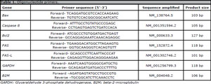
Table 1. Oligonucleotide primers.
Annexin V and propidium iodide staining
To investigate the cellular apoptosis induction, double staining was carried out using Annexin V-FITC and Propidium iodide (PI) to measure membrane PS exposure according to kit instructions. One of the key indicators of early apoptosis is the exposure of PS on the external side of the plasma membrane. Considering annexin V’s high affinity to PS binding, it was used to identify early apoptotic rate. PI, a red fluorescent intercalating dye, was used as a DNA stain to study dead cells or late apoptotic rate (Hamzeloo-Moghadam et al., 2018).The suspension of 1-5 x 10⁵ cells was centrifuged at 1,500 rpm for 3 min. Then, the supernatant was removed. Next, phosphate-buffered saline (PBS) was added to the cell pellet and centrifuged at 1,500 rpm for 3 min. The supernatant was removed and the cell pellet was re-suspended in 100-500 μl of 1X binding buffer. After that, 10 μl of annexin V - FITC was added to the cell suspension. The cell suspension was then incubated for 15 min at room temperature (RT) and in a dark place. Next, 1-5 μl of the PI solution was added and the cells were incubated for 1 to 5 min at RT and in a dark place. Cell analysis was performed with fluorescence microscopy. The percentage of green sperm cells with early apoptosis (Annexin+) and red sperm with final apoptosis (PI+ cells) was obtained.
Statistical analysis
For data analysis, SPSS version 20 (SPSS Inc., Chicago, IL, USA) was used. Data were presented as mean ± SEM. Following data normalization, Student’s t-test and the Kruskal-Wallis test were run to compare gene expression levels between the study groups. Statistically significant values had a p<0.05 for both cellular and molecular assessments. The association between apoptosis and protamine deficiency was assessed using Pearson’s and Spearman’s correlation coefficients.
RESULTS
A total of 75 men were included in the three study groups, which featured infertile patients with abnormal sperm parameters (experimental group I, n=25); infertile patients with normal sperm parameters diagnosed with UMI (experimental group II, n=25); and fertile men with normal sperm parameters (control group, n=25).
Sperm parameters
As expected, no significant difference was observed between the three study groups in terms of sperm volume (p=0.05). Experimental group I had significantly lower progressive motility in comparison with experimental group II and controls (p<0.0001 and 0.0005, respectively). Sperm concentration was significantly lower in experimental group I compared to experimental group II (p<0.0002). However, no significant difference was detected between experimental group I and controls regarding sperm concentration (p=0.08). In contrast, sperm samples of experimental group I showed significantly higher rates of immotile sperm than the samples from controls and experimental group II (p=0.002 and 0.0001, respectively). Non-progressive motility of sperm was significantly higher in experimental group I than in controls (p=0.008). Data analysis showed a significantly lower rate of normal sperm morphology in experimental group I than in experimental group II and controls (p<0.0001 and p<0.0001, respectively). We detected a significant increase in round cells in both experimental groups compared to controls (p<0.0001 vs. p<0.0001, respectively) (Table 2).
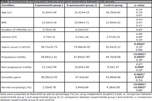
Table 2. Comparison of demographic features and sperm parameters between study groups.
Gene expression
No significant change was found in the three study groups regarding Fas and caspase 8 mRNA levels (p=0.05). A significantly higher expression of Fas ligand was found in experimental group I compared to controls. The gene expression profile of Bax and Bcl2 led to a significant difference between the experimental group I and experimental group II and controls. The expression level of Bax, Bcl2, and the Bax/Bcl2 ratio was significantly higher in experimental group I than in experimental group II and controls (Table 3).
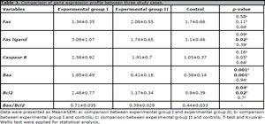
Table 3. Comparison of gene expression profile between three study cases.
In addition, data analysis showed a significant direct correlation between Fas and caspase 8 gene expression (r=0.32, p=0.01). The strongest correlation was seen between Fas ligand and caspase 8 gene expression (r=0.62, p=0.00). Furthermore, a significant positive correlation was observed between Bax and Bcl2 gene expression (r=0.47, p=0.00).
Annexin V and propidium iodide assay
No significant change was seen in terms of early apoptotic rates (ANXV+) of sperm samples between the three study groups. The highest number of necrotic sperm cells (PI+) was detected in experimental group I (Figure 1).
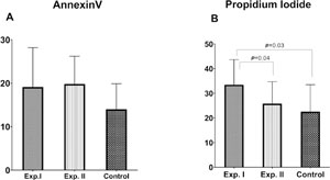
Figure 1. Detection of apoptosis using Annexin V-FITC/PI staining A) Annexin V (+) and B) PI (+) sperm cells indicated early and late apoptosis rates, respectively. Note: Significant differences between the study groups were presented by p<0.05.
There was a significant positive correlation between Fas as well as Fas ligand mRNA levels and ANXV+ sperm (r=0.31, p=0.01 vs. r=0.4, p=0.03, respectively).
DISCUSSION
The etiology of male infertility seems to be closely related to sperm concentration, motility, and morphology (Wei et al., 2015). Nevertheless, semen analysis is usually not considered a comprehensive method for semen assessment due to its limitations in the evaluation of molecular and cellular levels. On the other hand, sperm DNA integrity is necessary for sperm-egg interactions, fertilization, and early embryonic development (Van Blerkom, 1996).
We detected a significantly higher rates of Bax and Bcl2 gene expression in male partners with abnormal parameters compared to controls. The expression profile of Bcl-2 family members, due to their role in the regulation of apoptotic pathways, is required to assess the survival rate of Sertoli cells, spermatogonia, and spermatocytes. The Bax/Bcl-xl ratio also affects the fate of these cells (Yan et al., 2000). Selective expression of Bax and Bcl-2 proteins in germ cells strongly indicates that these proteins are involved in different phases of spermatogenesis, differentiation, and maturation (Oldereid et al., 2001). Bcl-2 proteins can be either pro-apoptotic, such as Bax and Bak, or anti-apoptotic, such as BCL-2, Bcl-xL, and Bcl-w. A fine balance between pro and anti-apoptotic gene function modulates the incidence of apoptosis. For instance, a high ratio of Bax/Bcl-2 reflects a pro-apoptotic tendency (Li et al., 2013). Irregular expression patterns of these proteins lead to cell death, fragmentation, and embryo mortality during the early stages of development (Giritharan et al., 2007). Dogan et al. (2013) demonstrated no significant correlation between male infertility and Bax gene expression or PS membrane translocation. They concluded that apoptosis could not be considered as one of the main fertility indicators. As for our findings, it seems that a Bax/Bcl2 expression ratio of less than 1 might not play a key role in male infertility. Furthermore, given that male partners of infertile couples with UMI showed no significant change in mRNA levels of bax, blc2, and bax/bcl2, one might conclude that apoptosis induction was more associated with abnormalities in sperm motility and/or morphology.
Caspases play a prominent role in the regulation of apoptosis in the human seminiferous epithelium. Regarding receptor-mediated programmed cell death, caspase 8 has the most important role in death signal transduction (Said et al., 2004). According to the results of the annexin V assay, there was no difference between the study groups with respect to the expression of this gene.
We did not find a significant change in Fas and Fas ligand or caspase 8 in external apoptotic pathways. One of the important apoptosis-related systems in the development of human testicular germ cells is the Fas system. Sertoli cells express FasL, binding to Fas on Fas-positive germ cells, and consequently starting apoptosis and restricting the size of the germ cell population to numbers that can be supported. Fas ligation induces the trimerization of the Fas receptor, activating caspase 8 by an adaptor Fas-associating protein with death domain (Passadaki et al., 2013). Since Fas upregulation is an external apoptotic trigger, it is possible that the sperm of infertile men have been in a more “hostile” environment than the sperm of fertile men, and that the sperm of infertile men have kept subapoptotic damage (Wang & Su, 2018). Multiple endogenous and exogenous factors are responsible for poor sperm quality and apoptosis, leading to infertility through the production of a surplus of ROS targeted towards healthy spermatozoa. For instance, genital tract infections, varicocele disease, spinal cord damage, diabetes, obesity, smoking, alcohol drinking and recreational drug use, ionizing radiation, psychological stress, freezing and thawing of sperm, strenuous exercise, or air pollutants lead to elevation of ROS (Said et al., 2010; Agarwal & Bui, 2017). Following our exclusion and inclusion criteria, we tried to eliminate the external and/or internal factors affecting ROS production to inhibit the stimulation of apoptosis pathways.
A previous study conducted by our group on a similar population about the impact of abnormal sperm parameters on the mRNA level of some sperm functional genes, oxidative stress, and SDF found significantly higher chromatin anomalies not only in experimental group I with abnormal parameters but also in experimental group II with UMI compared to controls. Moreover, we did not observe a significant change in malondialdehyde levels, a byproduct of oxidation, or total antioxidants among the study groups (Afsari et al., 2021).
Haghpanah et al. (2016) studied the impact of SDF on the developmental competence and the incidence of apoptosis after blastomeric biopsy by evaluating both TUNEL and apoptotic gene expression (BAX and bcl2). They showed that the incidence of apoptosis was not affected by SDF or by blastomeric biopsy. Taheri et al. (2015) investigated the association between Bax gene expression and the SDF index in sperm cells of infertile men. They showed no significant difference between the two groups with or without SDF in terms of Bax expression (Taheri et al., 2015). Sakkas et al. (2002) investigated the possible involvement of apoptosis in ejaculated human spermatozoa and found that TUNEL positivity and apoptotic markers were not always present in unison in spermatozoa. Nevertheless, they observed that semen samples with low sperm concentration and poor morphology were more likely to have high levels of TUNEL positivity and Fas and p53 expression. In line with our findings, they found that the presence of DNA damage was not directly related to an apoptotic process happening in spermatozoa and that it was due to problems in the nuclear remodeling process. Accordingly, the existence of apoptotic proteins in ejaculated spermatozoa may be associated with defects in cytoplasmic remodeling during the later stages of spermatogenesis (Sakkas et al., 2002).
As expected, the highest number of PI+ was found in experimental group I, with a significantly higher frequency of immotile sperm. Similarly, Januskauskas et al. (2003) showed a reverse correlation between PI+ sperm cells and sperm motility as well as viability. This finding can be attributed to the fact that PI labels all immotile cells with compromised plasma membranes.
CONCLUSION
The findings showed that the external pathways of apoptosis were not activated in the absence of external stimuli of sperm apoptosis in ejaculated sperm. Regardless of fertility status, apoptosis gene induction in the internal pathway was associated with abnormalities in sperm motility and/or morphology in men with abnormal parameters.
ACKNOWLEDGEMENTS
The authors would like to thank the Reproductive Immunology Research Center for the research facilities.
Agarwal A, Bui AD. Oxidation-reduction potential as a new marker for oxidative stress: Correlation to male infertility. Investig Clin Urol. 2017;58:385-99. PMID: 29124237 DOI: 10.4111/icu.2017.58.6.385 Medline
Agarwal A, Mulgund A, Hamada A, Chyatte MR. A unique view on male infertility around the globe. Reprod Biol Endocrinol. 2015;13:37. PMID: 25928197 DOI: 10.1186/s12958-015-0032-1 Medline
Agarwal A, Said TM. Oxidative stress, DNA damage and apoptosis in male infertility: a clinical approach. BJU Int. 2005;95:503-7. PMID: 15705068 DOI: 10.1111/j.1464-410X.2005.05328.x Medline
Carrell DT, Emery BR, Hammoud S. Altered protamine expression and diminished spermatogenesis: what is the link? Hum Reprod Update. 2007;13:313-27. PMID: 17208950 DOI: 10.1093/humupd/dml057 Medline
Dogan S, Mason MC, Govindaraju A, Belser L, Kaya A, Stokes J, Rowe D, Memili E. Interrelationships between apoptosis and fertility in bull sperm. J Reprod Dev. 2013;59:18-26. PMID: 22986927 DOI: 10.1262/jrd.2012-068 Medline
Erkkilä K, Henriksén K, Hirvonen V, Rannikko S, Salo J, Parvinen M, Dunkel L. Testosterone regulates apoptosis in adult human seminiferous tubules in vitro. J Clin Endocrinol Metab. 1997;82:2314-21. PMID: 9215313 DOI: 10.1210/jc.82.7.2314 Medline
Haghpanah T, Salehi M, Ghaffari Novin M, Masteri Farahani R, Fadaei-Fathabadi F, Dehghani-Mohammadabadi M, Azimi H. Does sperm DNA fragmentation affect the developmental potential and the incidence of apoptosis following blastomere biopsy? Syst Biol Reprod Med. 2016;62:1-10. PMID: 26678043 DOI: 10.3109/19396368.2015.1103324 Medline
Hamzeloo-Moghadam M, Aghaei M, Abdolmohammadi MH, Khalaj A, Fallahian F. Cytotoxic effect of Drimia maritima bulb extract and induction of mitochondrial apoptotic signaling in human breast cancer cells, MCF-7 and MDA-MB-468. Onco Targets Ther. 2018;11:7669-77. PMID: 30464515 DOI: 10.2147/OTT.S182786 Medline
Januskauskas A, Johannisson A, Rodriguez-Martinez H. Subtle membrane changes in cryopreserved bull semen in relation with sperm viability, chromatin structure, and field fertility. Theriogenology. 2003;60:743-58. PMID: 12832022 DOI: 10.1016/S0093-691X(03)00050-5 Medline
Kiraz Y, Adan A, Kartal Yandim M, Baran Y. Major apoptotic mechanisms and genes involved in apoptosis. Tumour Biol. 2016;37:8471-86. PMID: 27059734 DOI: 10.1007/s13277-016-5035-9 Medline
Li L, Wu W, Huang W, Hu G, Yuan W, Li W. NF-κB RNAi decreases the Bax/Bcl-2 ratio and inhibits TNF-α-induced apoptosis in human alveolar epithelial cells. Inflamm Res. 2013;62:387-97. PMID: 23334076 DOI: 10.1007/s00011-013-0590-7 Medline
Majtnerová P, Roušar T. An overview of apoptosis assays detecting DNA fragmentation. Mol Biol Rep. 2018;45:1469-78. PMID: 30022463 DOI: 10.1007/s11033-018-4258-9 Medline
Oldereid NB, Angelis PD, Wiger R, Clausen OP. Expression of Bcl-2 family proteins and spontaneous apoptosis in normal human testis. Mol Hum Reprod. 2001;7:403-8. PMID: 11331661 DOI: 10.1093/molehr/7.5.403 Medline
Panner Selvam MK, Agarwal A, Pushparaj PN, Baskaran S, Bendou H. Sperm Proteome Analysis and Identification of Fertility-Associated Biomarkers in Unexplained Male Infertility. Genes (Basel). 2019;10:522. PMID: 31336797 DOI: 10.3390/genes10070522 Medline
Passadaki T, Asimakopoulos B, Zeginiadou T, Nikolettos N. Soluble FAS and FAS ligand levels in seminal plasma: association with basic parameters of semen analysis. In Vivo. 2013;27:285-7. PMID: 23422491 Medline
Sadeghian-Nodoushan F, Aflatoonian R, Borzouie Z, Akyash F, Fesahat F, Soleimani M, Aghajanpour S, Moore HD, Aflatoonian B. Pluripotency and differentiation of cells from human testicular sperm extraction: An investigation of cell stemness. Mol Reprod Dev. 2016;83:312-23. PMID: 27077675. DOI: 10.1002/mrd.22620. Medline
Said TM, Gaglani A, Agarwal A. Implication of apoptosis in sperm cryoinjury. Reprod Biomed Online. 2010;21:456-62. PMID: 20800544 DOI: 10.1016/j.rbmo.2010.05.011 Medline
Said TM, Paasch U, Glander HJ, Agarwal A. Role of caspases in male infertility. Hum Reprod Update. 2004;10:39-51. PMID: 15005463 DOI: 10.1093/humupd/dmh003 Medline
Sakkas D, Moffatt O, Manicardi GC, Mariethoz E, Tarozzi N, Bizzaro D. Nature of DNA damage in ejaculated human spermatozoa and the possible involvement of apoptosis. Biol Reprod. 2002;66:1061-7. PMID: 11906926 DOI: 10.1095/biolreprod66.4.1061 Medline
Shukla KK, Mahdi AA, Rajender S. Apoptosis, spermatogenesis and male infertility. Front Biosci (Elite Ed). 2012;4:746-54. PMID: 22201910 DOI: 10.2741/e415 Medline
Talebi AR, Fesahat F, Mangoli E, Ghasemzadeh J, Nayeri M, Sadeghian-Nodoshan F. Relationship between sperm protamine deficiency and apoptosis in couples with unexplained repeated spontaneous abortions. Int J Reprod Biomed. 2016;14:199-204. PMID: 27294219 Medline
Van Blerkom J. Sperm centrosome dysfunction: a possible new class of male factor infertility in the human. Mol Hum Reprod. 1996;2:349-54. PMID: 9238702 DOI: 10.1093/molehr/2.5.349 Medline
Wang M, Su P. The role of the Fas/FasL signaling pathway in environmental toxicant-induced testicular cell apoptosis: An update. Syst Biol Reprod Med. 2018;64:93-102. PMID: 29299971 DOI: 10.1080/19396368.2017.1422046 Medline
Wei X, Li Q, Han Z, Lin D, Yu P. Differences in caspase-8 and-9 activity and sperm motility in infertile males of Li nationality in China. Int J Clin Exp Med. 2015;8:4721-6.PMID: 26064412 Medline
Yan W, Samson M, Jégou B, Toppari J. Bcl-w forms complexes with Bax and Bak, and elevated ratios of Bax/Bcl-w and Bak/Bcl-w correspond to spermatogonial and spermatocyte apoptosis in the testis. Mol Endocrinol. 2000;14:682-99. PMID: 10809232 DOI: 10.1210/mend.14.5.0443 Medline