JBRA Assist. Reprod. 2025;29(1):150-159
REVIEW
doi: 10.5935/1518-0557.20240088
Defining a panel of principal bacteria associated with endometritis
1Applied Molecular Biology Lab - LAPLIC. Biochemistry Department. Federal University of Rio Grande do Norte - UFRN. Natal, RN, Brazil
CONFLICT OF INTERESTS
The authors declare that there is no conflict of interest.
ABSTRACT
The aim of this study is to present a panel that includes the main bacterial genera associated with endometritis. We conducted a search using the terms “endometritis women” OR “female endometritis” OR “pelvic inflammatory disease” AND bacteria* OR “uterine microbiome” in two databases: PubMed and Web of Science, without language or publication year restrictions. The panel is based on an analysis of 40 studies published over the past 38 years. We identified 31 bacterial genera, with the following five being the most frequently cited: Chlamydia and Ureaplasma with 11.03% each, Streptococcus and Mycoplasma with 9.56% each, and Enterococcus with 8.09%. Regarding its etiological aspects, we found that bacterial infection is the most prevalent cause of the disease, occurring because of invasive procedures such as curettage, cesarean section, or insertion of intrauterine devices (IUDs), among others. These events facilitate the entry of pathogenic microorganisms into the uterus, resulting in an inflammatory response and subsequent development of endometritis. The main techniques used to detect these pathogens were microbial culture, Polymerase Chain Reaction (PCR), and Next-Generation Sequencing, with microbial culture being the most employed, followed by PCR or a combination of both techniques. This diversity of techniques has significantly expanded our understanding of the presence and identification of microorganisms associated with the pathophysiology of endometritis. Therefore, it is understood that these findings serve as a foundation for further investigations of microorganisms related to endometritis, and such analyses will help to clarify the relationship between endometritis and the bacteria that cause it..
Keywords: endometrial dysbiosis, endometrium, bacterial, IVF, fertilization
INTRODUCTION
Endometritis is characterized by a continuous inflammatory process of the endometrium and is histopathologically subdivided into acute and chronic categories (Moreno & Simon, 2018;Kitaya et al., 2018). Acute Endometritis (AE) is characterized by microabscess formation and neutrophil invasion on the endometrial epithelium surface due to the presence of aerobic and anaerobic bacteria (Singh & Sethi, 2022). Chronic Endometritis (CE) is marked by plasma cell infiltration in the endometrial stroma, causing prolonged inflammation. CE is also commonly caused by bacterial infection (Kitaya & Yasuo, 2011).
For a long time, it was believed that a healthy uterine cavity was devoid of microorganisms, as cervical mucus was considered an impermeable barrier against ascending bacteria from the vagina (Baker et al., 2018;Quayle, 2002). This hypothesis has been refuted by several studies that identified the presence of microorganisms in the uterine cavity of healthy women, noting that the mucus plug incompletely blocks the ascent of bacteria from the vagina and that uterine peristaltic movement assists in translocating particles from the vagina to the uterus (Heinonen et al., 1985;Kunz et al., 1997;Hansen et al., 2014;Chen et al., 2017).
In recent years, some studies have indicated that CE might be associated with an altered endometrial microbiome, potentially influencing the implantation process and pregnancy success (Moreno et al., 2018;Liu et al., 2019). CE is common in patients with unexplained infertility, and when diagnosed and treated, pregnancy rates are higher (Cicinelli et al., 2018;Ravel et al., 2021;Singh & Sethi, 2022). Kushnir et al. (2016) reported that about 46% of infertile patients had CE, especially those with recurrent implantation failure. However, the causal relationship between CE and reproductive failure has not been clearly established to date (Lozano et al., 2021).
The prevalence of CE is often underestimated, primarily due to diagnostic difficulties, with approximately 25% of patients showing no symptoms (Singh & Sethi, 2022;Kitaya et al., 2018). Kimura et al. (2019) report that the prevalence of CE varies from 8% to 72% in the reproductive-age women population, suggesting that this wide variation may be associated with different diagnostic criteria applied to the disease.
Given the current absence of a definitive understanding of the microbiota associated with endometritis, our objective was to formulate an inaugural panel comprising the key bacterial genera implicated in this inflammation. This carefully curated panel is poised to be instrumental in associative studies, aimed at elucidating the intricate relationship between endometritis and its bacterial provocateurs.
METHODS
We initially identified all studies published up to December 19, 2023, available in the PubMed and Web of Science databases, without language restriction. The search was conducted using the following parameters: endometritis women OR endometritis female OR “Pelvic Inflammatory Disease” AND bacteria* OR uterine microbiome.
To identify the main bacteria associated with endometritis, all titles, abstracts, and full texts of articles without publication date restriction were individually examined. Meta-analyses, and other publications that did not report original clinical data and/or did not have the term “endometritis” in the title or abstract were excluded, as were studies conducted on animal models, book chapters, and articles with unavailable full texts.
Once the main bacteria related to endometritis were identified, the search was expanded to include other already published articles about these target bacteria, as well as review studies to deepen the discussion of this study.
RESULTS
We identified 2,539 articles, with 346 in PubMed and 2,193 in Web of Science. Considering the number of publications from 1972, we observed that the number of studies citing the term endometritis increased annually over time, becoming more evident from the 90s (Figure 1).
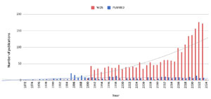
Figure 1. Publication Trends of Articles Mentioning ‘Endometritis’ (1972-2023). This graph depicts the number of articles citing ‘endometritis’ each year, based on data from Web of Science (WOS) and PubMed. The X-axis shows the year of publication, while the Y-axis represents the total number of articles published annually.
After screening by title and abstract, 68 articles were duplicates, 20 articles unavailable, 1 book chapter, and 2,340 articles that did not present the term “endometritis” or were conducted on other animals, were excluded for not meeting the inclusion criteria. After analyzing 110 articles in full, 70 of them were excluded for not being directly related to the topic. Thus, we considered only studies with women, describing the microbiota related to CE focusing on bacteria, including 40 articles of the initial total (~1.8%) published between 1985 and 2023. The detailed flowchart we used for selecting the articles is presented in Figure 2.
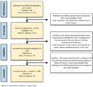
Figure 2. Literature Review and Article Selection Process Flowchart. This flowchart illustrates the methodology followed for the literature review and the criteria for selecting articles for this study PubMed: U. S. National Library of Medicine; n: sample number
Most studies were conducted in the United States (19 studies), but we also identified works from Finland and Italy with 3 studies each; Israel, Switzerland, Japan, and China with 2 studies each; Germany, Puerto Rico, Turkey, Belgium, Egypt, Kuwait, Spain, and Wales with one study each. Among the age ranges of the patients who participated in the studies, those aged between 20 and 30 years were the most represented (~28% of the works), but the set of works also included studies with younger patients (from 15 years) or older patients (40 to 69 years).
Among all the evaluated works, 16 used microbial culture techniques to investigate the presence of microorganisms related to the condition under study, 9 studies used PCR techniques to identify specific genetic material from relevant bacteria, and 4 articles combined both techniques, leveraging the strengths of each. Notably, only three studies evaluated the endometrial microbiota through next-generation sequencing, including the 16S ribosomal RNA sequencing approach. The details about each of the studies analyzed also considering the main outcomes related are available in Table 1.
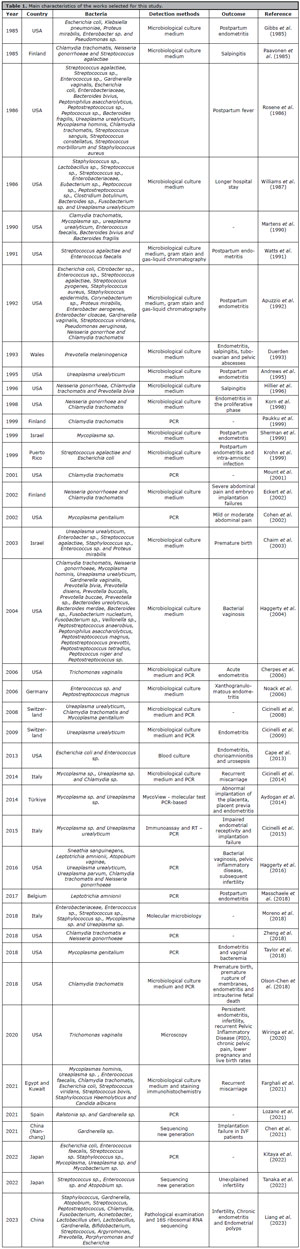
Table 1. Main characteristics of the works selected for this study.
We identified 75 bacterial genera in the endometrial microbiome of patients with endometritis. Among these, 31 genera associated with the disease were observed in 3 or more articles, with Streptococcus sp., Enterococcus sp., Chlamydia sp., Mycoplasma sp., and Ureaplasma sp. being the 5 most recurrent genera, all cited in more than 10 works (Figure 3A). Although endometritis has subtle symptomatology, the selected studies highlight the main symptoms associated with the disease: pelvic and abdominal pain, abnormal vaginal discharge, vaginal bleeding, fever, dyspareunia, purulent lochia, pyelonephritis, preterm labor, and amnionitis (Figure 3B).
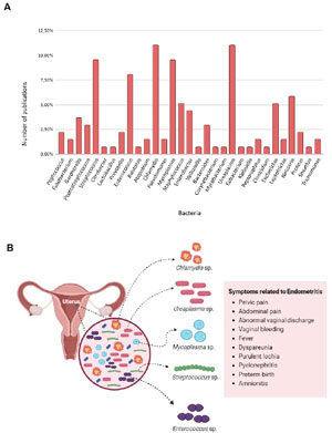
Figure 3. Bacteria and Common Symptoms Associated with Endometritis. (A) Bacteria Linked to Endometritis with the Highest Number of Mentions in Articles Published from 1985 to 2023. (B) Common Symptoms Caused by Dysbiosis Due to the Proliferation of These Bacteria in the Endometrial Tissue.
DISCUSSION
Bacterial colonization of the endometrium can be considered a key factor in the pathogenesis of endometritis. Our comprehensive analysis underscores that various bacterial genera can reach the endometrium via different routes, with the ascending pathway being the most common, where bacteria present in the vagina or cervix migrate to the uterus. Under these circumstances, during the menstrual period when the cervix is open, the risk of bacterial contamination is believed to be higher. Additionally, invasive medical procedures, such as the insertion of IUDs or dilation, curettage, and delivery methods, can facilitate the entry of bacteria into the endometrium.
As expected, we identified a significant increase in the number of studies related to endometritis over time, with the 1990s marked by a rise in publications mentioning “endometritis.” The selected studies provided important information about the symptoms associated with endometritis, as well as a growing trend in the use of molecular biology techniques to aid in characterizing the endometrial microbiome.
The collective analysis of the 40 studies selected in this research presents the most comprehensive bacterial profile of endometritis available to date, encompassing 75 genera associated with the disease. Of these, 31 genera have been frequently cited in publications over the past 38 years (Figure 3). Among them, Chlamydia sp., Ureaplasma sp., Mycoplasma sp., Streptococus sp., and Enterococcus sp. can be validated as a reduced panel for initial screenings at a lower cost.
While some authors have pointed to Gardnerella sp. and Neisseria sp. as the main pathogens associated with endometritis (Singh & Sethi, 2022;Qin & Xiao, 2022), these genera were not the most frequently mentioned in publications mapping the endometrial microbiota in patients with the disease, being cited in 5 and 8 publications, respectively.
In the study conducted by Leoni et al. (2019), bacterial genera that make up the endometrial microbiome of healthy patients were identified. Among these discoveries, the presence of Cutibacterium sp., Escherichia sp., Staphylococcus sp., Acinetobacter sp., Streptococcus sp., and Corynebacterium sp., identified in most samples, stand out. Although these genera are commonly associated with a healthy microbiota, it is intriguing to note that Escherichia sp., Staphylococcus sp., and Streptococcus sp. also appear associated with endometritis (Table 1). This duality suggests a complex interaction between bacterial composition and endometrial health, opening further avenues for investigation using more modern and comprehensive methodologies, which allow for a more precise and individualized assessment of all bacterial communities present in the sample.
Indeed, we identified only 3 studies that used next-generation sequencing for endometritis evaluation (Chen et al., 2021;Tanaka et al., 2022;Liang et al., 2023). Considering only the study by Liang et al. (2023), 5 new bacterial genera were associated with endometritis (Atopobium sp., Acinetobacter sp., Bifidobacterium sp., Argyromonas sp., and Porphyromonas sp.). This discovery suggests that the complexity of the interaction between the microbiome and endometritis is broader than previously thought and may have crucial implications for understanding the etiology of endometritis, potentially influencing more effective diagnostic and therapeutic strategies. The absence of mention of these genera in the last 38 years highlights the importance of continuous and updated research in the field of microbiology and reproductive health.
Based on our revision, we believe it is possible to establish a relationship between acute endometritis and chronic endometritis. Although the two subtypes of the disease have distinct microbial profiles, infections caused by Neisseria gonorrhoeae or Chlamydia trachomatis may initially be associated with acute endometritis but can persist in a chronic endometritis scenario, especially within the context of pelvic inflammatory disease (Kitaya et al., 2018;Singh & Sethi, 2022). In these cases, the progression of the bacterial infection may result in chronic inflammation of the endometrium, characteristic of chronic endometritis. However, this association still requires further validation, and we believe that the foundation provided here may support future studies in this area.
Given that this work also intends to establish an association between clinical practice and endometrial microbiota, we have listed a set of primary symptoms, possibly resulting from the local inflammatory response, which can be used as a baseline criterion for anamnesis. These are: pelvic and abdominal pain, abnormal vaginal discharge, vaginal bleeding, fever, dyspareunia, purulent lochia, pyelonephritis, premature birth, and amnionitis (Figure 3B). We believe that the application of this panel of symptoms, in conjunction with the bacterial genera panel presented here, can be used for associative studies, allowing for standardization in the diagnosis and indication of treatments for endometritis.
CONCLUSION
Our study offers insight into the intricate relationship between endometritis, its bacterial causatives, and consequent clinical impacts. By unveiling 31 bacterial genera, this research paves the way for an innovative diagnostic framework for endometritis, aiding in clinical diagnosis since this disease is usually asymptomatic and related to adverse outcomes in female fertility. A specialized subset, comprising Chlamydia sp., Ureaplasma sp., Mycoplasma sp., Streptococcus sp., and Enterococcus sp., stands out for cost-effective preliminary screening. These findings underscore the importance of identifying and monitoring these bacteria for a more effective approach in treating endometritis, while also providing a valuable foundation for future research aimed at developing more targeted and efficient therapies.
ACKNOWLEDGMENTS
This work did not receive specific funding for its execution. We thank the Coordination for the Improvement of Higher Education Personnel. for the financial support provided to the postgraduate program to which D.O.F. is affiliated.
REFERENCES