JBRA Assist. Reprod. 2025;29(2):338-350
REVIEW
doi: 10.5935/1518-0557.20250019
Use of time-lapse technology and artificial intelligence in the embryology laboratory: an updated review
1Fertility Medicine and Gynaecological Endocrinology Unit, Department Woman Mother Child, Lausanne University Hospital, 1011 Lausanne, Switzerland
2Département de Gynécologie-Obstétrique, Réseau Hospitalier Neuchâtelois, 2000 Neuchâtel, Switzerland
3Department of Obstetrics and Gynaecology, Villa Sofia Cervello Hospital, University of Palermo, Italy
CONFLICT OF INTEREST
No conflict of interest was reported by the author
ABSTRACT
During human in-vitro culture, morphological microscope analysis is routinely used to select embryos with the highest implantation potential for transfer, aiming for successful pregnancy and healthy live birth. This evaluation includes blastomere number, size, fragmentation, multinucleation, blastocyst (BL) expansion, and the inner-cell mass and trophectoderm appearance. However, this method requires removing embryos from the incubator, exposing them to non-physiological conditions such as fluctuations in pH, temperature, gases concentrations, as well as significant inter-observer variability. Continuous embryo culture using time-lapse monitoring (TLM) has revolutionized embryo evaluation by allowing continuous, real-time tracking of embryo development from fertilisation to blastocyst formation. This reduces the need to remove embryos from the incubator and helps maintain stable culture conditions. The monitoring system typically includes a standard incubator with an integrated microscope coupled to a digital camera, capturing images at regular intervals that are processed into a video for analysis. Despite its advantages, accurately predicting implantation rates in humans remains challenging. Recently, artificial intelligence (AI) has emerged as promising tool to objectively evaluate human embryos. AI can analyse large datasets, including embryological, clinical, and genetic information, and assist in individualizing treatment protocols. Integrating AI with TLM could improve embryo selection and enhance overall success rates. This paper explores the potential benefits of combining TLM and AI in reproductive and embryology laboratories, highlighting their potential to improve the outcomes of human ART.
Keywords: time-lapse monitoring, medically assisted reproduction, embryo culture and selection, embryo evaluation, artificial intelligence, machine learning, ploidy prediction
INTRODUCTION
Assisted reproductive technologies (ART) has advanced significantly over the past four decades, marked by several key milestones and historical achievements (Steptoe & Edwards, 1978). Since the birth of the first in-vitro fertilization (IVF) baby in 1978, over 10 million children have been born through IVF (European IVF Monitoring Consortium (EIM) for the European Society of Human Reproduction and Embryology (ESHRE), 2023). In recent years, the number of couples facing infertility has steadily increased, many of whom will eventually require IVF treatments. Worldwide, approximately 2.5 million ART cycles are performed each year, leading to over 500,000 births annually. In the UK, IVF babies account for about 3% of all babies born in 2016 (De Geyter et al., 2018; Human Fertilisation & Embryology Authority, 2018).
Despite these advancements, in-vitro embryo development remains suboptimal, and many high-quality embryos fail to implant, resulting in no pregnancy (Zhao et al., 2011; Niederberger et al., 2018; European IVF Monitoring Consortium (EIM) for the European Society of Human Reproduction and Embryology (ESHRE), 2023). The development of more complex culture media has improved embryo quality, allowing for embryo transfer at the blastocyst (BL) stage. Extending embryo culture enables the selection of embryos at a more advanced stage, and improves both uterine and embryonic synchronicity, and thus enhances pregnancy success rates (Gardner & Schoolcraft, 1999; De Vos et al., 2016; Gullo et al., 2022). Furthermore, transferring a single BL minimizes the medical risks associated with multiple pregnancies for both the mother and the baby (Sullivan et al., 2012).
However, the ability of embryologists to select the best embryo for transfer has not significantly changed since the birth of Louise Brown (Steptoe & Edwards, 1978). From the onset of IVF, it was recognized that the grade of embryo development was correlated with successful pregnancy (Edwards et al., 1984). Traditionally, embryos are selected for transfer based on morphological evaluation, which provides a snapshot of their development. For cleavage stage embryos, factors such as the number and size of blastomeres, fragmentation, and multinucleation are examined (ESHRE Guideline Group on Good Practice in IVF Labs, 2016; Gullo et al., 2023). For blastocyst assessment, the most widely used grading system was originally proposed by Gardner & Schoolcraft (1999). This alphanumeric system, while not covering all aspects of BL morphology, effectively classifies the appearance and compactness of the inner cell mass (ICM), the cohesiveness of the trophectoderm (TE) cells, and the degree of expansion of the blastocoels cavity (Gardner & Schoolcraft, 1999). However, morphological assessment has limitations in predicting implantation potential due to significant inter-observer variability (Braude, 2013).
Embryo development is a dynamic process, with morphology changing rapidly over short periods (Lemmen et al., 2008). Although TLM was first reported by Lewis & Gregory (1929), and Payne et al. (1997), it has only recently been introduced into embryology laboratory. This technology allows scientists to analyse the entire sequence of embryo development. A time-lapse system comprises three key components: an incubator, a microscope, and software. Together, these elements provide continuous monitoring while maintaining a stable and uninterrupted culture environment, which avoids the need to move embryos out of the incubator (Meseguer et al., 2012; Basile et al., 2014; Aparicio-Ruiz et al., 2016), preventing exposure to non-physiological conditions such as fluctuating temperature, humidity, pH and gas concentrations (Zhang et al., 2010). Wong et al. (2010) found that the development of human embryos to the BL stage was linked to key timing events during early divisions, such as the duration of the first cleavage and the interval between the second and third divisions. In 2011, Meseguer et al. (2011) introduced the “morphokinetics” to describe the timing of cell division events, showing that embryo implantation was associated with specific cell timing parameters.
This review paper does not aim to provide scientific evidence of the TLM, as this has been recently investigated by Armstrong et al. (2018). Instead, the primary goal is to illustrate the various TLM systems available, discuss their potential benefits in the embryology laboratory, and advise IVF clinics on selecting the most suitable system based on their specific conditions.
APPLICATION OF TIME-LAPSE TECHNOLOGY IN ART
Morphology has been the primary method of embryo assessment for over 40 years and remains the main approach for embryo selection during ART cycles. However, standard evaluations at specific time points have limitations, primarily due to the subjectivity of the embryologist and the inability to capture critical events that may affect embryo viability. Morphological assessment provides only a snapshot of embryo development at a specific moment, missing what occurs during the intervals between observations (Cruz et al., 2012). Additionally, the embryo’s grade can change significantly over a short period (Wong et al., 2010; Meseguer et al., 2011). In contrast, the TLM not only allows for the assessment of embryo morphology and dynamic changes during the in-vitro development but also provides stable culture conditions (Wong et al., 2010; Meseguer et al., 2012; Basile et al., 2014; Aparicio-Ruiz et al., 2016).
Although pioneering research on TLM began in the late 1990s (Payne et al., 1997), the technology became commercially available to embryology laboratories in 2009. The growing body of published articles on TLM in human embryology suggests its active application in embryology laboratories worldwide. However, there is limited data on its global use. Scotland is unique in this regard, as the government provided funding for all public assisted conception units (National Health Service - Scotland) to invest in TLM. Apart from this case, there are few studies reporting the worldwide use and implementation of TLM in ART cycles. One such study, by Dolinko et al. (2017), surveyed 294 IVF units in the USA. Of the 162 responding units, 35 laboratories reported using at least one time-lapse system. A similar report by Boueilh et al. (2018) from France found that among 78 respondents, 30 centres were using TLM clinically. While these surveys provide valuable insights into TLM use in two different countries, they are insufficient to draw conclusions about its global adoption. A comprehensive global survey would be valuable to assess the current use of time-lapse technology in IVF practices.
TIME-LAPSE MONITORING AND CONTINUOUS EMBRYO ASSESSMENT FROM FERTILIZATION TO BLASTOCYST FORMATION
The identification of the embryos with the highest implantation potential and viability, leading to successful pregnancies, remains a challenging goal in ART cycles. This paragraph investigates whether the use of the TLM and morphokinetic embryo assessment can aid in achieving this goal.
Time-lapse observations have been used in defining new or poorly understood aspects of human embryology, such as the fertilization process (Coticchio et al., 2018), the duration of the first three cell cycles (Wong et al., 2010; Meseguer et al., 2011), early compaction (Iwata et al., 2014) and BL formation (Marcos et al., 2015; Sciorio et al., 2020a; 2020b). Coticchio et al. (2018) conducted an in-depth analysis of the fertilization event, identifying previously unknown characteristics, including the cytoplasmic halo (its appearance and disappearance), the fading of the pronuclei (PN), the time from PN fading (tPNf), and the first cleavage. These new features were used to predict embryo quality on day-3 (Coticchio et al., 2018). A subsequent prospective study analysed the correlation between tPNf and live birth outcomes in 159 embryos. The pronuclei morphology of 46 embryos resulting in live birth was compared with that of 113 embryos that did not lead to live births. The results indicated that tPNf occurred significantly later in embryos that resulted in live births, never occurring earlier than 20 hours and 45 minutes (Azzarello et al., 2012). Further study found that erratic PN movement and delayed PN fading were indicative of compromised embryo development (Athayde Wirka et al., 2014). In a retrospective multicentre trial, the authors identified four atypical phenotypes, including abnormal syngamy, abnormal first cytokinesis, abnormal cleavage, and chaotic cleavage, which were correlated with embryo viability and implantation potential. The study concluded that embryos exhibiting these atypical phenotypes had significantly lower developmental potential compared to the control group (Athayde Wirka et al., 2014). Wong et al. (2010) suggested that the BL stage could be predicted with high sensitivity based on the timing of early developmental events, including the first cytokinesis (0-33 minutes), the time interval between the end of the first mitosis and the initiation of the second (duration of the two-cell stage: 7.8-14.3 hours), and the time interval between the second and third mitoses (duration of the three-cell stage: 0-5.8 hours). Lemmen et al. (2008) found that embryos resulting in successful pregnancies exhibited significantly higher cleavage synchrony and better synchrony in nuclear appearance at the two-cell stage compared to non-implanting embryos. Using morphokinetics assessment, associations have been demonstrated between various cleavage stage events and an embryo’s ability to reach the blastocyst stage (Wong et al., 2010; Cruz et al., 2012). Meseguer et al. (2011) analysed large datasets from embryos generated by ICSI and found that the timing of cleavage to five cells was the most predictive parameters for embryo viability and implantation. In a retrospective multicentre study conducted across ten IVF clinics, they compared pregnancy outcomes between embryos cultured using TLM (n=1,390 cycles) and standard incubators (n=5,915). The study reported a 20% improvement in pregnancy rates in the TLM group, which was attributed to a combination of both stable culture conditions and the use of morphokinetic parameters for embryo selection (Meseguer et al., 2012). Similar results were confirmed in a prospective randomized controlled trial conducted by the same group two years later (Rubio et al., 2014).
The introduction of more physiological culture conditions for in-vitro human embryos has led to the routine culture and transfer of embryos at the BL stage (Gardner & Schoolcraft, 1999; De Vos et al., 2016). In countries following a policy of single embryo transfer, there has been a notable reduction in the number of embryos being transferred. Additionally, the transfer of a single BL prevents the adverse medical conditions associated with multiple pregnancies (Sullivan et al., 2012; De Vos et al., 2016). Furthermore, blastocyst transfers result in higher implantation rates compared to transfers at the cleavage stage, although this outcome needs to be considered alongside potential detrimental epigenetic effects associated with extended in vitro culture (Kirkegaard et al., 2012). In this context, TLM has been used to predict BL formation and implantation potential based on novel morphokinetic parameters observed during the cleavage stage (Dal Canto et al., 2012). Kirkegaard et al. (2013) reported that cleavage from the two-to eight-cell stage occurs progressively earlier in embryos that will form a BL and successfully implant. They suggested high-quality blastocysts could be predicted within the first two days of in-vitro culture based on a short duration of the first cleavage and the duration of the three-cell stage (Kirkegaard et al., 2013). Similarly, Hashimoto et al. (2012) showed that higher-quality blastocysts exhibited significantly shorter times for synchrony between the three and four cell stages. More recently, Motato et al. (2016) assessed the morphokinetic parameters in 7,483 embryos and identified two features linked to BL formation: the time of morula formation (81.28-96.0 hours after ICSI) and the transition from five to eight cell embryos (≤8.78 hours). Finally, spontaneous BL collapse during in-vitro embryo development has been suggested as a novel marker of embryo viability and implantation potential. Retrospective studies have shown that BL exhibiting collapse during development are less likely to implant and result in a pregnancy compared to those that do not (Marcos et al., 2015; Sciorio et al., 2020a; 2020b). Annotation of collapse events may improve embryo assessment at blastocyst stage.
A summary of the main atypical features identified with the TLM and some published papers are presented in Tables 1 and 2.
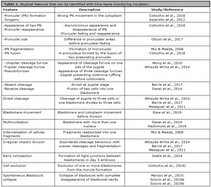
Table 1. Atypical features that can be identified with time-lapse monitoring incubator.
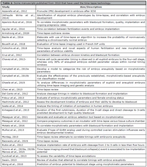
Table 2. Some manuscripts published from 2010 that have used the time-lapse technology.
DIFFERENT TLM SYSTEMS
At present, several commercially available time-lapse systems are used in reproductive laboratories for embryo selection. When selecting a TLM model, the clinic should consider various practical aspects, including the size and space requirements of each system, the cost, and the laboratory workload. In general, all systems require the use of a digital inverted microscope coupled with a camera to capture embryo images at specific time intervals. Some models feature an incubator that is equipped with a built-in camera, while other systems utilize a camera placed inside a traditional large-box incubator (Kirkegaard et al., 2012; Chen et al., 2013; Campbell & Fishel, 2015).
While all currently available TLM systems use an oil overlay on culture microdrops to prevent evaporation, there are differences in how embryos are cultured, and each system requires a specific culture dish supplied by the manufacturer. Some models offer individual culture set-up, where each dish has a designed number of wells, with each well holding one embryo (Chen et al., 2013; Campbell & Fishel, 2015; Racowsky et al., 2015). In contrast, other culture dishes allow for the sharing of culture media between compartments, making them suitable for group culture, which enables exchange of soluble components between embryos. This design may have implications for embryo development and could be an important consideration when selecting a specific model to purchase.
Additionally, each system uses a different light source and differs in the method used to bring embryos into the field of view. Some systems do not move the embryos during imaging, while others involve constant movement of the culture dish. The choice of light source also impacts the type of imaging technology used. A few systems use bright-field technology, which allows for the assessment of both kinetic parameters and embryo morphology. Other models use dark-field technology, which supports the determination of kinetic parameters but provides limited information on the morphological features of the embryos.
Other factors influencing the selection of a TLM system include the nature of the computer software used for visualisation and analysis, as well as the options for annotation. Annotations can be added either manually or automatically, which may affect the efficiency and accuracy of embryo assessment.
Finally, the selection of a time-lapse system is a critical decision that involves considering numerous factors such as system configuration, imaging technology, and software features to ensure optimal embryo evaluation and selection.
POTENTIAL BENEFIT OF TLM AND ITS IMPACT ON EMBRYO CULTURE
Human embryo culture involves various physical and chemical stressors (Wale & Gardner, 2016), which can create a hostile environment for the pre-implantation developing embryo. TLM provides consistent culture conditions, minimizing exposure to non-physiologic values, and enabling more reliable observation and assessment of the embryo development, thus improving the chances of a successful pregnancy. The culture media used to grow human embryos is another critical factor influencing embryo development. Over the past few decades, considerable improvements have been made in the composition of culture media, aimed at optimizing the conditions for embryonic growth. Two main approaches have been suggested: sequential media and single-step media.
Sequential media are designed to mimic the in vivo environment by providing different chemical compounds to the developing embryo, similar to its natural transition from the oviduct to the uterus. This media is typically tailored to meet the changing needs of the embryo as it progresses through different stages of development (Barnes et al., 1995). In contrast, the single step media is based on the concept that providing all the necessary metabolic nutrients from the outset allows the embryo to utilize these nutrients as needed, according on its specific developmental demand (Summers et al., 1995). While both methods have been widely used, the debate over which approach is superior in terms of supporting embryo development remains inconclusive (Sfontouris et al., 2016; Werner et al., 2016).
An additional layer of complexity arises with the introduction of TLM, which raises the questions of whether it can detect subtle differences in embryo development between sequential and single-step media. One of the first study to explore this was conducted by Ciray et al. (2012), who performed a randomized study on 446 oocytes. These oocytes were divided into two groups, culture in either single step or sequential media produced by the same manufacturer, and cultured in the same time-lapse incubator. The study found that embryos cultured in single-step media exhibited earlier fading of the PN and earlier cleavage up to the five-cell stage compared to those cultured in sequential media. Furthermore, in embryo that implanted, the time t2 to t4 were significantly shorter when cultured in single-step media. However, the clinical pregnancy rates did not differ significantly between the two groups (Ciray et al., 2012). A similar finding was reported by Kazdar et al. (2017), who also observed no significant differences in clinical outcomes between embryos cultured in single-step and sequential media. On the other hand, other studies have failed to identify any notable morphokinetic differences between the two approaches (Basile et al., 2013; Sfontouris et al., 2017). As a result, current data have not shown a clear advantage of either single-step or sequential media in terms of clinical pregnancies outcomes, whether embryos are cultured in standard incubators or using TLM.
As previously mentioned, TLM offers the advantage of preventing embryos from exposure to environmental fluctuations, including changes in pH, temperature, and gases concentrations (CO2 and O2), thus more closely emulating the conditions found in vivo. One of the key factors influencing embryo development is the concentration of oxygen, and an ongoing debate persists regarding culturing embryos at low oxygen levels versus ambient levels (Sciorio & Smith, 2019). The oxygen concentration within the mammalian female reproductive tract is between 2% and 8% (Fischer & Bavister, 1993). When embryos are exposed to atmospheric oxygen levels, there is a risk of increased production of reactive oxygen species, which can disrupt normal embryo metabolism and gene expression (Fischer & Bavister, 1993; Rinaudo et al., 2006; Wale & Gardner, 2012). There is considerable evidence suggesting that culturing embryo in lower oxygen concentration, such as 5% improves pregnancy outcomes. Studies have shown that embryos cultured in 5% oxygen display better developmental progression and higher implantation rates (Meintjes et al., 2009; Bontekoe et al., 2012). A large-scale prospective randomized multicentre study involving 1,563 oocytes confirmed that adding antioxidants to the culture media significantly improves embryo viability, implantation rates, and ongoing pregnancy rates. This improvement is likely due to the reduction of oxidative stress, which is harmful to developing embryos (Gardner et al., 2020).
Furthermore, TLM helps mitigate the effects of environmental stressors, allowing for stable culture conditions that more closely resemble the in vivo environment. The control of oxygen concentration and the use of antioxidants have proven beneficial in reducing oxidative stress, thus enhancing embryo development and improving clinical outcomes. As research continues, the use of TLM and optimized culture conditions promises to refine embryo selection and improve the chances of successful pregnancies in ART cycles.
EVOLUTION OF ARTIFICIAL INTELLIGENCE (AI) AND TLM
Although TLM has been proposed since the 1929 (Lewis & Gregory, 1929), the technology became commercially available only about a decade ago. In comparison to other advancements in cell biology, TLM can still be considered in its early stages, with much room for improvement. Looking ahead, it is expected that developments related to image collection will continue. The integration of fluorescence and confocal microscopy with TLM, allowing for the morphokinetic observation of organelles and chromosomes, has been already proposed (Holubcová et al., 2015; Patel et al., 2015), along with fluorescence live-cell imaging of human embryos (Hashimoto et al., 2016).
However, one of the challenges of TLM is the difficulty assessing and interpreting the vast amount of data it generates. This challenge presents an opportunity for the evolution of artificial intelligence (AI) and the use of more powered computer to analyse the large volume of image, with the goal of identifying specific parameters that correlate with embryo viability and pregnancy outcomes. In that context, software programs are being developed as automatic alternatives to standardize time-lapse annotations (Yeung et al., 2018). Unlike in other medical fields, ART has yet to fully explore the advantages of AI for automated embryo evaluation and selection.
It has been hypothesized that an AI system trained on thousands of embryo images and videos would later be able to identify and predict embryo quality autonomously. This could help reduce human error, standardize annotations, and allow embryologists to focus on other critical tasks. A study conducted by Khosravi et al. (2019) used AI in conjunction with TLM to analyse clinical data from 2,182 embryos and about 50,000 images. Their model achieved an area under the curve (AUC) of >0.98 when predicting BL quality (Khosravi et al., 2019). In another retrospective study, a deep learning approach has applied to automatically annotate 10,638 embryos videos from eight different IVF units across four countries. The results demonstrated that the deep learning model could predict foetal heartbeat and pregnancy outcomes from TLM videos with an AUC of 0.93 (Tran et al., 2019). Although these studies were retrospective, they highlight the promising potential of AI in predicting embryo viability and implantation success.
However, further prospective randomized controlled trials are required to evaluate the clinical significance of AI in IVF (Khosravi et al., 2019; Tran et al., 2019). Before AI based approaches can be clinically implemented, they will need to undergo rigours validation to ensure their reliability and accuracy.
AI: EMBRYO ASSESSMENT AND AUTOMATIC ANNOTATION
Imaging is one of the most prominent applications of AI, particularly in the field of human embryology. AI has proven effective in object identification, shape prediction, and texture analysis, with successful applications in other medical fields. In clinical embryology, AI can be applied to automatic embryo annotation, embryo grading, and embryo selection for implantation. These advancements have the potential to standardize and optimize embryo evaluation, especially with the integration TLM, which enables precise tracking of cellular divisions and detection of both normal and abnormal embryo development. AI applications in TLM can revitalize this technology and improve embryo assessment.
Commercial TLM systems, such as Geri, and ESCO, claim to incorporate machine learning (ML) into their evaluation software, although the manufacturers have not disclosed platform details or performance accuracy. Currently, embryo annotation during TLM incubation is a manual task performed by embryologists, who must annotate every features. However, this process is subject to high intraand inter-operator variability. As a result, AI-based automatic annotation systems with high accuracy and reliability are essential to reduce variability and standardize embryo assessment. The EmbryoScope by Vitrolife using AI technology, has developed an algorithm called iDAScore, that automatically analyses full time-lapse sequences to support embryo evaluation and identify embryos with the highest likelihood of implantation within a cohort. Recent studies have demonstrated the success of automatic, non-human-mediated embryo annotation systems (Dirvanauskas et al., 2019; Raudonis et al., 2019; Feyeux et al., 2020; Zaninovic & Rosenwaks, 2020). These systems must meet specific criteria: they should be fast, accurate, reproducible, and capable of distinguishing between normal and abnormal developmental features. Furthermore, these systems need to detect morphological features such as uneven size, vacuoles, and granularity, along with nuclear abnormalities like multinuclear blastomeres. The ideal system would assign appropriate weight to each characteristic for accurate prediction.
To achieve automatic annotation, several image-processing techniques, such as cell shape extraction, segmentation, can be integrated with AI, typically using convolutional neural networks (CNNs). One challenge is to recognize and detect embryos within the culture well and create an automatic region of interest (ROI). This can be accomplished using cascade classifiers or segmentation. Research has shown that the Inception V3 CNN model can perform automatic cell annotation without preprocessing the ROI, achieving high accuracy (93.9%) on human TLM images up to the eight-cell stage. Recent studies, such as those by Feyeux et al. (2020), have successfully used segmentation tools to automatically annotate embryos up to the BL stage These systems rely on quantifying features like zona pellucida thickness to identify BL expansion.
The automated tool was validated with annotations from embryologists, demonstrating the effectiveness of these automated tools and highlighting the potential of AI to improve embryo evaluation in ART (Feyeux et al., 2020).
AI AND EMBRYO GRADING
Advancements in AI within embryology have primarily focused on embryo grading, particularly at the blastocyst stage, as this is strongly correlated with implantation success. Despite the widespread use of the Gardner grading system (Gardner & Schoolcraft, 1999), its combination of numerical and letter grades often leads to inconsistencies. To address this, a simplified numerical BL score has been proposed, incorporating quantitative features of the blastocyst, such as expansion, ICM and TE (Zhan et al., 2020). However, grading variability persists even with the use of TLM. A study of ten embryologists revealed significant grading differences, while AI algorithms demonstrated superior performance in embryo evaluation and selection (Bormann et al., 2020).
The main challenge in automating this process lies in ensuring the availability of high-quality training data, as AI learns from images that have been manually graded by embryologists. Ideally, ML systems should function autonomously, without human intervention. Early attempts to automate BL grading involved differentiating the ICM from the TE through image segmentation, using support vector machines applied to two-dimensional images. AI applications have also extended to animal research. For example, a study by Matos et al. (2014) demonstrated that automatic morphological classification of mouse blastocysts could achieve 95% accuracy. The same research group applied an AI tool to successfully assess bovine embryos (Rocha et al., 2017), which was a critical step for applying AI methods to human embryo quality evaluation and prediction (Saeedi et al., 2017). For instance, CNN-based systems have been developed to automatically grade BL features, including ICM and TE morphology.
One approach, using TLM data, involved preprocessing images with cropping and applying a CNN combined with recurrent neural networks. This method predicted BL components with high accuracy of 97.8% for ICM and 98.1% for TE (Kragh et al., 2019). Another method, which used static images, achieved 96% accuracy in predicting BL expansion, 91% for ICM, and 84% for TE (Chen et al., 2019). The debate between using static images versus videos for embryo assessment continues. Videos, providing dynamic data, may offer a more comprehensive view of embryo development, while static images capture critical time points, such as 66 or 110 hours post-ICSI, that can accurately assess developmental competence. However, static images are limited in evaluating dynamic events, like blastocoel collapse, which could interfere with assessment. These approaches require further testing on similar datasets to determine which method is more effective for embryo selection (Manna et al., 2013). Recent work by Khosravi et al. (2019) suggests that AI can be clinically applied for embryo classification. Their study, focusing on blastocysts at 110 hours post-ICSI, achieved over 98% accuracy in distinguishing high grade from low grade embryos. An innovative approach involves using smartphone-based systems to evaluate embryos, with AI algorithms trained on high-quality images and applied to lower-quality images captured by portable devices. This approach achieved around 90% accuracy in distinguishing blastocysts from non-blastocysts, demonstrating AI adaptability and potential for clinical use in diverse settings (Kanakasabapathy et al., 2019).
Several AI startups are now developing algorithms to predict embryo development and implantation across various stages. Despite claims of high predictive accuracy, these AI platforms are still evolving, with CNNs proving particularly effective for image analysis. However, many studies have been based on limited training data, raising concerns about the generalizability of AI models. A significant challenge in ML is the use of unbalanced datasets, which can skew predictions towards the majority class, highlighting the need for larger and more diverse data sets for training.
EMBRYO SELECTION TO IMPROVE IMPLANTATION
Embryo selection in ART represents a critical aspect of improving implantation and pregnancy success. Traditional methods rely on morphological evaluation, but this technique suffers from significant variability and inconsistency between operators. AI offers a promising solution, aiming to improve embryo selection and predict implantation potential more objectively. However, AI models focused on predicting live birth rates, may overlook other essential factors, such as uterine conditions, which also influence pregnancy success.
Several statistical models have been developed to predict implantation success by analysing morphokinetic embryos parameters using TLM. These parameters are linked to embryo quality and can provide valuable insights into implantation potential. Some AI-based systems use TLM data to predict implantation at both the cleavage and blastocyst stages. For instance, a deep learning model developed by Tran et al. (2019) aimed to predict foetal heart rate by analysing TLM videos, claiming to offer a fully automated system for embryo assessment. While the study used a large dataset, concerns have been raised about the inclusion of abnormal or discarded embryos, which could lead to unbalanced training data and skew the results. Additional study (Chavez-Badiola et al., 2020), which used a relatively small size of approximately 200 transferred blastocysts from two clinics, applied static images to predict implantation rate and achieved an accuracy of about 70%. However, the predictive value of the model remained unclear, especially when compared to traditional grading methods. A further multicenter study using a large retrospective dataset tested an AI platform for predicting implantation rate using single blastocyst images (VerMilyea et al., 2020). The model showed an accuracy of 67.7%, with sensitivity (true positive) of 71.1% and specificity (true negative) of 65.3%. The model’s predictions were compared to those made by embryologists across eleven different IVF units, resulting in an overall improvement of 30.8%. However, the study’s methodology for grading embryos and the variation in grading practices across laboratories could influence the results.
Despite the promise of AI, the question remains whether a universal AI system for embryo evaluation and selection will be effective across different IVF laboratories. Local conditions, such as culture media, incubators, and assessment methods, could significantly impact data interpretation. To improve the generalizability of AI systems, it is essential to use diverse datasets from multiple labs during training. Additionally, incorporating other clinical parameters, such as patient age, ovarian reserve, and stimulation protocols, is necessary to enhance AI’s predictive accuracy. Finally, while AI offers significant potential in embryo selection, more research is needed to address challenges like data quality, model generalizability, and the integration of multiple clinical factors (Zaninovic et al., 2019). A comprehensive AI model incorporating both embryo imaging and morphokinetics, along with clinical data, could ultimately lead to better outcomes in ART cycles.
POTENTIAL CORRELATION BETWEEN TLM AND EMBRYOS ANEUPLOIDY
Aneuploidy refers to the presence of an incorrect number of chromosomes in a cell, such as 45 or 47 chromosomes instead of the normal 46. Aneuploidy is a significant concern in in-vitro human embryos obtained through ART treatments. The transfer of aneuploid embryos may result in implantation failure, miscarriages, or the birth of offspring with a range of potential abnormalities (Sciorio & Dattilo, 2020). The conventional procedure to investigate aneuploidy in human embryos is called preimplantation genetic testing for aneuploidy (PGT-A), previously known as preimplantation genetic screening (PGS). This procedure involves an IVF cycle in which embryos are biopsied and screened for chromosomal abnormalities before being transferred to the uterus. The procedure was first introduced by Handyside et al. (1990). However, PGT-A is an expensive technology, is not permitted in some countries, and there is ongoing debate regarding its cost-effectiveness, the invasiveness of the procedure, and its clinical efficiency (Sermon et al., 2016; Sciorio & Dattilo, 2020).
It has been hypothesized that TLM could be used to identify embryo aneuploidy, offering a cheaper, faster, and less invasive evaluation approach. Several studies have correlated morphokinetic parameters observed through TLM with the likelihood of selecting chromosomally normal embryos. It has been suggested that cell division timing need to be within an optimum range to ensure the proper execution of cellular processes before cytokinesis (Davies et al., 2012; Campbell et al., 2013a; 2013b; Montag, 2013; Swain, 2013; Chawla et al., 2015). Davies et al. (2012) found that aneuploid embryos exhibited delays in the first two cleavages and prolonged transitions between 2-cell and 4-cell stages. The authors also observed that abnormal embryos had higher rates of irregular divisions and asynchronous PN disappearance compared to normal embryos. Chavez et al. (2012) investigated the relationship between genetic status and morphokinetic parameters, demonstrating that euploid embryos have distinct timing for the first cell divisions up to the 4-cell stage. Chawla et al. (2015) assessed several morphokinetic features, including the timing of second polar body extrusion, pronuclei appearance and fading, and the duration of first, second, and third cleavages in 460 embryos to distinguish abnormal embryos. The results revealed significant differences in morphokinetic parameters between euploid and aneuploidy embryos (Chawla et al., 2015). Campbell et al. (2013a; 2013b) used TLM to develop a model for identifying embryo aneuploidies. They found the timing of early blastulation and full blastocyst formation as a key features related to embryo euploidy. Basile et al. (2014) investigated the differences in cleavage timing between chromosomally normal and abnormal embryos to help identify normal embryos. They found that normal and abnormal embryos exhibited different kinetic behaviours and proposed an algorithm as a non-invasive tool to improve the likelihood of selecting genetically normal embryos (Basile et al., 2014). A comprehensive review on the use of TLM to identify and select euploid embryos was recently published by Reignier et al. (2018). They concluded that, although several studies demonstrated significant differences in morphokinetic parameters between euploid and aneuploid embryos, none provided adequate evidence to recommend the clinical use of TLM for identifying embryo aneuploidies. Consequently, the use of time-lapse technology should not be considered as a replacement for PGT-A (Reignier et al., 2018).
CONCLUSIVE REMARKS
Despite significant progress in ART across the glove, many IVF clinics still rely on standard morphological evaluation for embryo selection, a method that has inherent limitations. To improve the selection process, it is crucial to integrate novel, objective criteria that can better predict which embryos should be selected for transfer in IVF cycles. In that context, the introduction of TLM offers exciting new morphokinetic features throughout in-vitro culture. This technology provides embryologists with deeper insights into key developmental stages of embryos, ultimately enhancing the embryo selection process and improving the chances of a successful pregnancy. Detection of atypical embryo phenotypes has proven to be essential for eliminating embryos with poor prognoses, which could lead to unsuccessful pregnancies. With the current advancements in TLM technology, a continuous and stable embryo culture environment can be maintained, allowing embryologists to detect previously unnoticed or undetectable aspects of embryonic development. These include abnormal events like direct cleavage into three cells, which have been shown to negatively impact clinical pregnancy outcomes. Looking forward, with the further advancement of AI over the next decade, it is expected that TLM will become a well-established and indispensable tool for embryo selection. By integrating TLM with non-invasive analytical methods, it is likely to be routinely utilized in IVF clinics. As a result, TLM and AI will become a vital part of the toolkit for embryologists, ensuring better embryo culture conditions and more successful outcomes in ART treatments.
REFERENCES
Aguilar J, Motato Y, Escribá MJ, Ojeda M, Muñoz E, Meseguer M. The human first cell cycle: impact on implantation. Reprod Biomed Online. 2014;28:475-84. PMID: 24581982 DOI: 10.1016/j.rbmo.2013.11.014 Medline
Aparicio-Ruiz B, Basile N, Pérez Albalá S, Bronet F, Remohí J, Meseguer M. Automatic time-lapse instrument is superior to single-point morphology observation for selecting viable embryos: retrospective study in oocyte donation. Fertil Steril. 2016;106:1379-85.e10. PMID: 27530063 DOI: 10.1016/j.fertnstert.2016.07.1117 Medline
Armstrong S, Bhide P, Jordan V, Pacey A, Farquhar C. Time-lapse systems for embryo incubation and assessment in assisted reproduction. Cochrane Database Syst Rev. 2018;5:CD011320. PMID: 31140578 DOI: 10.1002/14651858.CD011320.pub4 Medline
Athayde Wirka K, Chen AA, Conaghan J, Ivani K, Gvakharia M, Behr B, Suraj V, Tan L, Shen S. Atypical embryo phenotypes identified by time-lapse microscopy: high prevalence and association with embryo development. Fertil Steril. 2014;101:1637-48.e1-5. PMID: 24726214 DOI: 10.1016/j.fertnstert.2014.02.050 Medline
Azzarello A, Hoest T, Mikkelsen AL. The impact of pronuclei morphology and dynamicity on live birth outcome after time-lapse culture. Hum Reprod. 2012;27:2649-57. PMID: 22740496 DOI: 10.1093/humrep/des210 Medline
Barnes FL, Crombie A, Gardner DK, Kausche A, Lacham-Kaplan O, Suikkari AM, Tiglias J, Wood C, Trounson AO. Blastocyst development and birth after in-vitro maturation of human primary oocytes, intracytoplasmic sperm injection and assisted hatching. Hum Reprod. 1995;10:3243-7. PMID: 8822452 DOI: 10.1093/oxfordjournals.humrep.a135896 Medline
Barrie A, Homburg R, McDowell G, Brown J, Kingsland C, Troup S. Examining the efficacy of six published time-lapse imaging embryo selection algorithms to predict implantation to demonstrate the need for the development of specific, in-house morphokinetic selection algorithms. Fertil Steril. 2017;107:613-21. PMID: 28069186 DOI: 10.1016/j.fertnstert.2016.11.014 Medline
Basile N, Morbeck D, García-Velasco J, Bronet F, Meseguer M. Type of culture media does not affect embryo kinetics: a time-lapse analysis of sibling oocytes. Hum Reprod. 2013;28:634-41. PMID: 23315059 DOI: 10.1093/humrep/des462 Medline
Basile N, Nogales M del C, Bronet F, Florensa M, Riqueiros M, Rodrigo L, García-Velasco J, Meseguer M. Increasing the probability of selecting chromosomally normal embryos by time-lapse morphokinetics analysis. Fertil Steril. 2014;101:699-704. PMID: 24424365 DOI: 10.1016/j.fertnstert.2013.12.005 Medline
Bontekoe S, Mantikou E, Wely M van, Seshadri S, Repping S, Mastenbroek S. Low oxygen concentrations for embryo culture in assisted reproductive technologies. Cochrane Database Syst Rev. 2012;2012:CD008950. PMID: 22786519 DOI: 10.1093/humupd/dms055 Medline
Bormann CL, Thirumalaraju P, Kanakasabapathy MK, Kandula H, Souter I, Dimitriadis I, Gupta R, Pooniwala R, Shafiee H. Consistency and objectivity of automated embryo assessments using deep neural networks. Fertil Steril. 2020;113:781-7.e1. PMID: 32228880 DOI: 10.1016/j.fertnstert.2019.12.004 Medline
Boueilh T, Reignier A, Barriere P, Freour T. Time-lapse imaging systems in IVF laboratories: a French national survey. J Assist Reprod Genet. 2018;35:2181-6. PMID: 30187427 DOI: 10.1007/s10815-018-1302-6 Medline
Braude P. Selecting the “best” embryos: prospects for improvement. Reprod Biomed Online. 2013;27:644-53. PMID: 24145119 DOI: 10.1016/j.rbmo.2013.08.009 Medline
Campbell A, Fishel S. Atlas of Time Lapse Embryology. Boca Raton: CRC Press; 2015.
Campbell A, Fishel S, Bowman N, Duffy S, Sedler M, Hickman CF. Modelling a risk classification of aneuploidy in human embryos using non-invasive morphokinetics. Reprod Biomed Online. 2013a;26:477-85. PMID: 23518033 DOI: 10.1016/j.rbmo.2013.02.006 Medline
Campbell A, Fishel S, Bowman N, Duffy S, Sedler M, Thornton S. Retrospective analysis of outcomes after IVF using an aneuploidy risk model derived from time-lapse imaging without PGS. Reprod Biomed Online. 2013b;27:140-6. PMID: 23683847 DOI: 10.1016/j.rbmo.2013.04.013 Medline
Chavez SL, Loewke KE, Han J, Moussavi F, Colls P, Munne S, Behr B, Reijo Pera RA. Dynamic blastomere behaviour reflects human embryo ploidy by the four-cell stage. Nat Commun. 2012;3:1251. PMID: 23212380 DOI: 10.1038/ncomms2249 Medline
Chavez-Badiola A, Flores-Saiffe Farias A, Mendizabal-Ruiz G, Garcia-Sanchez R, Drakeley AJ, Garcia-Sandoval JP. Predicting pregnancy test results after embryo transfer by image feature extraction and analysis using machine learning. Sci Rep. 2020;10:4394. PMID: 32157183 DOI: 10.1038/s41598-020-61357-9 Medline
Chawla M, Fakih M, Shunnar A, Bayram A, Hellani A, Perumal V, Divakaran J, Budak E. Morphokinetic analysis of cleavage stage embryos and its relationship to aneuploidy in a retrospective time-lapse imaging study. J Assist Reprod Genet. 2015;32:69-75. PMID: 25395178 DOI: 10.1007/s10815-014-0372-3 Medline
Chen AA, Tan L, Suraj V, Reijo Pera R, Shen S. Biomarkers identified with time-lapse imaging: discovery, validation, and practical application. Fertil Steril. 2013;99:1035-43. PMID: 23499001 DOI: 10.1016/j.fertnstert.2013.01.143 Medline
Ciray HN, Aksoy T, Goktas C, Ozturk B, Bahceci M. Time-lapse evaluation of human embryo development in single versus sequential culture media--a sibling oocyte study. J Assist Reprod Genet. 2012;29:891-900. PMID: 22714134 DOI: 10.1007/s10815-012-9818-7 Medline
Coticchio G, Lagalla C, Sturmey R, Pennetta F, Borini A. The enigmatic morula: mechanisms of development, cell fate determination, self-correction and implications for ART. Hum Reprod Update. 2019;25:422-38. PMID: 30855681 DOI: 10.1093/humupd/dmz008 Medline
Coticchio G, Mignini Renzini M, Novara PV, Lain M, De Ponti E, Turchi D, Fadini R, Dal Canto M. Focused time-lapse analysis reveals novel aspects of human fertilization and suggests new parameters of embryo viability. Hum Reprod. 2018;33:23-31. PMID: 29149327 DOI: 10.1093/humrep/dex344 Medline
Cruz M, Garrido N, Herrero J, Pérez-Cano I, Muñoz M, Meseguer M. Timing of cell division in human cleavage-stage embryos is linked with blastocyst formation and quality. Reprod Biomed Online. 2012;25:371-81. PMID: 22877944 DOI: 10.1016/j.rbmo.2012.06.017 Medline
Dal Canto M, Coticchio G, Mignini Renzini M, De Ponti E, Novara PV, Brambillasca F, Comi R, Fadini R. Cleavage kinetics analysis of human embryos predicts development to blastocyst and implantation. Reprod Biomed Online. 2012;25:474-80. PMID: 22995750 DOI: 10.1016/j.rbmo.2012.07.016 Medline
De Geyter C, Calhaz-Jorge C, Kupka MS, Wyns C, Mocanu E, Motrenko T, Scaravelli G, Smeenk J, Vidakovic S, Goossens V; European IVF-monitoring Consortium (EIM) for the European Society of Human Reproduction and Embryology (ESHRE). ART in Europe, 2014: results generated from European registries by ESHRE: The European IVF-monitoring Consortium (EIM) for the European Society of Human Reproduction and Embryology (ESHRE). Hum Reprod. 2018 S;33:1586-601. PMID: 30032255 DOI: 10.1093/humrep/dey242 Medline
De Vos A, Van Landuyt L, Santos-Ribeiro S, Camus M, Van de Velde H, Tournaye H, Verheyen G. Cumulative live birth rates after fresh and vitrified cleavage-stage versus blastocyst-stage embryo transfer in the first treatment cycle. Hum Reprod. 2016;31:2442-9. PMID: 27619768 DOI: 10.1093/humrep/dew219 Medline
Desai N, Ploskonka S, Goodman LR, Austin C, Goldberg J, Falcone T. Analysis of embryo morphokinetics, multinucleation and cleavage anomalies using continuous time-lapse monitoring in blastocyst transfer cycles. Reprod Biol Endocrinol. 2014;12:54. PMID: 24951056 DOI: 10.1186/1477-7827-12-54 Medline
Dirvanauskas D, Maskeliunas R, Raudonis V, Damasevicius R. Embryo development stage prediction algorithm for automated time lapse incubators. Comput Methods Programs Biomed. 2019;177:161-74. PMID: 31319944 DOI: 10.1016/j.cmpb.2019.05.027 Medline
Dolinko AV, Farland LV, Kaser DJ, Missmer SA, Racowsky C. National survey on use of time-lapse imaging systems in IVF laboratories. J Assist Reprod Genet. 2017;34:1167-72. PMID: 28600620 DOI:10.1007/s10815-017-0964-9 Medline
Edwards RG, Fishel SB, Cohen J, Fehilly CB, Purdy JM, Slater JM, Steptoe PC, Webster JM. Factors influencing the success of in vitro fertilization for alleviating human infertility. J In Vitro Fert Embryo Transf. 1984;1:3-23. PMID: 6242159 DOI: 10.1007/BF01129615 Medline
ESHRE Guideline Group on Good Practice in IVF Labs; De los Santos MJ, Apter S, Coticchio G, Debrock S, Lundin K, Plancha CE, Prados F, Rienzi L, Verheyen G, Woodward B, Vermeulen N. Revised guidelines for good practice in IVF laboratories (2015). Hum Reprod. 2016;31:685-6. PMID: 26908842 DOI: 10.1093/humrep/dew016 Medline
European IVF Monitoring Consortium (EIM) for the European Society of Human Reproduction and Embryology (ESHRE); Smeenk J, Wyns C, De Geyter C, Kupka M, Bergh C, Cuevas Saiz I, De Neubourg D, Rezabek K, Tandler-Schneider A, Rugescu I, Goossens V. ART in Europe, 2019: results generated from European registries by ESHRE†. Hum Reprod. 2023;38:2321-38. PMID: 37847771 DOI: 10.1093/humrep/dead197 Medline
Ezoe K, Ohata K, Morita H, Ueno S, Miki T, Okimura T, Uchiyama K, Yabuuchi A, Kobayashi T, Montag M, Kato K. Prolonged blastomere movement induced by the delay of pronuclear fading and first cell division adversely affects pregnancy outcomes after fresh embryo transfer on Day 2: a time-lapse study. Reprod Biomed Online. 2019;38:659-68. PMID: 30853350 DOI: 10.1016/j.rbmo.2018.12.014 Medline
Feyeux M, Reignier A, Mocaer M, Lammers J, Meistermann D, Barrière P, Paul-Gilloteaux P, David L, Fréour T. Development of automated annotation software for human embryo morphokinetics. Hum Reprod. 2020;35:557-64. PMID: 32163566 DOI: 10.1093/humrep/deaa001 Medline
Fischer B, Bavister BD. Oxygen tension in the oviduct and uterus of rhesus monkeys, hamsters and rabbits. J Reprod Fertil. 1993;99:673-9. PMID: 8107053 DOI: 10.1530/jrf.0.0990673 Medline
Fréour T, Dessolle L, Lammers J, Lattes S, Barrière P. Comparison of embryo morphokinetics after in vitro fertilization-intracytoplasmic sperm injection in smoking and nonsmoking women. Fertil Steril. 2013;99:1944-50. PMID: 23465820 DOI: 10.1016/j.fertnstert.2013.01.136 Medline
Gardner DK, Kuramoto T, Tanaka M, Mitzumoto S, Montag M, Yoshida A. Prospective randomized multicentre comparison on sibling oocytes comparing G-Series media system with antioxidants versus standard G-Series media system. Reprod Biomed Online. 2020;40:637-44. PMID: 32299733 DOI: 10.1016/j.rbmo.2020.01.026 Medline
Gardner DK, Schoolcraft WB. Culture and transfer of human blastocysts. Curr Opin Obstet Gynecol. 1999;11:307-11. PMID: 10369209 DOI: 10.1097/00001703-199906000-00013 Medline
Gullo G, Basile G, Cucinella G, Greco ME, Perino A, Chiantera V, Marinelli S. Fresh vs. frozen embryo transfer in assisted reproductive techniques: a single center retrospective cohort study and ethical-legal implications. Eur Rev Med Pharmacol Sci. 2023;27:6809-23. PMID: 37522692 DOI: 10.26355/eurrev_202307_33152 Medline
Gullo G, Scaglione M, Cucinella G, Perino A, Chiantera V, D’Anna R, Laganà AS, Buzzaccarini G. Impact of assisted reproduction techniques on the neuro-psycho-motor outcome of newborns: a critical appraisal. J Obstet Gynaecol. 2022;42:2583-7. PMID: 35959847 DOI: 10.1080/01443615.2022.2109953 Medline
Handyside AH, Kontogianni EH, Hardy K, Winston RM. Pregnancies from biopsied human preimplantation embryos sexed by Y-specific DNA amplification. Nature. 1990;344:768-70. PMID: 2330030 DOI: 10.1038/344768a0 Medline
Hashimoto S, Kato N, Saeki K, Morimoto Y. Selection of high-potential embryos by culture in poly(dimethylsiloxane) microwells and time-lapse imaging. Fertil Steril. 2012;97:332-7. PMID: 22217963 DOI: 10.1016/j.fertnstert.2011.11.042 Medline
Hashimoto S, Nakano T, Yamagata K, Inoue M, Morimoto Y, Nakaoka Y. Multinucleation per se is not always sufficient as a marker of abnormality to decide against transferring human embryos. Fertil Steril. 2016;106:133-9.e6. PMID: 27060728 DOI: 10.1016/j.fertnstert.2016.03.025 Medline
Holubcová Z, Blayney M, Elder K, Schuh M. Human oocytes. Error-prone chromosome-mediated spindle assembly favors chromosome segregation defects in human oocytes. Science. 2015;348:1143-7. PMID: 26045437 DOI: 10.1126/science.aaa9529 Medline
Iwata K, Yumoto K, Sugishima M, Mizoguchi C, Kai Y, Iba Y, Mio Y. Analysis of compaction initiation in human embryos by using time-lapse cinematography. J Assist Reprod Genet. 2014;31:421-6. PMID: 24610095 DOI: 10.1007/s10815-014-0195-2 Medline
Kanakasabapathy MK, Thirumalaraju P, Bormann CL, Kandula H, Dimitriadis I, Souter I, Yogesh V, Kota Sai Pavan S, Yarravarapu D, Gupta R, Pooniwala R, Shafiee H. Development and evaluation of inexpensive automated deep learning-based imaging systems for embryology. Lab Chip. 2019;19:4139-45. PMID: 31755505 DOI: 10.1039/c9lc00721k Medline
Kazdar N, Brugnon F, Bouche C, Jouve G, Veau S, Drapier H, Rousseau C, Pimentel C, Viard P, Belaud-Rotureau MA, Ravel C. Comparison of human embryomorphokinetic parameters in sequential or global culture media. Ann Biol Clin. 2017;75:403-10. PMID: 28751285 DOI: 10.1684/abc.2017.1259 Medline
Khosravi P, Kazemi E, Zhan Q, Malmsten JE, Toschi M, Zisimopoulos P, Sigaras A, Lavery S, Cooper LAD, Hickman C, Meseguer M, Rosenwaks Z, Elemento O, Zaninovic N, Hajirasouliha I. Deep learning enables robust assessment and selection of human blastocysts after in vitro fertilization. NPJ Digit Med. 2019;2:21. PMID: 31304368 DOI:10.1038/s41746-019-0096-y Medline
Kirkegaard K, Agerholm IE, Ingerslev HJ. Time-lapse monitoring as a tool for clinical embryo assessment. Hum Reprod. 2012;27:1277-85. PMID: 22419744 DOI: 10.1093/humrep/des079 Medline
Kirkegaard K, Kesmodel US, Hindkjær JJ, Ingerslev HJ. Time-lapse parameters as predictors of blastocyst development and pregnancy outcome in embryos from good prognosis patients: a prospective cohort study. Hum Reprod. 2013;28:2643-51. PMID: 23900207 DOI: 10.1093/humrep/det300 Medline
Kragh MF, Rimestad J, Berntsen J, Karstoft H. Automatic grading of human blastocysts from time-lapse imaging. Comput Biol Med. 2019;115:103494. PMID: 31630027 DOI: 10.1016/j.compbiomed.2019.103494 Medline
Lemmen JG, Agerholm I, Ziebe S. Kinetic markers of human embryo quality using time-lapse recordings of IVF/ICSI-fertilized oocytes. Reprod Biomed Online. 2008;17:385-91. PMID: 18765009 DOI: 10.1016/s1472-6483(10)60222-2 Medline
Lewis WH, Gregory PW. Cinematographs of living developing rabbit-eggs. Science. 1929;69:226-9. PMID: 17789322 DOI: 10.1126/science.69.1782.226-a Medline
Manna C, Nanni L, Lumini A, Pappalardo S. Artificial intelligence techniques for embryo and oocyte classification. Reprod Biomed Online. 2013;26:42-9. PMID: 23177416 DOI: 10.1016/j.rbmo.2012.09.015 Medline
Marcos J, Pérez-Albalá S, Mifsud A, Molla M, Landeras J, Meseguer M. Collapse of blastocysts is strongly related to lower implantation success: a time-lapse study. Hum Reprod. 2015;30:2501-8. PMID: 26355116 DOI: 10.1093/humrep/dev216 Medline
Matos FD, Rocha JC, Nogueira MF. A method using artificial neural networks to morphologically assess mouse blastocyst quality. J Anim Sci Technol. 2014;56:15. PMID: 26290704 DOI: 10.1186/2055-0391-56-15 Medline
Meintjes M, Chantilis SJ, Douglas JD, Rodriguez AJ, Guerami AR, Bookout DM, Barnett BD, Madden JD. A controlled randomized trial evaluating the effect of lowered incubator oxygen tension on live births in a predominantly blastocyst transfer program. Hum Reprod. 2009;24:300-7. PMID: 18927130 DOI: 10.1093/humrep/den368 Medline
Meseguer M, Herrero J, Tejera A, Hilligsøe KM, Ramsing NB, Remohí J. The use of morphokinetics as a predictor of embryo implantation. Hum Reprod. 2011;26:2658-71. PMID: 21828117 DOI: 10.1093/humrep/der256 Medline
Meseguer M, Rubio I, Cruz M, Basile N, Marcos J, Requena A. Embryo incubation and selection in a time-lapse monitoring system improves pregnancy outcome compared with a standard incubator: a retrospective cohort study. Fertil Steril. 2012;98:1481-9.e10. PMID: 22975113 DOI: 10.1016/j.fertnstert.2012.08.016 Medline
Mio Y, Maeda K. Time-lapse cinematography of dynamic changes occurring during in vitro development of human embryos. Am J Obstet Gynecol. 2008;199:660.e1-5. PMID: 18823872 DOI: 10.1016/j.ajog.2008.07.023 Medline
Montag M. Morphokinetics and embryo aneuploidy: has time come or not yet? Reprod Biomed Online. 2013;26:528-30. PMID: 23602679 DOI: 10.1016/j.rbmo.2013.03.011 Medline
Muñoz M, Cruz M, Humaidan P, Garrido N, Pérez-Cano I, Meseguer M. The type of GnRH analogue used during controlled ovarian stimulation influences early embryo developmental kinetics: a time-lapse study. Eur J Obstet Gynecol Reprod Biol. 2013;168:167-72. PMID: 23351671 DOI: 10.1016/j.ejogrb.2012.12.038 Medline
National Health Service: Scotland’s Health on the Web. Available from: https://www.scot.nhs.uk/
Niederberger C, Pellicer A, Cohen J, Gardner DK, Palermo GD, O’Neill CL, Chow S, Rosenwaks Z, Cobo A, Swain JE, Schoolcraft WB, Frydman R, Bishop LA, Aharon D, Gordon C, New E, Decherney A, Tan SL, Paulson RJ, Goldfarb JM, et al. Forty years of IVF. Fertil Steril. 2018;110:185-324.e5. PMID: 30053940 DOI: 10.1016/j.fertnstert.2018.06.005 Medline
Otsuki J, Iwasaki T, Tsuji Y, Katada Y, Sato H, Tsutsumi Y, Hatano K, Furuhashi K, Matsumoto Y, Kokeguchi S, Shiotani M. Potential of zygotes to produce live births can be identified by the size of the male and female pronuclei just before their membranes break down. Reprod Med Biol. 2017;16:200-5. PMID: 29259470 DOI: 10.1002/rmb2.12032 Medline
Patel J, Tan SL, Hartshorne GM, McAinsh AD. Unique geometry of sister kinetochores in human oocytes during meiosis I may explain maternal age-associated increases in chromosomal abnormalities. Biol Open. 2015;5:178-84. PMID: 26718930 DOI: 10.1242/bio.016394 Medline
Payne D, Flaherty SP, Barry MF, Matthews CD. Preliminary observations on polar body extrusion and pronuclear formation in human oocytes using time-lapse video cinematography. Hum Reprod. 1997;12:532-41. PMID: 9130755 DOI: 10.1093/humrep/12.3.532 Medline
Racowsky C, Kovacs P, Martins WP. A critical appraisal of time-lapse imaging for embryo selection: where are we and where do we need to go? J Assist Reprod Genet. 2015;32:1025-30. PMID: 26126876 DOI: 10.1007/s10815-015-0510-6 Medline
Raudonis V, Paulauskaite-Taraseviciene A, Sutiene K, Jonaitis D. Towards the automation of early-stage human embryo development detection. Biomed Eng Online. 2019;18:120. PMID: 31830988 DOI: 10.1186/s12938-019-0738-y Medline
Reignier A, Lammers J, Barriere P, Freour T. Can time-lapse parameters predict embryo ploidy? A systematic review. Reprod Biomed Online. 2018;36:380-7. PMID: 29398421 DOI: 10.1016/j.rbmo.2018.01.001 Medline
Rinaudo PF, Giritharan G, Talbi S, Dobson AT, Schultz RM. Effects of oxygen tension on gene expression in preimplantation mouse embryos. Fertil Steril. 2006;86:1252-65,e1-36. PMID: 17008149 DOI: 10.1016/j.fertnstert.2006.05.017 Medline
Rocha JC, Passalia FJ, Matos FD, Takahashi MB, Ciniciato DS, Maserati MP, Alves MF, de Almeida TG, Cardoso BL, Basso AC, Nogueira MFG. A Method Based on Artificial Intelligence To Fully Automatize The Evaluation of Bovine Blastocyst Images. Sci Rep. 2017;7:7659. PMID: 28794478 DOI: 10.1038/s41598-017-08104-9 Medline
Rubio I, Galán A, Larreategui Z, Ayerdi F, Bellver J, Herrero J, Meseguer M. Clinical validation of embryo culture and selection by morphokinetic analysis: a randomized, controlled trial of the EmbryoScope. Fertil Steril. 2014;102:1287-94.e5. PMID: 25217875 DOI: 10.1016/j.fertnstert.2014.07.738 Medline
Rubio I, Kuhlmann R, Agerholm I, Kirk J, Herrero J, Escribá MJ, Bellver J, Meseguer M. Limited implantation success of direct-cleaved human zygotes: a time-lapse study. Fertil Steril. 2012;98:1458-63. PMID: 22925687 DOI: 10.1016/j.fertnstert.2012.07.1135 Medline
Saeedi P, Yee D, Au J, Havelock J. Automatic Identification of Human Blastocyst Components via Texture. IEEE Trans Biomed Eng. 2017;64:2968-78. PMID: 28991729 DOI: 10.1109/TBME.2017.2759665 Medline
Sciorio R, Dattilo M. PGT-A preimplantation genetic testing for aneuploidies and embryo selection in routine ART cycles: Time to step back? Clin Genet. 2020;98:107-15. PMID: 32141057 DOI: 10.1111/cge.13732 Medline
Sciorio R, Herrer Saura R, Thong KJ, Esbert Algam M, Pickering SJ, Meseguer M. Blastocyst collapse as an embryo marker of low implantation potential: a time-lapse multicentre study. Zygote. 2020a;1-9. PMID: 31928572 DOI: 10.1017/S0967199419000819 Medline
Sciorio R, Smith GD. Embryo culture at a reduced oxygen concentration of 5%: a mini review. Zygote. 2019;27:355-61. PMID: 31544720 DOI: 10.1017/S0967199419000522 Medline
Sciorio R, Thong KJ, Pickering SJ. Spontaneous blastocyst collapse as an embryo marker of low pregnancy outcome: A Time-Lapse study. JBRA Assist Reprod. 2020b;24:34-40. PMID: 31397550 DOI: 10.5935/1518-0557.20190044 Medline
Sermon K, Capalbo A, Cohen J, Coonen E, De Rycke M, De Vos A, Delhanty J, Fiorentino F, Gleicher N, Griesinger G, Grifo J, Handyside A, Harper J, Kokkali G, Mastenbroek S, Meldrum D, Meseguer M, Montag M, Munné S, Rienzi L, et al. The why, the how and the when of PGS 2.0: current practices and expert opinions of fertility specialists, molecular biologists, and embryologists. Mol Hum Reprod. 2016;22:845-57. PMID: 27256483 DOI: 10.1093/molehr/gaw034 Medline
Sfontouris IA, Kolibianakis EM, Lainas GT, Venetis CA, Petsas GK, Tarlatzis BC, Lainas TG. Blastocyst utilization rates after continuous culture in two commercial single-step media: a prospective randomized study with sibling oocytes. J Assist Reprod Genet. 2017;34:1377-83. PMID: 28718081 DOI: 10.1007/s10815-017-0997-0 Medline
Sfontouris IA, Martins WP, Nastri CO, Viana IGR, Navarro PA, Raine-Fenning N, Poel S van der, Rienzi L, Racowsky C. Blastocyst culture using single versus sequential media in clinical IVF: a systematic review and meta-analysis of randomized controlled trials. J Assist Reprod Genet. 2016;33:1261-72. PMID: 27491772 DOI: 10.1007/s10815-016-0774-5 Medline
Steptoe PC, Edwards RG. Birth after the reimplantation of a human embryo. Lancet. 1978;2:366. PMID: 79723 DOI: 10.1016/s0140-6736(78)92957-4 Medline
Sullivan EA, Wang YA, Hayward I, Chambers GM, Illingworth P, McBain J, Norman RJ. Single embryo transfer reduces the risk of perinatal mortality, a population study. Hum Reprod. 2012;27:3609-15. PMID: 22990515 DOI: 10.1093/humrep/des315 Medline
Summers MC, Bhatnagar PR, Lawitts JA, Biggers JD. Fertilization in vitro of mouse ova from inbred and outbred strains: complete preimplantation embryo development in glucose-supplemented KSOM. Biol Reprod. 1995;53:431-7. PMID: 7492697 DOI: 10.1095/biolreprod53.2.431 Medline
Sundvall L, Ingerslev HJ, Breth Knudsen U, Kirkegaard K. Interand intra-observer variability of time-lapse annotations. Hum Reprod. 2013;28:3215-21. PMID: 24070998 DOI: 10.1093/humrep/det366 Medline
Swain JE. Could time-lapse embryo imaging reduce the need for biopsy and PGS? J Assist Reprod Genet. 2013;30:1081-90. PMID: 23842747 DOI: 10.1007/s10815-013-0048-4 Medline
Tran D, Cooke S, Illingworth PJ, Gardner DK. Deep learning as a predictive tool for fetal heart pregnancy following time-lapse incubation and blastocyst transfer. Hum Reprod. 2019;34:1011-8. PMID: 31111884 DOI: 10.1093/humrep/dez064 Medline
VerMilyea M, Hall JMM, Diakiw SM, Johnston A, Nguyen T, Perugini D, Miller A, Picou A, Murphy AP, Perugini M. Development of an artificial intelligence-based assessment model for prediction of embryo viability using static images captured by optical light microscopy during IVF. Hum Reprod. 2020;35:770-84. PMID: 32240301 DOI: 10.1093/humrep/deaa013 Medline
Wale PL, Gardner DK. Oxygen regulates amino acid turnover and carbohydrate uptake during the preimplantation period of mouse embryo development. Biol Reprod. 2012;87:24, 1-8. PMID: 22553221 DOI: 10.1095/biolreprod.112.100552 Medline
Wale PL, Gardner DK. The effects of chemical and physical factors on mammalian embryo culture and their importance for the practice of assisted human reproduction. Hum Reprod Update. 2016;22:2-22. PMID: 26207016 DOI: 10.1093/humupd/dmv034 Medline
Werner MD, Hong KH, Franasiak JM, Forman EJ, Reda CV, Molinaro TA, Upham KM, Scott RT Jr. Sequential versus Monophasic Media Impact Trial (SuMMIT): a paired randomized controlled trial comparing a sequential media system to a monophasic medium. Fertil Steril. 2016;105:1215-21. PMID: 26826274 DOI: 10.1016/j.fertnstert.2016.01.005 Medline
Wong CC, Loewke KE, Bossert NL, Behr B, De Jonge CJ, Baer TM, Reijo Pera RA. Non-invasive imaging of human embryos before embryonic genome activation predicts development to the blastocyst stage. Nat Biotechnol. 2010;28:1115-21. PMID: 20890283 DOI: 10.1038/nbt.1686 Medline
Yeung S, Downing NL, Fei-Fei L, Milstein A. Bedside Computer Vision - Moving Artificial Intelligence from Driver Assistance to Patient Safety. N Engl J Med. 2018;378:1271-3. PMID: 29617592 DOI: 10.1056/NEJMp1716891 Medline
Zaninovic N, Nohales M, Zhan Q, de Los Santos ZMJ, Sierra J, Rosenwaks Z, Meseguer M. A comparison of morphokinetic markers predicting blastocyst formation and implantation potential from two large clinical data sets. J Assist Reprod Genet. 2019;36:637-46. PMID: 30671702 DOI: 10.1007/s10815-018-1396-x Medline
Zaninovic N, Rosenwaks Z. Artificial intelligence in human in vitro fertilization and embryology. Fertil Steril. 2020;114:914-20. PMID: 33160513 DOI: 10.1016/j.fertnstert.2020.09.157 Medline
Zhan Q, Sierra ET, Malmsten J, Ye Z, Rosenwaks Z, Zaninovic N. Blastocyst score, a blastocyst quality ranking tool, is a predictor of blastocyst ploidy and implantation potential. F S Rep. 2020;1:133-41. PMID: 34223229 DOI: 10.1016/j.xfre.2020.05.004 Medline
Zhang JQ, Li XL, Peng Y, Guo X, Heng BC, Tong GQ. Reduction in exposure of human embryos outside the incubator enhances embryo quality and blastulation rate. Reprod Biomed Online. 2010;20:510-5. PMID: 20129824 DOI: 10.1016/j.rbmo.2009.12.027 Medline
Zhao Y, Brezina P, Hsu CC, Garcia J, Brinsden PR, Wallach E. In vitro fertilization: four decades of reflections and promises. Biochim Biophys Acta. 2011;1810:843-52. PMID: 21605628 DOI: 10.1016/j.bbagen.2011.05.001 Medline