JBRA Assist. Reprod. 2025;29(4):721-737
REVIEW
doi: 10.5935/1518-0557.20250043
Biomarker candidates for oocyte and embryo quality assessment in assisted reproduction: A systematic review
1Programa de Pós-graduação em Biologia Celular e do Desenvolvimento, Universidade Federal de Santa Catarina, Florianópolis, SC, Brasil
2Laboratório de Reprodução e Desenvolvimento Animal (LRDA), Centro de Ciências Biológicas, Universidade Federal de Santa Catarina, Florianópolis, SC, Brasil
3Departamento de Biologia Celular, Embriologia e Genética, Centro de Ciências Biológicas, Universidade Federal de Santa Catarina, Florianópolis, SC, Brasil
CONFLICT OF INTEREST
The authors declare that they have no competing interests.#These authors contributed equally to this work
ABSTRACT
In assisted reproductive technologies, a major challenge is the relatively low overall success rate, mainly due to the absence of reliable markers for assessing oocyte and embryo quality. This study focuses on identifying new biomarkers in follicular fluid and granulosa cells to better understand the factors essential for oocyte maturation. We followed the 2020 PRISMA guidelines to review recent studies from PubMed, Scopus, and Embase, dated 2014-2024. Our review included 3062 participants from 29 studies, which identified key biomarkers in follicular fluid, such as melatonin and interleukin-10, and in granulosa cells, including mitochondrial DNA and specific transcriptomic signatures. These studies also highlighted the importance of morphological variables and DNA methylation in oocyte quality assessment. Our findings suggest that oxidative stress evaluation in follicular fluid and transcriptomic analysis of granulosa cells are promising approaches for determining oocyte quality in assisted reproduction. This knowledge has the potential to enhance patient care and optimize treatment outcomes in this field.
Keywords: assisted reproductive technology, follicular fluid, granulosa cell.
INTRODUCTION
Since the birth of the first child conceived through in vitro fertilization (IVF) in 1978, the field of reproductive medicine has experienced significant advancements (Severino & Póvoa, 2021). The global demand for assisted reproductive technology (ART) continues to surge (Beilby et al., 2023), as evidenced by the European Society of Human Reproduction and Embryology (ESHRE) reporting the delivery of over 10 million babies through ART (Feuer et al., 2014). Despite these achievements, the success rate of IVF remains relatively low, with live birth rates ranging from 30%-40% (Sang et al., 2021). Within this context, these diminished success rates can be partially attributed to the absence of reliable molecular markers to assess oocyte and embryo quality, along with recurring challenges such as interruption of oocyte maturation, fertilization failure, impaired embryonic formation, and implantation difficulties (Patrizio et al., 2007; Sang et al., 2021; Sciorio et al., 2022).
Failures in human oocyte competence arise from multifaceted factors, including intrinsic oocyte quality (Sciorio et al., 2022). In this context, dynamic interactions in the follicular microenvironment play a pivotal role. Cumulus cells (CC) or granulosa cells (GC), a subset of the follicular cells, intricately orchestrate essential cytoplasmic processes that are crucial for oocyte maturation and quality (Sciorio et al., 2022). These activities are vital for carbohydrate-protein production, organelle positioning, and metabolic pathways necessary for oocyte maturation, competence, and successful fertilization (Patrizio et al., 2007; Sciorio et al., 2022).
While oocyte morphological classification predominantly relies on microscopy techniques, it is important to note that some techniques might introduce a degree of evaluator subjectivity and possess inherent limitations (Ruvolo et al., 2013; Sirait et al., 2021; Lemseffer et al., 2022; Sciorio et al., 2022). Certain methods may not identify critical nuclear and cytoplasmic processes as well as signaling pathways in the cumulus-oocyte complex (COC) regulatory loop (Ruvolo et al., 2013; Sirait et al., 2021). Consequently, there is a need to explore non-invasive methods that can complement morphological analysis and identify quality predictors, thereby enhancing oocyte evaluation and improving ART outcomes (Fischer et al., 2021). In this context, the identification of candidate biomarkers capable of evaluating oocyte quality is welcome. Recent studies have focused on investigating non-invasive preditors associated with follicular fluid (FF) and GC, as they offer valuable insights into the molecular and cellular environment of oocytes (Wyse et al., 2020; 2021). These potential biomarkers hold significant promise in augmenting the success rates of ART procedures by enabling a more comprehensive and integrative assessment of oocyte quality.
In this study, we conducted a systematic review of the literature published from 2014 to 2024, The primary goal of this review is to evaluate oocyte quality by identifying potential biomarkers in the literature. This effort seeks to enhance the precision of oocyte assessment, which may lead to improved oocyte selection and better ART outcomes, including higher fertilization rates, increased blastocyst implantation success, and improved overall gestational outcomes. By integrating recent scientific advancements, this study aims to provide relevant findings that could impact future research and clinical practices in reproductive technologies.
METHODS
In this study, we followed the recommended review framework outlined in the 2020 Preferred Reporting Items for Systematic Reviews and Meta-Analyses (PRISMA) guidelines for the process of literature search, study selection, and data extraction (Page et al., 2021).
Literature Search
To identify articles investigating biomarkers of oocyte quality in ART cases, we conducted a search in major databases related to reproductive medicine, including PubMed, Scopus, and Embase. The search was conducted in March and April 2024, focusing on articles written in English and published between 2014-2024. This ten-year period was chosen to capture the most recent advancements in methodologies and technologies related to biomarker detection and oocyte quality assessment. To optimize our search strategy, we focused on more targeted MeSH terms that are directly related to known predictors and aspects of oocyte quality. These included: (‘oocyte’ AND ‘biomarker’ AND ‘quality’), (‘oocyte biomarker’ AND ‘oocyte quality’ OR ‘oocyte competence’ OR ‘oocyte maturity’) AND (‘polar body’ OR ‘oocyte morphology’ OR ‘meiotic spindle’ OR ‘follicular fluid’ OR ‘granulosa cells’ OR ‘cumulus cells’ OR ‘mitochondria’) as part of our query.
Literature Selection
Following the search strategy, we excluded ineligible articles and duplicates using Mendeley Reference Manager. Articles not available in full-text or not in English were also omitted. In addition, we filtered out studies on non-human species or those not aligned with our research scope. Additionally, studies involving specific clinical diagnoses such as severe endometriosis and polycystic ovary syndrome (PCOS) were excluded to align with our research objectives. Notably, our study did not specifically select for any particular age group of patients, Body Mass Index (BMI), or specific causes of infertility, including male factors, as inclusion or exclusion criteria. This decision was influenced by the diversity of the studies analyzed. Furthermore, we deliberately opted to include various study designs to encompass a broader spectrum of evidence. Our broader approach enabled us to encompass a wider array of studies, allowing for a more comprehensive evaluation of oocyte quality predictors across various study designs and patient profiles.
After evaluating titles and abstracts, we selected articles that met inclusion criteria for a full-text review. To ensure an assessment of the quality and risk of bias, we utilized the widely accepted Newcastle-Ottawa Scale (NOS), designed for evaluating the methodological quality of non-randomized studies. In the 9-scoring system, we consider studies with an overall score greater than 7 as high quality, followed by scores between 5 and 7 as fair quality and below 5 as low quality. Independently, two reviewers (E.A.D.M., V.M.L.) assessed the quality of the studies based on the NOS criteria. Articles of low quality were excluded from the review, while articles of fair or high quality were analyzed. Discrepancies or disagreements between reviewers were resolved by discussion and consensus.
Data Extraction
For the studies included in the analysis, data points were collected that included study design, publication date, sample size (number of subjects and/or samples), candidate predictors of oocyte and/or embryo quality, main results, p-value, and additional outcomes, if available.
After applying the NOS, an evaluation of the remaining studies was carried out. To enhance the understanding of the methods utilized for oocyte quality evaluation and their implications, we categorized the studies based on the biological samples examined. These categories encompassed parameters of FF, GC, and additional predictors, such as extracellular vesicle-derived microRNAs (EV-miRNAs), morphological assessment, and DNA methylation.
Although some studies have mentioned CC (Scarica et al., 2019; Shen et al., 2020; Wyse et al., 2020; Daei-Farshbaf et al., 2021) or granulosa mural cells (Karabulut et al., 2020), we chose to standardize GC to improve understanding, given that both are follicular cells.
RESULTS
Literature Search and Characteristics
The databases search resulted in 2725 results. After removing 277 duplicates and identifying articles as ineligible or unavailable for full-text reading using reference management software Mendeley, an additional 807 records were excluded at the title screening phase. These records were eliminated due to various reasons not encompassed by the previous categories, such as encountering animal studies or articles including conditions not pertinent to our inclusion criteria (severe endometriosis and PCOS), as discerned from their titles. We screened 1539 articles based on their titles/abstracts. We excluded 1208 studies due to irrelevance, misaligned objectives, unavailability in full text, or wrong parameters of the subjects. Further evaluation of 287 full-text screens for eligibility resulted in the exclusion of 258 studies, leaving a set of 29 studies (Figure 1).
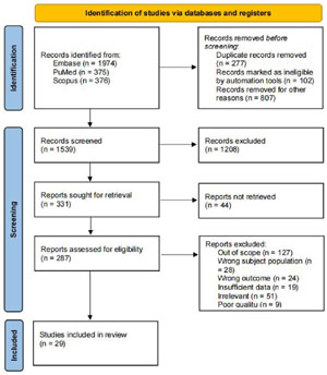
Figure 1. PRISMA 2020 flow diagram for a systematic review investigating biomarkers of oocyte quality.
According to the NOS, 27 out of the 29 articles included in the analysis demonstrated a classification of good quality. However, two articles (Ekapatria et al., 2022; Machtinger et al., 2023) were classified as fair quality due to an imbalance in the sample sizes between the analyzed groups and the lack of vitamin D data, respectively. The final set of studies encompassed various study designs, which are detailed in Tables 1 to 4.
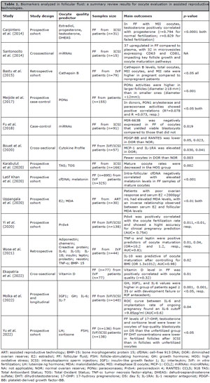
Table 1. Biomarkers analyzed in follicular fluid: a summary review results for oocyte evaluation in assisted reproductive technologies.
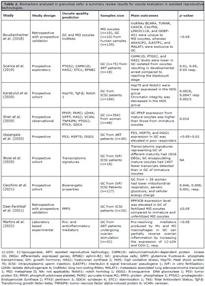
Table 2. Biomarkers analyzed in granulosa cells: a summary review results for oocyte evaluation in assisted reproductive technologies.
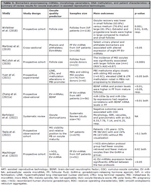
Table 3. Biomarkers encompassing miRNAs, morphology parameters, DNA methylation, and patient characteristics: a summary of review results for oocyte evaluation in assisted reproductive technologies.
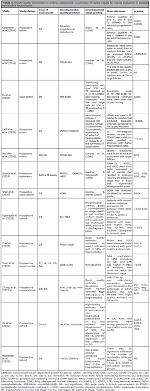
Table 4. Oocyte quality biomarkers in embryo assessment: a summary of review results for oocyte evaluation in assisted reproductive technologies.
Patient Profile
Our systematic review encompassed 3,062 individuals from 29 studies, highlighting substantial heterogeneity in participant inclusion and exclusion criteria, particularly regarding age, infertility causes, and BMI.
Age Criteria
Age-based inclusion criteria varied widely across studies. Some studies exclusively focused on younger participants, such as Ekapatria et al. (2022), Santonocito et al. (2014), Meijide et al. (2017), McCulloh et al. (2020), Uppangala et al. (2020), and Yi et al. (2020), which limited inclusion to participants under 35 years. Others imposed stricter or broader limits: Karabulut et al. (2020) included only those aged 25-37, Machtinger et al. (2023) set the upper age limit at 36, and Yu et al. (2022) excluded individuals above 38 years. Broader age ranges were observed in studies like Shen et al. (2020) (22-42 years), Molka et al. (2022) (19-42 years), and Martinez et al., (2019) (19-38 years). Conversely, some studies focused on older populations, such as Scarica et al. (2019), Cecchino et al. (2021), and Tepla et al. (2022), which primarily included women over 35 years. Additionally, some studies did not specify age criteria (Bouckenheimer et al., 2018; Latif Khan et al., 2020; Daei-Farshbaf et al., 2021; Yuan et al., 2021; Zhang et al., 2021a), while others only reported mean participant ages, such as 33-35 years (Wyse et al., 2020; 2021) and 32 years (Martins et al., 2022).
Causes of Infertility
The reviewed studies also varied in how male infertility was considered. Some studies applied strict exclusion criteria for male infertility, explicitly excluding participants with severe male factor infertility (Martinez et al., 2019; Scarica et al., 2019; Zhang et al., 2021a; Molka et al., 2022; Yu et al., 2022), while Fu et al. (2018) specifically excluded azoospermia, severe oligozoospermia, and teratozoospermia. In contrast, other studies imposed no restrictions on male factor infertility, including Carpintero et al. (2014), Bastu et al. (2015), Meijide et al. (2017), and Wirleitner et al. (2018).
Some studies explicitly focused on male infertility factors (Santonocito et al., 2014; Latif Khan et al., 2020; Shen et al., 2020; Yi et al., 2020; Cecchino et al., 2021; Martins et al., 2022; Machtinger et al., 2023). Shen et al. (2020) identified asthenozoospermia and oligozoospermia as primary infertility causes, while Machtinger et al. (2023) reported that 41.8% of cases were due to male factors. Similarly, Latif Khan et al. (2020) found that male factor infertility accounted for 57.5% of cases, and Meijide et al. (2017) observed that some type of male infertility- often in combination with other causes-was present in 58% of cases. Yi et al. (2020) and Latif Khan et al. (2020) further noted that male age and BMI did not significantly differ between pregnant and non-pregnant groups. On the other hand, some studies provided limited or no information on male infertility, such as Wyse et al. (2020; 2021) and Tepla et al. (2022), while Bouckenheimer et al. (2018) and Bouet et al. (2020) did not specify infertility causes.
BMI
Several studies applied BMI-based exclusions, such as He et al. (2023) and Yi et al. (2020), which excluded participants with BMI ≥24 kg/m2, while Sheng et al. (2017) and Ekapatria et al. (2022) only included individuals with BMI <25kg/m2. Similarly, (Machtinger et al., 2023) restricted inclusion to women with a normal or high-normal BMI (BMI < 28 kg/m2), and Yu et al. (2022) analyzed a cohort with BMI ranging from 17.2 to 27.7kg/m2. Other studies, such as Meijide et al. (2017), Daei-Farshbaf et al. (2021), Karabulut et al. (2020), and Uppangala et al. (2020), focused on participants within specific BMI ranges, excluding those above 26kg/m2.
Beyond eligibility criteria, BMI was also linked to molecular variations in CC and FF composition. Shen et al. (2020) reported that patient age and BMI influenced gene expression patterns in CC, while Wyse et al. (2021) identified a consistent correlation between TNF-α and BMI, with a disruption of leptin regulation at BMI ≥28kg/m2. Additionally, Wyse et al. (2021) found that, when stratified by BMI, the anti-inflammatory cytokine IL-10 and the pro inflammatory cytokine IL-18 were predictive of oocyte maturation in women with normal BMI, with IL-10 remaining significant even after controlling for BMI.
Biomarkers Found in Follicular Fluid
Among the 29 articles included in the analysis, 14 evaluated various aspects of the FF environment, such as the levels of Vitamin D, growth hormone (GH) or melatonin supplementation, cell-free DNA (cfDNA) fragments, as well as inflammatory parameters, biomarkers of oxidative stress, steroid levels, and regulation of immune responses within the FF (Table 1).
Biomarkers Within Granulosa Cells
We identified 9 articles that focused on several parameters associated with GC, including the evaluation of diverse aspects such as bioenergetic properties, apoptotic indices, enzymatic regulation, mitochondrial DNA (mtDNA) copy numbers, proand anti-inflammatory mediators, and the transcriptomic profile of GC (Table 2).
Additional Parameters in Oocyte Quality Assessment: EV-miRNAs, Morphological Features, and DNA Methylation
Our investigation uncovered three studies (Martinez et al., 2019; Zhang et al., 2021a; Machtinger et al., 2023) that utilized advanced in silico analyses to explore EV-miRNA activities and two (McCulloh et al., 2020; Bartolacci et al., 2022) that assessed oocyte morphology parameters’ impact on oocyte development. Additionally, we found one study Tepla et al. (2022) that examined the relationship between meiotic spindle (MS) morphology and its positioning relative to the polar body, shedding light on its critical role in oocyte maturation. We also identified one study (Zhang et al., 2021a) that focused on DNA methylation changes in metaphase II (MII) oocytes with diminished potential for embryonic development, unraveling the epigenetic complexities that underlie oocyte competence. Furthermore, Wirleitner et al. (2018) focused on the relationship between follicle size and oocyte quality, blastocyst development, and live-birth rate, identifying the optimal follicle size for oocyte retrieval (Table 3).
Impact of Oocyte Quality Biomarkers on Further Outcomes
Among the 29 articles analyzed, 17 investigated the correlation between their findings and the potential for oocyte fertilization and/or fertilization rates (Carpintero et al., 2014; Bastu et al., 2015; Meijide et al., 2017; Fu et al., 2018; Wirleitner et al., 2018; Karabulut et al., 2020; Latif Khan et al., 2020; McCulloh et al., 2020; Shen et al., 2020; Uppangala et al., 2020; Yi et al., 2020; Daei-Farshbaf et al., 2021; Wyse et al., 2021; Yuan et al., 2021; Ekapatria et al., 2022; Molka et al., 2022; Yu et al., 2022).
In the context of fertilization rates and FF biomarkers, Karabulut et al. (2020) found a correlation between the oxidative status of FF and fertilization rates (p=0.001, r=0.518). Similarly, Yi et al. (2020) demonstrated a positive correlation between FF superoxide dismutase (SOD) levels and the fertilization rate (r=0.307; p=0.020); Yu et al. (2022) indicated that FF dihydrotestosterone (DHT) levels may predict unsuccessful fertilization (sensitivity of 0.82, specificity of 0.65), while Wyse et al. (2021) recognized prolactin in FF as the key predictor for fertilization rates (sensitivity of 0.77, specificity of 0.78).
Carpintero et al. (2014) reported higher FF progesterone levels during normal fertilization compared to failed fertilization (p=0.003). Additionally, Bastu et al. (2015) found that FF Cathepsin B levels correlated with fertilization rates (p=0.042). Moreover, Meijide et al. (2017) observed significant differences in FF PON3 activity between donors and patients in large follicles, with a 20.7% higher level in donors (16.3±1.0 vs. 13.5±0.5nmol/min/ml), remaining significant after adjusting for age, BMI, number of retrieved oocytes, and fertilization rate (p=0.020).
In terms of other biomarkers and fertilization rates, McCulloh et al. (2020) stated that the follicle diameter does not effectively predict fertilization. Yuan et al. (2021) noted significant increases in the expression of LINE and LTR post-fertilization in the first polar body (PB1) and MII oocyte pair. Meanwhile, Shen et al. (2020) reported a negative correlation between TNFAIP6 expression in GC and fertilization (p=0.044). Additionally, 5 other studies (Wirleitner et al., 2018; Latif Khan et al., 2020; Uppangala et al., 2020; Ekapatria et al., 2022; Molka et al., 2022) found no substantial link between their potential biomarkers and fertilization rates in the studied groups. For development, 13 articles examined biomarkers of embryo development parameters (Table 4).
In the domain of pregnancy outcomes related to oocyte biomarkers, 7 articles investigated this association (Carpintero et al., 2014; Bastu et al., 2015; Wirleitner et al., 2018; Latif Khan et al., 2020; Yi et al., 2020; Molka et al., 2022; Tepla et al., 2022). In this context, Tepla et al. (2022) observed that in patients over 35 years of age, the visualization of MS influenced the pregnancy rate. Molka et al. (2022) reported a significant association between IGF1 levels and the cumulative pregnancy rate (AUC=0.73, p=0.001). Furthermore, they identified correlations between Interleukin-6 (IL-6) and IGF1 levels (r=0.26, p=0.005), and between IGF1 levels and endometrial thickness (r=0.37; p=0.001).
Carpintero et al. (2014) found higher estradiol levels in patients who achieved pregnancy (p=0.02). Bastu et al. (2015) reported that the area under the curve for cathepsin B predicting pregnancy was 0.662 (p=0.024, 95% CI 0.528-0.797). Wirleitner et al. (2018) observed similar implantation, pregnancy, clinical pregnancy rates, and live birth rates across transfers of blastocysts from small, medium, or large follicles.
Latif Khan et al. (2020) explored cfDNA and melatonin concentrations within FF samples during IVF procedures. Their findings highlighted a marked reduction in cfDNA paired with an elevation in melatonin levels in samples from pregnant women, in contrast to those from non-pregnant subjects (p<0.001 for both metrics). Concurrently, Yi et al. (2020) elucidated disparities in oxidative stress marker levels, namely GSH-Px (Glutathione Peroxidase), SOD (Superoxide Dismutase), and Prdx4 (Peroxiredoxin 4) in FF samples between pregnant and non-pregnant cohorts (p=0.01). Notably, Prdx4 emerged as a prominent predictor of clinical pregnancy, achieving an AUC of 0.754.
DISCUSSION
The low rate of successful live births in ART poses a significant challenge in the field (Patrizio et al., 2007; Sang et al., 2021; Sciorio et al., 2022). This may be attributed to factors encompassing inadequate oocyte maturation, failures in fertilization, embryonic development, and/or implantation processes (Patrizio et al., 2007; Pirtea et al., 2021; Sang et al., 2021). As the fate of an embryo is determined at an early stage of development, the corresponding oocyte plays a crucial role in determining the potential for human embryo development (Yuan et al., 2021).
Within the processes of oocyte interaction with its surrounding cells, there appears to be a regulatory dependency on COC, modulating essential functions to establish the optimal microenvironment for oocyte development. Consequently, gene expression profiles in GC as well as FF analyses should also reflect oocyte developmental potential. Therefore, evaluating biomarkers that reflect oocyte quality and examining their influences in predicting embryonic quality are crucial for achieving overall positive outcomes in ART (Belli et al., 2021).
Biomarkers Within Granulosa Cells: Gene Expression Profiles
HSP70, a conserved member of the heat shock protein family, has been recognized for its potent antioxidant, anti-inflammatory, and anti-apoptotic properties (Uppangala et al., 2020). In this context, studies (Karabulut et al., 2020; Uppangala et al., 2020) have explored the association between HSP70 transcripts and oocyte quality. Karabulut et al. (2020) reported lower levels of HSP70 expression in groups experiencing high oxidative stress (HOS), while Uppangala et al. (2020) observed a higher expression of transcripts in poor responder patients, characterized by an elevated oxidative stress environment. Although both studies found a correlation between oxidative stress and HSP70 expression, the cause-and-effect relationship is still unclear. While Uppangala et al. (2020) propose that HSP70 modulates oxidative stress, Karabulut et al. (2020) suggest that oxidative stress suppresses HSP70, leading to a reduction in the oxidative tolerance of the cells.
The hyaluronic acid synthase 2 (HAS2) gene, integral to GC physiology, orchestrates the synthesis of hyaluronic acid, fundamental for the structural integrity of the mature cumulus oophorus (Scarica et al., 2019). In this domain, Scarica et al. (2019) correlated enhanced HAS2 transcriptional activity in GC with oocytes that achieved blastocyst morphogenesis, intimating its prospective utility as a prognostic biomarker. Conversely, Shen et al. (2020) did not corroborate this correlation across parameters including oocyte maturation, fertilization kinetics, embryonic grading, or implantation success.
Prostaglandin endoperoxide synthase 2 (PTGS2), an enzyme essential for prostaglandin biosynthesis and implicated in cumulus expansion Scarica et al. (2019), has yielded intriguing findings. While Shen et al. (2020) found no association between PTGS2 and outcomes like oocyte maturity, fertilization, embryo grade, or implantation, Scarica et al. (2019) linked PTGS2 strongly with blastocyst development. Additionally, Wyse et al. (2020) highlighted PTGS2 as a key factor in GC pivotal for oocyte maturation. These contrasting results highlight the importance of further research with larger cohorts and uniform methodologies to better understand the roles of such biomarkers in oocyte quality.
Biomarkers Identified in Follicular Fluid: Key Molecular Signatures
Studies investigating FF biomarkers have primarily analyzed the profile of proand anti-inflammatory cytokines within the FF environment. Among these cytokines, Molka et al. (2022) observed a significant increase in pro-inflammatory cytokine IL-6 levels among patients aged ≥ 35 or with a history of embryo development failure or recurrent implantation failure (RIF). It is notable that IL-6 not only regulates the proliferation and apoptosis of GC but also plays a role in follicular dynamics. Specifically, elevated IL-6 levels have been linked to increased preantral follicle survival (Yang et al., 2020), suggesting that higher IL-6 concentrations could enhance GC viability, thereby potentially influencing oocyte quality. Bouet et al. (2020) further identified variations in cytokine levels, such as IL-2, IL-8, and IL-10, in the follicular fluid of older women and poor ovarian responders, highlighting the complexity of cytokine interactions in ovarian ageing. Moreover, their study pointed out a significant decrease in PDGF-BB concentration in diminished ovarian reserve patients, which may be linked to increased oxidative stress and altered folliculogenesis.
Concurrently, tumor necrosis factor-alpha (TNF-α) has previously been shown to enhance folliculogenesis and ovulation by promoting vascularization and suppressing GC proliferation and spontaneous apoptosis (Wyse et al., 2021). In this way, inflammatory profiles seem to impact oocyte development, with IL-10 mediating communication between oocytes and GC, potentially affecting the intrafollicular environment during maturation (Jatesada et al., 2013; Kollmann et al., 2017; Lee, 2021). Consistently, Wyse et al. (2021) identified TNF-α as a reliable predictor of oocyte maturation rate across the examined cohort. However, when data was stratified by BMI, IL-10 showed a stronger predictive capacity for oocyte maturation in normal-weight patients. Such data suggest that their predictive efficacy may vary depending on patient characteristics.
Estradiol (E2) also plays a critical role in oocyte development, particularly during nuclear and cytoplasmic maturation Uppangala et al. (2020). Synthesized by GC, E2 influences the cellular redox state, exhibiting both pro and antioxidant properties. In this context, insufficient serum E2 levels may compromise the protection against oxidative damage to the COC in poor responders (Uppangala et al., 2020). Furthermore, an intra-follicular environment lacking both E2 and melatonin negatively impacts embryo quality and fragmentation rate Latif Khan et al. (2020). However, Uppangala et al. (2020) did not assess E2 levels in the FF, and they do not solely attribute oxidative stress to inadequate E2. Carpintero et al. (2014) emphasized the importance of a balanced hormonal environment in follicular fluid, noting that higher estradiol levels are associated with better embryo quality and higher pregnancy rates. This supports the notion that E2 plays a crucial role in creating an optimal environment for oocyte maturation and successful fertilization.
Similarly, Bastu et al. (2015) found that high levels of cathepsin B in follicular fluid correlated with higher pregnancy rates, suggesting its involvement in oocyte maturation and embryo development. Meijide et al. (2017) also highlighted the importance of antioxidant enzymes, such as PONs, in follicular development, noting increased PON activities in larger follicles. However, further studies are necessary to fully understand the role of PONs in human reproduction.
GH has been the subject of numerous studies (Ipsa et al., 2019; Scheffler et al., 2021; Stoecklein et al., 2021; Molka et al., 2022; He et al., 2023) due to its multifaceted role in enhancing oocyte quality. Specifically, GH has been shown to increase the FSH and LH receptors of follicles and improve oocyte mitochondrial function (Molka et al., 2022; Yu et al., 2022). Yu et al. (2022) established a significant correlation between FF LH, FSH, cortisone levels, and oocyte development. Additionally, GH positively regulates the expression of insulin growth factor I (IGF1), thereby promoting follicle growth in patients with suboptimal responses to treatment (Scheffler et al., 2021). IGF1, in turn, plays a crucial role in ovarian function, follicular development, estrogen production, and oocyte maturation (Scheffler et al., 2021). Notably, Molka et al. (2022) established a correlation between IL-6 and IGF1 levels, indicating potential crosstalk between inflammatory and growth factor signaling pathways in the follicular environment. Interestingly, IGF1 was also found to be linked to GH levels (Molka et al., 2022). These interconnections underscore the relationship between GH, IGF1, and the ovary, offering alternatives to improve strategies for patients with poor responses to ovarian stimulation.
Melatonin, a potential biomarker of oocyte maturation (Latif Khan et al., 2020; Zhang et al., 2021b), has been shown to maintain mitochondrial membrane potential, regulate Ca2+ levels, suppress oxidative stress, and preserve oolemma permeability (Liu et al., 2019). In this scope, Latif Khan et al. (2020) found a negative correlation between melatonin concentration and cfDNA levels. Similarly, Debbarh et al. (2023) established a correlation between higher levels of cfDNA and reduced oocyte maturity and embryo quality.
Vitamin D also plays a role in oocyte maturation, influencing follicular physiology through mechanisms such as promoting cell proliferation, modulating inflammatory responses, and increasing E2 and progesterone levels (Ekapatria et al., 2022). However, the relationship between follicular vitamin D concentrations and oocyte quality remains inconsistent across studies. Ekapatria et al. (2022) reported a significant positive correlation, with oocyte quality being higher in patients with vitamin D levels ≥13.7ng/mL (n=39) compared to those with lower levels. In contrast, Ciepiela et al. (2018) observed a negative correlation, categorizing patients into <20ng/mL (n=114) and ≥20ng/mL (n=84), and reporting lower follicular vitamin D concentrations in successfully fertilized oocytes compared to those that failed fertilization. These discrepancies may stem from differences in threshold values used to classify vitamin D status, as well as variations in patient selection criteria across studies.
Previous studies have demonstrated a correlation between serum and follicular vitamin D levels, consistently reporting that follicular concentrations tend to be higher than in serum (Ozkan et al., 2010; Aleyasin et al., 2011; Firouzabadi et al., 2014). Importantly, these studies did not exclude patients with PCOS as a cause of infertility, differing from the selection criteria applied in this review. However, Aleyasin et al. (2011) reported no significant variation in serum and follicular vitamin D levels across causes of infertility. More recently, Baldini et al. (2024) investigated the impact of vitamin D supplementation combined with myo-inositol, folic acid, and melatonin, demonstrating that increasing serum and follicular vitamin D levels prior to ART was associated with improved embryo quality. These findings suggest that vitamin D supplementation may represent a promising therapeutic approach in ART; however, further investigations are warranted to clarify its role in follicular physiology and oocyte and embryo quality (Ciepiela et al., 2018; Ciepiela, 2019).
Additional Parameters of Oocyte Quality Assessment
Patient-Related Factors
Several physiological factors, including age, ovarian reserve, and BMI, influence both oocyte and embryo quality. Age-related declines in mitochondrial function and alterations in GC gene expression have been well-documented (Shen et al., 2020; Cecchino et al., 2021), while antral follicle count (AFC) and anti-Müllerian hormone (AMH) remain established indicators of oocyte competence (Broekmans et al., 2006; Jayaprakasan et al., 2010; La Marca et al., 2010; Scantamburlo et al., 2021). BMI, however, has a more complex association with reproductive outcomes. Some studies suggest that it modulates inflammatory cytokine activity (Wyse et al., 2021) and affects oocyte metabolism and maturation, with gene expression variations in cumulus cells influencing proliferation and glycolytic activity (Shen et al., 2020). Others, however, have found no significant impact of BMI on oocyte quality or ART success (Banker et al., 2017; Sheng et al., 2017; Maged et al., 2019), emphasizing the need for further investigation into these mechanisms (Snider & Wood, 2019).
Beyond female factors, sperm quality also plays a critical role in fertilization and embryo development. Male infertility has been associated with oxidative stress, DNA fragmentation, and epigenetic alterations, all of which may compromise embryonic viability (Takeshima et al., 2020). However, comparing outcomes across studies remains challenging due to variability in patient selection criteria, as some apply strict inclusion criteria by setting different limits for BMI and age, as well as varying inclusion and exclusion criteria for male infertility, while others adopt a more inclusive approach without these restrictions, potentially increasing heterogeneity in reported fertilization rates and embryo quality. These inconsistencies highlight the need for standardized selection criteria in future research to improve comparability and refine biomarker analysis.
Advancements in Non-Invasive Technologies
Numerous studies reaffirm that morphological assessment plays an essential role in optimizing ART outcomes (Figueira et al., 2010; Lasiene et al., 2011; Sciorio et al., 2024). Oocyte morphology has been evaluated based on attributes such as size, cytoplasm, zona pellucida, perivitelline space, and polar body (Ekapatria et al., 2022). Additionally, Bartolacci et al. (2022) highlighted the relevance of COC morphology, vacuoles, smooth endoplasmic reticulum aggregates, and granularity in predicting developmental competence. However, despite its clinical relevance, morphological assessment may be influenced by subjective interpretation Manna et al. (2013). To address this limitation, data automation tools have been introduced as complementary methods to refine oocyte evaluation (Fischer et al., 2021; Chéles et al., 2022), enhancing objectivity and reproducibility in clinical practice.
Integrating molecular and imaging-based techniques, non-invasive approaches have emerged as promising tools for evaluating oocyte competence (Sciorio et al., 2022). Optical-based methods, such as polarized light microscopy and refractive index measurements, have been applied to assess oocyte structures, while time-lapse embryo culture technology enables continuous imaging of embryo development (Marquet et al., 2005; Zeng et al., 2024). Additionally, artificial intelligence-driven models integrate imaging data with clinical factors to enhance predictive accuracy (Zeng et al., 2024).
At a molecular level, EV-miRNAs have been identified as key players in ovarian follicle communication, influencing granulosa cell function, follicular development, and oocyte maturation (Andronico et al., 2019; Martinez et al., 2019; Qasemi & Amidi, 2020; Shen et al., 2020; Zhang et al., 2021a). Their profiles are modulated by stimulation protocols, suggesting potential implications for IVF success (Machtinger et al., 2023). These molecular biomarkers candidates, alongside imaging-based approaches, contribute to a more comprehensive and non-invasive evaluation of oocyte quality. Although these techniques offer valuable insights into oocyte and embryo viability, further validation is required to establish their clinical applicability and standardization.
Strengths and Limitations
Our study has certain limitations related to the variability among the selected studies. Differences in inclusion and exclusion criteria led to heterogeneity in patient demographics, infertility management protocols, and the consideration of male factor infertility, which may affect the generalizability of the findings. Beyond demographic factors, variability in ovarian stimulation protocols, timing of human chorionic gonadotropin administration, and criteria related to hormonal disorders or mitochondrial function further contributed to methodological heterogeneity. These differences reflect the diverse approaches used to assess biomarkers of oocyte and embryo quality, highlighting the complexity of consolidating findings across studies. Additionally, small sample sizes and study design biases, such as retrospective analyses and methodological inconsistencies, may have influenced the reported associations between biomarkers and reproductive outcomes.However, one of the key strengths of this review is the minimized clinical impact of PCOS and severe endometriosis on oocyte evaluation. By excluding studies involving patients with these diagnoses, we ensured that the results were not biased by these clinical conditions. Furthermore, the predominant inclusion of prospective studies reduces reliance on tertiary data, enhancing the reliability of the findings. Despite the inherent variability across studies, this review provides valuable insights for reproductive medicine professionals by refining biomarker-based assessments. Future research should focus on larger, well-controlled prospective studies to validate these findings and establish standardized clinical applications, ultimately contributing to the improvement of ART outcomes.
CONCLUSION AND FUTURE DIRECTIONS
Most articles identified significant associations between predictors in FF and GC with oocyte quality. In FF, levels of specific cytokines, melatonin, GH, IGF1, and TNF-α were considered relevant biomarkers (Latif Khan et al., 2020; Wyse et al., 2021; Molka et al., 2022; Yu et al., 2022). In GC, bioenergetic properties, proand anti-inflammatory mediators, and specific transcriptomic signatures have been identified as influential predictors in the assessment of oocyte quality (Martinez et al., 2019; Scarica et al., 2019; Karabulut et al., 2020; Shen et al., 2020; Uppangala et al., 2020; Wyse et al., 2020; Daei-Farshbaf et al., 2021; Martins et al., 2022). Additionally, morphological evaluations (McCulloh et al., 2020; Bartolacci et al., 2022; Ekapatria et al., 2022; Tepla et al., 2022) and EV-miRNA expression profiles (Martinez et al., 2019; Zhang et al., 2021a; Machtinger et al., 2023) have emerged as promising tools for assessing oocyte maturity, quality, and overall reproductive potential.
The identification of multiple factors as potential predictors of oocyte quality highlights the complexity of this process. Our review underscores FF oxidative stress evaluation and GC transcriptomic signatures as promising biomarkers for oocyte assessment, with potential relevance in clinical settings. Advances in non-invasive technologies, including genomics, molecular profiling and artificial intelligence-assisted evaluation, may further enhance biomarker-based assessments, refining oocyte selection criteria and optimizing ART success rates. To ensure their effective clinical implementation, additional research is needed to validate these approaches and establish standardized protocols.
Declaration of Generative AI and AI-Assisted Technologies
During the preparation of this work, the authors used GPT-4 by OpenAI to improve textual flow and correct English language usage. A proofreading service was also engaged and following the utilization of these services, the authors meticulously reviewed and edited the content to ensure its accuracy and relevance. The authors take full responsibility for the content of the publication.
Funding
The Foundation for Research and Innovation of the State of Santa Catarina - FAPESC CP 48/2021. EM Nazari received a productivity fellowship from Conselho Nacional de Desenvolvimento Científico e Tecnológico/CNPq, Brazil (number 311570/2020-8).
REFERENCES
Aleyasin A, Hosseini MA, Mahdavi A, Safdarian L, Fallahi P, Mohajeri MR, Abbasi M, Esfahani F. Predictive value of the level of vitamin D in follicular fluid on the outcome of assisted reproductive technology. Eur J Obstet Gynecol Reprod Biol. 2011;159:132-7. PMID: 21835540 DOI: 10.1016/j.ejogrb.2011.07.006 Medline
Andronico F, Battaglia R, Ragusa M, Barbagallo D, Purrello M, Di Pietro C. Extracellular Vesicles in Human Oogenesis and Implantation. Int J Mol Sci. 2019;20:2162. PMID: 31052401 DOI: 10.3390/ijms20092162 Medline
Baldini GM, Russo M, Proietti S, Forte G, Baldini D, Trojano G. Supplementation with vitamin D improves the embryo quality in in vitro fertilization (IVF) programs, independently of the patients’ basal vitamin D status. Arch Gynecol Obstet. 2024;309:2881-90. PMID: 38580857 DOI: 10.1007/s00404-024-07473-7 Medline
Banker M, Sorathiya D, Shah S. Effect of Body Mass Index on the Outcome of In-Vitro Fertilization/Intracytoplasmic Sperm Injection in Women. J Hum Reprod Sci. 2017;10:37-43. PMID: 28479754 DOI: 10.4103/jhrs.JHRS_75_16 Medline
Bartolacci A, Intra G, Coticchio G, dell’Aquila M, Patria G, Borini A. Does morphological assessment predict oocyte developmental competence? A systematic review and proposed score. J Assist Reprod Genet. 2022;39:3-17. PMID: 34993709 DOI: 10.1007/s10815-021-02370-3 Medline
Bastu E, Gokulu SG, Dural O, Yasa C, Bulgurcuoglu S, Karamustafaoglu Balci B, Celik C, Buyru F. The association between follicular fluid levels of cathepsin B, relaxin or AMH with clinical pregnancy rates in infertile patients. Eur J Obstet Gynecol Reprod Biol. 2015;187:30-4. PMID: 25739053 DOI: 10.1016/j.ejogrb.2015.02.009 Medline
Beilby KH, Kneebone E, Roseboom TJ, Marrewijk IM van, Thompson JG, Norman RJ, Robker RL, Mol BWJ, Wang R. Offspring physiology following the use of IVM, IVF and ICSI: a systematic review and meta-analysis of animal studies. Hum Reprod Update. 2023;29:272-90. PMID: 36611003 DOI: 10.1093/humupd/dmac043 Medline
Belli M, Palmerini MG, Bianchi S, Bernardi S, Khalili MA, Nottola SA, Macchiarelli G. Ultrastructure of mitochondria of human oocytes in different clinical conditions during assisted reproduction. Arch Biochem Biophys. 2021;703:108854. PMID: 33794190 DOI: 10.1016/j.abb.2021.108854 Medline
Bouckenheimer J, Fauque P, Lecellier CH, Bruno C, Commes T, Lemaître JM, De Vos J, Assou S. Differential long non-coding RNA expression profiles in human oocytes and cumulus cells. Sci Rep. 2018;8:2202. PMID: 29396444 DOI: 10.1038/s41598-018-20727-0 Medline
Bouet PE, Boueilh T, de la Barca JMC, Boucret L, Blanchard S, Ferré-L’Hotellier V, Jeannin P, Descamps P, Procaccio V, Reynier P, May-Panloup P. The cytokine profile of follicular fluid changes during ovarian ageing. J Gynecol Obstet Hum Reprod. 2020;49:101704. PMID: 32028036 DOI: 10.1016/j.jogoh.2020.101704 Medline
Broekmans FJ, Kwee J, Hendriks DJ, Mol BW, Lambalk CB. A systematic review of tests predicting ovarian reserve and IVF outcome. Hum Reprod Update. 2006;12:685-718. PMID: 16891297 DOI: 10.1093/humupd/dml034 Medline
Carpintero NL, Suárez OA, Mangas CC, Varea CG, Rioja RG. Follicular steroid hormones as markers of oocyte quality and oocyte development potential. J Hum Reprod Sci. 2014;7:187-93. PMID: 25395744 DOI: 10.4103/0974-1208.142479 Medline
Cecchino GN, García-Velasco JA, Rial E. Reproductive senescence impairs the energy metabolism of human luteinized granulosa cells. Reprod Biomed Online. 2021;43:779-87. PMID: 34600856 DOI: 10.1016/j.rbmo.2021.08.006 Medline
Ciepiela P. Is it realistic to consider vitamin D as a follicular and serum marker of human oocyte quality? J Assist Reprod Genet. 2019;36:175-6. PMID: 30374731 DOI: 10.1007/s10815-018-1344-9 Medline
Ciepiela P, Dulęba AJ, Kowaleczko E, Chełstowski K, Kurzawa R. Vitamin D as a follicular marker of human oocyte quality and a serum marker of in vitro fertilization outcome. J Assist Reprod Genet. 2018;35:1265-76. PMID: 29774457 DOI: 10.1007/s10815-018-1179-4 Medline
Daei-Farshbaf N, Aflatoonian R, Amjadi FS, Nikniyaz H, Taleahmad S, Bakhtiyari M. Identification of calcineurin as a predictor of oocyte quality and fertilization competence based on microarray data. Comput Biol Chem. 2021;94:107561. PMID: 34461466 DOI: 10.1016/j.compbiolchem.2021.107561 Medline
Debbarh H, Jamil M, Jelloul H, Zakaria A, Louanjli N, Cadi R. Assessment of cell-free DNA and apoptosis in an oocyte microenvironment: promising biomarkers to predict intracytoplasmic sperm injection outcomes. Zygote. 2023;31:296-302. PMID: 37066898 DOI: 10.1017/S0967199423000126 Medline
Ekapatria C, Hartanto B, Wiryawan P, Tono D, Maringan Diapari Lumban T, Meita D, Arief B, Johanes Cornelius M. The Effects of Follicular Fluid 25(OH)D Concentration on Intrafollicular Estradiol Level, Oocyte Quality, and Fertilization Rate in Women Who Underwent IVF Program. J Obstet Gynaecol India. 2022;72:313-8. PMID: 35928067 DOI: 10.1007/s13224-021-01615-6 Medline
Feuer SK, Liu X, Donjacour A, Lin W, Simbulan RK, Giritharan G, Piane LD, Kolahi K, Ameri K, Maltepe E, Rinaudo PF. Use of a mouse in vitro fertilization model to understand the developmental origins of health and disease hypothesis. Endocrinology. 2014;155:1956-69. PMID: 24684304 DOI: 10.1210/en.2013-2081 Medline
Figueira RDCS, de Almeida Ferreira Braga DP, Semião-Francisco L, Madaschi C, Iaconelli A Jr, Borges E Jr. Metaphase II human oocyte morphology: contributing factors and effects on fertilization potential and embryo developmental ability in ICSI cycles. Fertil Steril. 2010;94:1115-7. PMID: 20045105 DOI: 10.1016/j.fertnstert.2009.11.039 Medline
Firouzabadi RD, Rahmani E, Rahsepar M, Firouzabadi MM. Value of follicular fluid vitamin D in predicting the pregnancy rate in an IVF program. Arch Gynecol Obstet. 2014;289:201-6. PMID: 23880888 DOI: 10.1007/s00404-013-2959-9 Medline
Fu J, Qu RG, Zhang YJ, Gu RH, Li X, Sun YJ, Wang L, Sang Q, Sun XX. Screening of miRNAs in human follicular fluid reveals an inverse relationship between microRNA-663b expression and blastocyst formation. Reprod Biomed Online. 2018;37:25-32. PMID: 29703434 DOI: 10.1016/j.rbmo.2018.03.021 Medline
Gardner DK, Lane M, Stevens J, Schlenker T, Schoolcraft WB. Blastocyst score affects implantation and pregnancy outcome: towards a single blastocyst transfer. Fertil Steril. 2000;73:1155-8. PMID: 10856474 DOI: 10.1016/S0015-0282(00)00518-5 Medline
Gardner DK, Sakkas D. Assessment of embryo viability: the ability to select a single embryo for transfer--a review. Placenta. 2003;24:5-12. PMID: 14559024 DOI: 10.1016/S0143-4004(03)00136-X Medline
Gardner DK, Schoolcraft WB. Culture and transfer of human blastocysts. Curr Opin Obstet Gynecol. 1999;11:307-11. PMID: 10369209 DOI: 10.1097/00001703-199906000-00013 Medline
He F, Wang F, Yang Y, Yuan Z, Sun C, Zou H, Chen H, Yi H, Gao SH, Zhang S, Hu L, Han TL, et al. The effect of growth hormone on the metabolome of follicular fluid in patients with diminished ovarian reserve. Reprod Biol Endocrinol. 2023;21:21. PMID: 36849898 DOI: 10.1186/s12958-023-01073-x Medline
Ipsa E, Cruzat VF, Kagize JN, Yovich JL, Keane KN. Growth Hormone and Insulin-Like Growth Factor Action in Reproductive Tissues. Front Endocrinol. 2019;10:777. PMID: 31781044 DOI: 10.3389/fendo.2019.00777 Medline
Jatesada J, Elisabeth P, Anne-Marie D. Seminal plasma did not influence the presence of transforming growth factor-β1, interleukine-10 and interleukin-6 in porcine follicles shortly after insemination. Acta Vet Scand. 2013;55:66. PMID: 24020676 DOI: 10.1186/1751-0147-55-66 Medline
Jayaprakasan K, Campbell B, Hopkisson J, Johnson I, Raine-Fenning N. A prospective, comparative analysis of anti-Müllerian hormone, inhibin-B, and three-dimensional ultrasound determinants of ovarian reserve in the prediction of poor response to controlled ovarian stimulation. Fertil Steril. 2010;93:855-64. PMID: 19046583 DOI: 10.1016/j.fertnstert.2008.10.042 Medline
Karabulut S, Korkmaz O, Kutlu P, Gozel HE, Keskin I. Effects o follicular fluid oxidative status on human mural granulosa cells, oocyte competency and ICSI parameters. Eur J Obstet Gynecol Reprod Biol. 2020;252:127-36. PMID: 32599478 DOI: 10.1016/j.ejogrb.2020.06.021 Medline
Kollmann Z, Schneider S, Fux M, Bersinger NA, von Wolff M. Gonadotrophin stimulation in IVF alters the immune cell profile in follicular fluid and the cytokine concentrations in follicular fluid and serum. Hum Reprod. 2017;32:820-31. PMID: 28201504 DOI: 10.1093/humrep/dex005 Medline
La Marca A, Sighinolfi G, Radi D, Argento C, Baraldi E, Artenisio AC, Stabile G, Volpe A. Anti-Mullerian hormone (AMH) as a predictive marker in assisted reproductive technology (ART). Hum Reprod Update. 2010;16:113-30. PMID: 19793843 DOI: 10.1093/humupd/dmp036 Medline
Latif Khan H, Bhatti S, Latif Khan Y, Abbas S, Munir Z, Rahman Khan Sherwani IA, Suhail S, Hassan Z, Aydin HH. Cell-free nucleic acids and melatonin levels in human follicular fluid predict embryo quality in patients undergoing in-vitro fertilization treatment. J Gynecol Obstet Hum Reprod. 2020;49:101624. PMID: 31472269 DOI: 10.1016/j.jogoh.2019.08.007 Medline
Lee SH. Human Adipose-Derived Stem Cells’ Paracrine Factors in Conditioned Medium Can Enhance Porcine Oocyte Maturation and Subsequent Embryo Development. Int J Mol Sci. 2021;22:579. PMID: 33430095 DOI: 10.3390/ijms22020579 Medline
Lemseffer Y, Terret ME, Campillo C, Labrune E. Methods for Assessing Oocyte Quality: A Review of Literature. Biomedicines. 2022;10:2184. PMID: 36140285 DOI: 10.3390/biomedicines10092184 Medline
Liu YJ, Ji DM, Liu ZB, Wang TJ, Xie FF, Zhang ZG, Wei ZL, Zhou P, Cao YX. Melatonin maintains mitochondrial membrane potential and decreases excessive intracellular Ca2+ levels in immature human oocytes. Life Sci. 2019;235:116810. PMID: 31472147 DOI: 10.1016/j.lfs.2019.116810 Medline
Machtinger R, Racowsky C, Baccarelli AA, Bollati V, Orvieto R, Hauser R, Barnett-Itzhaki Z. Recombinant human chorionic gonadotropin and gonadotropin-releasing hormone agonist differently affect the profile of extracellular vesicle microRNAs in human follicular fluid. J Assist Reprod Genet. 2023;40:527-36. PMID: 36609942 DOI: 10.1007/s10815-022-02703-w Medline
Maged AM, Fahmy RM, Rashwan H, Mahmood M, Hassan SM, Nabil H, Hany A, Lotfy R, Lasheen YS, Dahab S, Darwish M. Effect of body mass index on the outcome of IVF cycles among patients with poor ovarian response. Int J Gynaecol Obstet. 2019;144:161-6. PMID: 30407618 DOI: 10.1002/ijgo.12706 Medline
Manna C, Nanni L, Lumini A, Pappalardo S. Artificial intelligence techniques for embryo and oocyte classification. Reprod Biomed Online. 2013;26:42-9. PMID: 23177416 DOI: 10.1016/j.rbmo.2012.09.015 Medline
Marquet P, Rappaz B, Magistretti PJ, Cuche E, Emery Y, Colomb T, Depeursinge C. Digital holographic microscopy: a noninvasive contrast imaging technique allowing quantitative visualization of living cells with subwavelength axial accuracy. Opt Lett. 2005;30:468-70. PMID: 15789705 DOI: 10.1364/OL.30.000468 Medline
Martinez RM, Hauser R, Liang L, Mansur A, Adir M, Dioni L, Racowsky C, Bollati V, Baccarelli AA, Machtinger R. Urinary concentrations of phenols and phthalate metabolites reflect extracellular vesicle microRNA expression in follicular fluid. Environ Int. 2019;123:20-8. PMID: 30481674 DOI: 10.1016/j.envint.2018.11.043 Medline
Martins TS, Fonseca BM, Rebelo I. The role of macrophages phenotypes in the activation of resolution pathways within human granulosa cells. Reprod Biol Endocrinol. 2022;20:116. PMID: 35948935 DOI: 10.1186/s12958-022-00983-6 Medline
McCulloh DH, Kutchukhidze N, Charkviani T, Zhorzholadze T, Barbakadze T, Munné S, Chkonia L. Follicle size indicates oocyte maturity and blastocyst formation but not blastocyst euploidy following controlled ovarian hyperstimulation of oocyte donors. Hum Reprod. 2020;35:545-56. PMID: 32142586 DOI: 10.1093/humrep/dez291 Medline
Meijide S, Pérez-Ruiz I, Hernández ML, Navarro R, Ferrando M, Larreategui Z, Ruiz-Sanz JI, Ruiz-Larrea MB. Paraoxonase activities in human follicular fluid: role in follicular maturation. Reprod Biomed Online. 2017;35:351-62. PMID: 28688923 DOI: 10.1016/j.rbmo.2017.06.008 Medline
Molka B, Gwladys B, Dorian B, Lucie M, Mustapha B, Rosalie C, Brigitte G, Hafida KC, Moncef B. Follicular Fluid Growth Factors and Interleukin Profiling as Potential Predictors of IVF Outcomes. Front Physiol. 2022;13:859790. PMID: 35770187 DOI: 10.3389/fphys.2022.859790 Medline
Ozkan S, Jindal S, Greenseid K, Shu J, Zeitlian G, Hickmon C, Pal L. Replete vitamin D stores predict reproductive success following in vitro fertilization. Fertil Steril. 2010;94:1314-9. PMID: 19589516 DOI: 10.1016/j.fertnstert.2009.05.019 Medline
Page MJ, McKenzie JE, Bossuyt PM, Boutron I, Hoffmann TC, Mulrow CD, Shamseer L, Tetzlaff JM, Akl EA, Brennan SE, Chou R, Glanville J, Grimshaw JM, Hróbjartsson A, Lalu MM, Li T, Loder EW, Mayo-Wilson E, McDonald S, McGuinness LA, Stewart LA, Thomas J, Tricco AC, Welch VA, Whiting P, Moher D. The PRISMA 2020 statement: An updated guideline for reporting systematic reviews. Int J Surg. 2021 88:105906. PMID: 33789826. DOI: 10.1016/j.ijsu.2021.105906 Medline
Patrizio P, Fragouli E, Bianchi V, Borini A, Wells D. Molecular methods for selection of the ideal oocyte. Reprod Biomed Online. 2007;15:346-53. PMID: 17854537 DOI: 10.1016/S1472-6483(10)60349-5 Medline
Pirtea P, Scott RT Jr, de Ziegler D, Ayoubi JM. Recurrent implantation failure: how common is it? Curr Opin Obstet Gynecol. 2021;33:207-12. PMID: 33896917 DOI: 10.1097/GCO.0000000000000698 Medline
Qasemi M, Amidi F. Extracellular microRNA profiling in human follicular fluid: new biomarkers in female reproductive potential. J Assist Reprod Genet. 2020;37:1769-80. PMID: 32642870 DOI: 10.1007/s10815-020-01860-0 Medline
Ruvolo G, Fattouh RR, Bosco L, Brucculeri AM, Cittadini E. New molecular markers for the evaluation of gamete quality. J Assist Reprod Genet. 2013;30:207-12. PMID: 23371558 DOI: 10.1007/s10815-013-9943-y Medline
Sang Q, Zhou Z, Mu J, Wang L. Genetic factors as potential molecular markers of human oocyte and embryo quality. J Assist Reprod Genet. 2021;38:993-1002. PMID: 33895934 DOI: 10.1007/s10815-021-02196-z Medline
Santonocito M, Vento M, Guglielmino MR, Battaglia R, Wahlgren J, Ragusa M, Barbagallo D, Borzì P, Rizzari S, Maugeri M, Scollo P, Tatone C, Valadi H, Purrello M, Di Pietro C. Molecular characterization of exosomes and their microRNA cargo in human follicular fluid: bioinformatic analysis reveals that exosomal microRNAs control pathways involved in follicular maturation. Fertil Steril. 2014;102:1751-61.e1. PMID: 25241362 DOI: 10.1016/j.fertnstert.2014.08.005 Medline
Scantamburlo VM, Linsingen R von, Centa LJR, Toso KFD, Scaraboto D, Araujo Júnior E, Kulak Junior J. Association between decreased ovarian reserve and poor oocyte quality. Obstet Gynecol Sci. 2021;64:532-9. PMID: 34663062 DOI: 10.5468/ogs.20168 Medline
Scarica C, Cimadomo D, Dovere L, Giancani A, Stoppa M, Capalbo A, Ubaldi FM, Rienzi L, Canipari R. An integrated investigation of oocyte developmental competence: expression of key genes in human cumulus cells, morphokinetics of early divisions, blastulation, and euploidy. J Assist Reprod Genet. 2019;36:875-87. PMID: 30710230 DOI: 10.1007/s10815-019-01410-3 Medline
Scheffler F, Cabry R, Soyez M, Copin H, Ben Khalifa M, Devaux A, Desailloud R. Growth hormone replacement improved oocyte quality in a patient with hypopituitarism: A study of follicular fluid. Ann Endocrinol (Paris). 2021;82:590-6. PMID: 34186075 DOI: 10.1016/j.ando.2021.05.003 Medline
Sciorio R, Miranian D, Smith GD. Non-invasive oocyte quality assessment. Biol Reprod. 2022;106:274-90. PMID: 35136962 DOI: 10.1093/biolre/ioac009 Medline
Sciorio R, Tramontano L, Greco PF, Greco E. Morphological assessment of oocyte quality during assisted reproductive technology cycle. JBRA Assist Reprod. 2024;28:511-20. PMID: 38801314 DOI: 10.5935/1518-0557.20240034 Medline
Severino AI, Póvoa AM. Frozen Embryo Transfer and Preeclampsia Risk. J Gynecol Obstet Hum Reprod. 2021;50:102167. PMID: 34015549 DOI: 10.1016/j.jogoh.2021.102167 Medline
Shen Q, Chen M, Zhao X, Liu Y, Ren X, Zhang L. Versican expression level in cumulus cells is associated with human oocyte developmental competence. Syst Biol Reprod Med. 2020;66:176-84. PMID: 32138539 DOI: 10.1080/19396368.2020.1725685 Medline
Sheng Y, Lu G, Liu J, Liang X, Ma Y, Zhang X, Zhang S, Sun Y, Sun Y, Chen W, Fang W, Chen Z. Effect of body mass index on the outcomes of controlled ovarian hyperstimulation in Chinese women with polycystic ovary syndrome: a multicenter, prospective, observational study. J Assist Reprod Genet. 2017;34:61-70. PMID: 27817037 DOI: 10.1007/s10815-016-0830-1 Medline
Sirait B, Wiweko B, Jusuf AA, Iftitah D, Muharam R. Oocyte Competence Biomarkers Associated With Oocyte Maturation: A Review. Front Cell Dev Biol. 2021;9:710292. PMID: 34527670 DOI: 10.3389/fcell.2021.710292 Medline
Snider AP, Wood JR. Obesity induces ovarian inflammation and reduces oocyte quality. Reproduction. 2019;158:R79-R90. PMID: 30999278 DOI: 10.1530/REP-18-0583 Medline
Stoecklein KS, Ortega MS, Spate LD, Murphy CN, Prather RS. Improved cryopreservation of in vitro produced bovine embryos using FGF2, LIF, and IGF1. PLoS One. 2021;16:e0243727. PMID: 33534866 DOI: 10.1371/journal.pone.0243727 Medline
Takeshima T, Usui K, Mori K, Asai T, Yasuda K, Kuroda S, Yumura Y. Oxidative stress and male infertility. Reprod Med Biol. 2020;20:41-52. PMID: 33488282 DOI: 10.1002/rmb2.12353 Medline
Tepla O, Topurko Z, Jirsova S, Moosova M, Fajmonova E, Cabela R, Komrskova K, Kratochvilova I, Masata J. Timing of ICSI with Respect to Meiotic Spindle Status. Int J Mol Sci. 2022;24:105. PMID: 36613547 DOI: 10.3390/ijms24010105 Medline
Uppangala S, Fernandes G, Salian SR, Kumar P, Talevi R, Kalthur G, Adiga SK. Reduced ovarian response to controlled ovarian stimulation is associated with increased oxidative stress in the follicular environment. Reprod Biol. 2020;20:402-7. PMID: 32387020 DOI: 10.1016/j.repbio.2020.04.005 Medline
Wirleitner B, Okhowat J, Vištejnová L, Králíčková M, Karlíková M, Vanderzwalmen P, Ectors F, Hradecký L, Schuff M, Murtinger M. Relationship between follicular volume and oocyte competence, blastocyst development and live-birth rate: optimal follicle size for oocyte retrieval. Ultrasound Obstet Gynecol. 2018;51:118-25. PMID: 29134715 DOI: 10.1002/uog.18955 Medline
Wyse BA, Fuchs Weizman N, Defer M, Montbriand J, Szaraz P, Librach C. The follicular fluid adipocytokine milieu could serve as a prediction tool for fertility treatment outcomes. Reprod Biomed Online. 2021;43:738-46. PMID: 34474976 DOI: 10.1016/j.rbmo.2021.07.001 Medline
Wyse BA, Fuchs Weizman N, Kadish S, Balakier H, Sangaralingam M, Librach CL. Transcriptomics of cumulus cells - a window into oocyte maturation in humans. J Ovarian Res. 2020;13:93. PMID: 32787963 DOI: 10.1186/s13048-020-00696-7 Medline
Yang J, Yang X, Yang H, Bai Y, Zha H, Jiang F, Meng Y. Interleukin 6 in follicular fluid reduces embryo fragmentation and improves the clinical pregnancy rate. J Assist Reprod Genet. 2020;37:1171-6. PMID: 32189182 DOI: 10.1007/s10815-020-01737-2 Medline
Yi Q, Meng C, Cai LB, Cui YG, Liu JY, Meng Y. Peroxiredoxin 4, a new oxidative stress marker in follicular fluid, may predict in vitro fertilization and embryo transfer outcomes. Ann Transl Med. 2020;8:1049. PMID: 33145268 DOI: 10.21037/atm-20-397 Medline
Yu L, Liu M, Xu S, Wang Z, Liu T, Zhou J, Zhang D, Dong X, Pan B, Wang B, Liu S, Guo W. Follicular fluid steroid and gonadotropic hormone levels and mitochondrial function from exosomes predict embryonic development. Front Endocrinol. 2022;13:1025523. PMID: 36440207 DOI: 10.3389/fendo.2022.1025523 Medline
Yuan P, Guo Q, Guo H, Lian Y, Zhai F, Yan Z, Long C, Zhu P, Tang F, Qiao J, Yan L. The methylome of a human polar body reflects that of its sibling oocyte and its aberrance may indicate poor embryo development. Hum Reprod. 2021;36:318-30. PMID: 33313772 DOI: 10.1093/humrep/deaa292 Medline
Zhang Q, Su J, Kong W, Fang Z, Li Y, Huang Z, Wen J, Wang Y. Roles of miR-10a-5p and miR-103a-3p, Regulators of BDNF Expression in Follicular Fluid, in the Outcomes of IVF-ET. Front Endocrinol. 2021a;12:637384. PMID: 34054723 DOI: 10.3389/fendo.2021.637384 Medline
Zhang Z, Mu Y, Ding D, Zou W, Li X, Chen B, Leung PC, Chang HM, Zhu Q, Wang K, Xue R, Xu Y, Zou H, Zhou P, Wei Z, Cao Y. Melatonin improves the effect of cryopreservation on human oocytes by suppressing oxidative stress and maintaining the permeability of the oolemma. J Pineal Res. 2021b;70:e12707. PMID: 33274466 DOI: 10.1111/jpi.12707 Medline