JBRA Assist. Reprod. 2025;29(4):823-826
CASE REPORT
doi: 10.5935/1518-0557.20250028
PLCZ1 gene mutation leads to fertilization disorder: a case report
1Department of Reproductive Medicine, Puren Hospital Affiliated to Wuhan University of Science and Technology,Wuhan, China
2School of Medicine, Medical Department, Wuhan University of Science and Technology, Wuhan, China
CONFLICT OF INTERESTS
The authors declared no conflict of interest.
ABSTRACT
Objective: Sperm-specific phospholipase C-zeta (PLCζ) is a sperm-derived oocyte activating factor, which can induce Ca2+ oscillation and initiate oocyte activation. The mutation of this gene will affect oocyte activation and lead to fertilization failure(FF). In this paper, we report a fertilization disorder caused by a PLCZ1 gene mutation. The patient’s peripheral blood was collected for whole exon detection. The results showed that the patient had heterozygous mutations in PLCZ1 gene c.1733 C > T (p.M578L) and c.471 G > C (p.M157I), and the male’s brother also carried heterozygous mutations in the gene. Finally, the patient obtained clinical pregnancy by in vitro fertilization with donor sperm. At the same time, the related literature on PLCZ1 gene mutation at home and abroad was reviewed and analyzed to improve the clinicians’ understanding of the PLCZ1 gene and fertilization disorders.
Keywords: fertilization disorders, PLCZ1 gene, intracytoplasmic sperm injection, assisted oocyte activation
INTRODUCTION
Infertility is a global reproductive health problem. More than 50 million couples worldwide suffer from infertility. Assisted reproductive technology (ART) has brought new hope to patients, but there are still some unexplained fertilization disorders.
The causes of fertilization disorders are complex and diverse. Oocyte activation defect (OAD) is widely regarded as the primary reason for fertilization failure. Intracytoplasmic sperm injection (ICSI) is commonly employed to enhance fertilization rates following in vitro fertilization (IVF) failure. However, fertilization failure (FF) still occurs in 1-5% of ICSI cycles. Artificial oocyte activation (AOA) is a technique for artificially increasing intracellular calcium. ICSI in conjunction with AOA (ICSI-AOA) enhances fertilization rates in patients with male factor-related oocyte activation issues. Genetic influences significantly contribute to oocyte activation issues, potentially resulting in fertilization failure post-ICSI-AOA. The PLCZ1 gene is the earliest discovered pathogenic gene that causes male factor fertilization failure. It is an oocyte activator that initiates oocyte activation by inducing Ca2+ oscillations. In this study, whole exome sequencing was utilized to identify the genetic factors behind recurrent fertilization failure following ICSI-AOA in a family with non-consanguineous marriage. We identified novel compound heterozygous mutations c.1733 C > T (p.M578L) and c.471 G > C (p.M157I) in the PLCZ1 gene of a male patient.
CASE PRESENTATION
The patient, a female, 33 years old, was admitted to the reproductive center in May 2021 due to 12 years of infertility without contraception. Usually irregular menstruation, cycle 5-7 days / 30-60 days, BMI: 19.33kg / m2. Gynecologic examination: vulva was normal, the vagina was smooth, the uterus was horizontal, the cervix was smooth and soft, no tenderness, mass was not touched in the double attachment area, no tenderness. Female basal endocrine: FSH: 7.98mIU / ml, LH: 7.38mIU / ml, E2: 27.94pg / ml, AMH: 4.97ng / ml, AFC: about 20. Hysterosalpingography in other hospitals showed that the uterine cavity was normal and bilateral fallopian tubes were blocked. The karyotype is 46, XX. In 2012, due to ‘endometrial polyps’, hysteroscopic endometrial lesion resection was performed in other hospitals, and bilateral fallopian tube plastic surgery was performed due to ‘bilateral fallopian tube obstruction’. The male was 32 years old, BMI: 23.74 kg / m2, no abnormality was found in the semen routine, and the chromosome karyotype was 46, XY. Both sides had no bad habits and were not engaged in reproductive toxicity-related work. The parents were healthy, and his brother had been infertile for many years, denying any familial history of infectious or hereditary diseases.
In 2017, IVF assisted pregnancy treatment was performed in other hospitals. In the first cycle, a long-acting protocol program was performed, and 14 oocytes were collected. No fertilization, no transferable embryos. In the second cycle, the antagonist regimen was performed, and 2 oocytes were collected. ICSI assisted pregnancy treatment was performed without fertilization, and the specific embryo situation was unknown.
The first cycle of our hospital in May 2021: natural luteal phase: Gn was used for a total of 8 days, trigger day E2:5524pg / mL, P:9.23ng / mL. Oocytes were collected 36 hours after the trigger, and 13 MII oocytes were collected. On the first day following ICSI, two 2PN were normally fertilized; on the second day, one each of 3PN, 4PN, and multinucleate were seen to form; on the third day, one 3PN and two 4PN were seen; and on the third day of normal fertilization, the embryos were graded 8-cell grade II and were not transferred in the fresh cycle. In July 2021, two Day3 embryos were transferred in the resuscitation cycle without pregnancy. In combination with the male brother’s infertility for many years and assisted reproductive IVF infertility, communication with patients is recommended to consider whole exon gene detection, and the next treatment is performed after the cause is clarified. The patient refused and asked for ICSI treatment again.
The second cycle of our hospital in April 2022: the antagonist regimen was used, Gn was used for a total of 8 days, E2:3107pg / ml, P:0.61ng / ml on the trigger day. At 36 h after the trigger, 24 oocytes were collected, including 21 MII, 1 MI and 2 GV. The patient failed in one IVF and two ICSI. For patients with low fertilization or no fertilization, AOA can significantly improve the fertilization rate and the success rate of embryonic development (Ferrer-Buitrago et al., 2018)in particular, regarding its impact on assisted reproduction technology (ART. As a result, the patient received AOA therapy. One 2PN was normally fertilized, one 2PN was observed on the second day, two unequally divided cells, two whole fragments, and one 3PN was observed on the third day. To continue to observe the embryo potential, a free blastocyst culture was performed for the patient, and 5 embryos were degraded. Finally, one Day3 available embryo was formed, and no pregnancy was transferred in the fresh cycle.
No clinical pregnancy was found in 1 IVF and 3 ICSI cycles. The MII oocytes obtained in each cycle were ideal, the oocyte maturity was acceptable, and the possibility of sperm abnormality was considered. AOA intervention has been carried out, but fertilization is still poor, and 3PN and 4PN have occurred many times, considering whether there are genetic abnormalities in oocytes or sperm. In May 2023, the patient returned to the hospital again, and the couple along with the male brother and his wife performed whole exon gene detection together. The male gene detection found heterozygous mutations in PL0D1 and PLCZ1 genes, and the clinical significance was unknown (Table 1, Figure 1). The male brother gene indicated a heterozygous mutation of PLCZ1 at the same site, and the clinical significance was unknown (Table 2). The MSHS gene heterozygous mutation was suspected to be pathogenic, and the female gene was not abnormal. Combined with the patient’s medical history and appeal, after discussion by the ethics committee, the patient chose to provide sperm for pregnancy. Comprehensive consideration, the woman’s ovarian function is better, bilateral fallopian tubes are unobstructed, and the patient does not get pregnant after 2 cycles of artificial insemination in other hospitals.
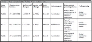
Table 1. Male exons tested for PLOD1, PLCZ1 mutation sites.
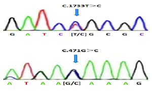
Figure 1. Localization of mutations in the PLCZ1 gene.

Table 2. Exons of male brother to detect MSH5, PLCZ1 mutation sites.
In May 2023, our hospital underwent donor IVF treatment, a short-acting long-term regimen, Gn was used for a total of 9 days, trigger day E2:8285pg / ml, P:0.88ng / ml. Thirty-six hours after the trigger, 13 oocytes were collected. Eleven 2PN were formed after in vitro fertilization of the donor sperm and two were unfertilized. The patient required all blastocysts to be cultured to form 6 blastocysts. The resuscitation cycle was started in July 2023, and two Day5 embryos were transplanted. The Serum human chorionic gonadotropin (HCG) was 328.1U / L on the 15th day after operation, and the HCG was 4490U / L on the 24th day after operation. B-ultrasound showed gestational sac and cardiac tube pulsation, which was determined as clinical pregnancy. By the time of writing this article, the patient had had a full-term cesarean section of a male live baby, now healthy, nearly one year old.
DISCUSSION
In this study, a novel PLCZ1 compound heterozygous mutation was found in an infertile male who experienced repeated fertilization failure even after ICSI-AOA by whole exome sequencing.
PLCZ1 is a sperm-specific protein and an important factor for oocyte activation. It is mainly located in the perinuclear sheath between the inner membrane and the nuclear membrane of the sperm head (Che et al., 2024)the gene encoding PLCζ, cause male infertility and intracytoplasmic sperm injection (ICSI. After sperm-oocyte plasma membrane fusion, the entry of PLCZ1 into oocytes can trigger the release of Ca2+ in the endoplasmic reticulum, and the continuous fluctuation of Ca2+ concentration induces the release of oocyte block and initiates embryonic development. The autosomal recessive mutation of the PLCZ1 gene is associated with type 17 spermatogenesis dysfunction, and its mutation or deletion affects oocyte activation, resulting in a low fertilization rate or complete fertilization failure (Jones et al., 2022). Kashir et al. (2012)with abnormal testicular gene expression considered to be a major cause. Certain types of male infertility are caused by failure of the sperm to activate the oocyte, a process normally regulated by calcium oscillations, thought to be induced by a sperm-specific phospholipase C, PLCzeta (PLCζ first discovered the first heterozygous mutation of the PLCZ1 gene in infertile men diagnosed with oocyte activation defects and complete fertilization failure. In recent years, related cases of PLCZ1 gene mutation have been reported. Yuan et al. (2020)there are still many unexplained aspects of FF. Here, we aimed to assess the clinical and genetic characteristics of two families experiencing primary infertility with FF.\nMETHODS: We have characterized two families from China. All of the infertile couples presented with similar clinical phenotypes, that is, partial or total fertilization failure in repeated cycles. We performed Sanger sequencing of their WEE2, TLE6, and PLCZ1 genes, and further bioinformatics and functional analyses were performed to identify the pathogenic elements of the variants.\nRESULTS: We identified novel compound heterozygous mutations c.1259C>T (p.P420L found new compound heterozygous mutations c.1259C > T (p.P420L) and c.1727T > C (p.L576P) in the PLCZ1 gene in a male patient with complete fertilization failure. Lin et al. (2023) found a compound heterozygous mutation of the PLCZ1 gene (p.C196X, p.c 403_404del) and a homozygous mutation of p.W536X in 2 patients with fertilization failure.
In this instance, the patient had a history of infertility for 12 years. After excluding other infertility factors, one cycle of IVF and three cycles of ICSI were performed, and the fertilization of the patient was not ideal. It was highly suspected that the patient’s husband and wife had genetic defects. In order to find out the reason, the patient’s husband and wife were subjected to whole exon detection. The results suggested that the man had a PLCZ1 heterozygous mutation with PLOD1 mutation. The first mutation site of PLCZ1 was c.1733C > T (p.M578L), and the second mutation site was c.471G > C (p.M157I), which was a new mutation site. PLCZ1 gene heterozygous mutation also exists in the same locus in the male brother. PLCZ1 has become an important gene that can not be ignored in the genetic screening and diagnosis of infertile men.
Although several pathogenic PLCZ1 gene mutation types have been found, there is still a lack of effective treatment. ICSI injects a single sperm into the cytoplasm of MII oocytes through a micromanipulation system to fertilize them, which helps to avoid most male infertility (Palermo et al., 2017). Although ICSI technology can improve the fertilization rate after IVF failure, there are still 1%-5% of challenging infertile men who have total fertilization failure (TFF) or low fertilization rate (fertilization rate < 30 %) after ICSI treatment, which is all in the category of fertilization disorders (Yeste et al., 2016). For patients with complete fertilization failure caused by PLCZ1 mutation, the AOA method is recommended for intervention. AOA can significantly improve the embryo fertilization rate of patients by increasing the level of calcium ions in oocytes to assist oocyte activation. It has been reported that ICSI combined with AOA is effective for infertile men with PLCZ1 mutations, which can partially rescue fertilization failure and contribute to successful pregnancy (Torra-Massana et al., 2019; Antonova et al., 2024)mainly due to oocyte activation failure (OAF. In this case, we found that not all patients could benefit from AOA even if fertilization failed under ICSI and AOA treatment (Nicholson et al., 2024)Dundee between January 2014 and December 2021 (n = 231. Wang et al. (2020)the infertile man with PLCZ1 mutation was treated with intracytoplasmic sperm injection (ICSI identified a homozygous PLCZ1 nonsense mutation c.588C > A (p.Cys196Ter), which had a low fertilization rate and poor embryo development after ICSI + AOA treatment. Consequently, it becomes clear that PLCZ1 is necessary for early embryonic development and fertilization.
CONCLUSION
In this paper, the patient finally obtained clinical pregnancy by in vitro fertilization with donor sperm. The causes of fertilization disorders are complex and diverse, which is a difficult problem for clinicians. A new PLCZ1 mutation site, c.471G > C (p.M157I), was discovered in a pair of brothers with a fertilization abnormality. This finding broadened the spectrum of PLCZ1 gene mutations associated with male infertility and further supported the crucial function PLCZ1 plays in human fertilization. For patients with primary infertility and multiple assisted reproductive failure, especially those with low fertilization rate or repeated fertilization failure, it is recommended to carry out genetic testing as soon as possible to clarify the genetic etiology. For patients with genetic problems in one of the spouses, if they have brothers and sisters, they should also improve the genetic testing of the family as soon as possible before giving birth, so as to achieve accurate diagnosis and treatment, improve the pregnancy outcome of patients, and avoid repeated fertilization failure to patients. Unnecessary economic losses and physical and mental injuries.
ACKNOWLEDGEMENT
The authors thank patients for allowing us to share their medical information in this case report.
REFERENCES
Antonova I, Yunakova M, Bochev I, Magunska N, Yaneva G, Ivanov D. Assisted oocyte activation significantly improves zygote formation, cleavage, and implantation rates in patients with a history of fertilization failures. Physiol Int. 2024;111:207-21. PMID: 39150772 DOI: 10.1556/2060.2024.00342 Medline
Che JF, Wu HX, Zeng SC, Wu YR, Dai J, Cheng DH, Gong F, Lu GX, Lin G, Dai C. Defects in phospholipase C zeta cause polyspermy and low fertilization after conventional IVF: not just ICSI failure. Asian J Androl. 2024;26:175-82. PMID: 38048167 DOI: 10.4103/aja202355 Medline
Ferrer-Buitrago M, Bonte D, De Sutter P, Leybaert L, Heindryckx B. Single Ca2+ transients vs oscillatory Ca2+ signaling for assisted oocyte activation: limitations and benefits. Reproduction. 2018;155:R105-19. PMID: 29122969 DOI: 10.1530/REP-17-0098 Medline
Jones C, Meng X, Coward K. SPERM FACTORS AND EGG ACTIVATION: Phospholipase C zeta (PLCZ1) and the clinical diagnosis of oocyte activation deficiency. Reproduction. 2022;164:F53-66. PMID: 35312629 DOI: 10.1530/REP-21-0458 Medline
Kashir J, Konstantinidis M, Jones C, Lemmon B, Lee HC, Hamer R, Heindryckx B, Deane CM, De Sutter P, Fissore RA, Parrington J, Wells D, Coward K. A maternally inherited autosomal point mutation in human phospholipase C zeta (PLCζ) leads to male infertility. Hum Reprod. 2012;27:222-31. PMID: 22095789 DOI: 10.1093/humrep/der384 Medline
Lin Y, Huang Y, Li B, Zhang T, Niu Y, Hu S, Ding Y, Yao G, Wei Z, Yao N, Yao Y, Lu Y, He Y, Zhu Q, Zhang L, Sun Y. Novel mutations in PLCZ1 lead to early embryonic arrest as a male factor. Front Cell Dev Biol. 2023;11:1193248. PMID: 37261077 DOI: 10.3389/fcell.2023.1193248 Medline
Nicholson CL, Dean M, Attia A, Milne PA, Martins da Silva S. Artificial oocyte activation improves ICSI outcomes following unexplained fertilization abnormalities. Reprod Biomed Online. 2024;49:104327. PMID: 39241689 DOI: 10.1016/j.rbmo.2024.104327 Medline
Palermo GD, O’Neill CL, Chow S, Cheung S, Parrella A, Pereira N, Rosenwaks Z. Intracytoplasmic sperm injection: state of the art in humans. Reproduction. 2017;154:F93-110. PMID: 29158352 DOI: 10.1530/REP-17-0374 Medline
Torra-Massana M, Cornet-Bartolomé D, Barragán M, Durban M, Ferrer-Vaquer A, Zambelli F, Rodriguez A, Oliva R, Vassena R. Novel phospholipase C zeta 1 mutations associated with fertilization failures after ICSI. Hum Reprod. 2019;34:1494-504. PMID: 31347677 DOI: 10.1093/humrep/dez094 Medline
Wang F, Zhang J, Kong S, Li C, Zhang Z, He X, Wu H, Tang D, Zha X, Tan Q, Duan Z, Cao Y, Zhu F. A homozygous nonsense mutation of PLCZ1 cause male infertility with oocyte activation deficiency. J Assist Reprod Genet. 2020;37:821-8. PMID: 32146562 DOI: 10.1007/s10815-020-01719-4 Medline
Yeste M, Jones C, Amdani SN, Patel S, Coward K. Oocyte activation deficiency: a role for an oocyte contribution? Hum Reprod Update. 2016;22:23-47. PMID: 26346057 DOI: 10.1093/humupd/dmv040 Medline
Yuan P, Zheng L, Liang H, Lin Q, Ou S, Zhu Y, Lai L, Zhang Q, He Z, Wang W. Novel mutations in the PLCZ1 gene associated with human low or failed fertilization. Mol Genet Genomic Med. 2020;8:e1470. PMID: 32840018 DOI: 10.1002/mgg3.1470 Medline