JBRA Assist. Reprod. 2026 2026;00(0):00-00
ORIGINAL ARTICLE
doi: 10.5935/1518-0557.20260072
Obesity and infertility: clinical associations and implications in wom-en from Goiânia - Brazil
1Graduate Program in Medicine, University Center of Mineiros (UNIFIMES), Trindade, Goiás, Brazil
2Department of Veterinary Medicine, University Center of Mineiros (UNIFIMES), Mineiros, Goiás, Brazil
3Department of Medicine, University Center of Mineiros (UNIFIMES), Trindade, Goiás, Brazil
4Department of Medicine, Federal University of Goiás, Goiânia, Goiás, Brazil
CONFLICT OF INTEREST
The authors declare no conflicts of interest related to this work.
ABSTRACT
Objective: Obesity is a major metabolic condition linked to reduced fertility. Excess adipose tissue disrupts the neuroendocrine axis and impairs several reproduc-tive processes. Evidence shows that, even when assisted reproductive technologies are used, women with obesity experience lower oocyte recovery, fewer viable embryos, and higher rates of miscarriage and gestational complica-tions. This study evaluated the relationship between obe-sity and female infertility by analyzing in vitro fertilization outcomes.
Methods: This quantitative, cross-sectional, retro-spective study included all in vitro fertilization cycles per-formed at a private assisted reproduction clinic in Goiânia between January and December 2022. Ethical approval was obtained from the Research Ethics Committee. After applying the inclusion and exclusion criteria, 226 cycles from women aged 24-46 years were analyzed. Logistic regression models were used to assess associations be-tween maternal and paternal body mass index and in vitro fertilization outcomes, as well as metabolic comorbidities, gestational, intrapartum, and neonatal complications, and miscarriage, prematurity, and newborn birth weight.
Results: Women with overweight (body mass index ≥25kg/m2) were 2.4 times more likely to develop gesta-tional complications (odds ratio, 2.42; 95% confidence in-terval, 1.048-5.598) and 2.8 times more likely to develop neonatal complications (odds ratio, 2.80; 95% confidence interval, 1.712-6.70) compared with eutrophic women. No other significant association was identified.
Conclusion: Higher maternal body mass index was as-sociated with increased gestational and neonatal compli-cations, but not with in vitro fertilization success. Further research using broader clinical and anthropometric indica-tors is needed to better characterize the impact of obesity and to guide preventive strategies.
Keywords: Body mass index, obesity, female infertility, in vitro fertilization, metabolic syndrome
INTRODUCTION
Obesity is defined by the World Health Organization (WHO) using body mass index (BMI), which is calculated from weight and height and used to classify nutritional status (Brazil, 2020). However, BMI is a limited measure because it estimates body fat percentage without accounting for factors such as muscle mass, age, sex, and ethnicity. As a result, metabolically healthy individuals may be misclassified as high risk for comorbidities, whereas others with higher adiposity may be categorized as eutrophic (Blundell et al., 2014). Assuming that eutrophic individuals are healthy solely because their BMI is less than 25kg/m2 can delay or prevent the diagnosis of clinically significant conditions. Although BMI is practical, accessible, and low-cost in clinical settings, relying on it alone can lead to adverse health consequences. Additional indicators, such as laboratory markers, can provide more accurate diagnoses and guide more effective care. When laboratory testing is unavailable, complementary measures that better reflect the metabolic profile-such as abdominal circumference, body fat percentage, physical activity intensity, and cardiorespiratory fitness-are recommended (Tomiyama et al., 2016).
According to the Brazilian Federation of Gynecology and Obstetrics Associations (FEBRASGO), infertility is defined as the inability to conceive after one year or more of regular, unprotected sexual intercourse (FEBRASGO, 2021). Couples are increasingly postponing parenthood, and age remains the factor with the greatest impact on female fertility. In addition, unhealthy lifestyle habits have contributed to the rising global prevalence of infertility. WHO estimates indicate that one in six people worldwide experiences fertility difficulties, representing approximately 17.5% of the global population (FEBRASGO, 2021).
Adipose tissue functions as an active endocrine organ and has a direct influence on female reproductive health. Therefore, when maternal obesity is present, addressing it should be the first step before initiating infertility treatment. Increased visceral fat promotes lipotoxicity, which disrupts the hypothalamic-pituitary-ovarian axis (HPO), triggers chronic systemic inflammation, increases hypercoagulability and insulin resistance, and contributes to endometrial and placental alterations. In vitro fertilization (IVF) and intracytoplasmic sperm injection are assisted reproductive technologies that provide hope for many couples experiencing infertility. However, obesity remains a barrier even with these interventions. Obese women generally require higher doses of ovarian stimulants and longer treatment courses, and they experience lower oocyte retrieval rates, reduced embryo production, higher rates of pregnancy failure, and an increased risk of spontaneous abortion (Zheng et al., 2024; Barbouni et al., 2025).
This study therefore aims to examine the relationship between obesity and female infertility, considering the epidemiological profile and IVF success rates.
MATERIALS AND METHODS
Projects and patients
This quantitative, cross-sectional, retrospective study was conducted using data from a private assisted reproduction clinic in Goiânia, Brazil. It included all IVF cycles performed between January and December 2022. The project was approved by the Research Ethics Committee via the Brazil Platform, under opinion no. 7,665,526, in conformity with the Declaration of Helsinki.During the study period, 385 cycles were recorded. Of these, 290 frozen embryo transfers using oocytes from the same patient were eligible. Cycles involving egg donation (n=80), surrogate gestation (n=15), medical records without maternal BMI data, and cases involving multiple pregnancies were excluded from the analysis. After applying the inclusion and exclusion criteria, the final sample comprised 226 cases.Participants ranged from 24 to 46 years of age. For comparative analyses, patients were stratified into three BMI categories, according to WHO criteria: underweight (n=3), eutrophic (n=127), and overweight (n=96).
Variables and data collection
Data were extracted from electronic medical records and included the variables shown in Figure 1.
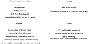
Figure 1. Representative image of the variables used in the study
The analysis included the following variables: maternal and paternal height (meters), maternal and paternal weight (kilograms), maternal and paternal BMI (kg/m2), gestational age at delivery (weeks), number of previous IVF attempts, history of miscarriage (present or absent), birth weight of the newborn (grams), complications during pregnancy (present or absent), maternal complications during delivery (present or absent), complications involving the newborn (present or absent), history of comorbidities (present or absent), IVF result (positive or negative), pregnancy outcome (positive or negative), and type of pregnancy (biochemical, single, twin, unidentified, or miscarriage).For the classification of nutritional status, the categorization established by the Clinical Protocol and Therapeutic Guidelines for Overweight and Obesity in Adults, approved by SCTIE/MS Ordinance No. 53 on November 11, 2020, was adopted. This classification is based on BMI and includes the following groups: underweight (<18.5kg/m2), eutrophic (18.5-24.99kg/m2), and overweight. The overweight group includes overweight (25.0-29.99kg/m2), obesity grade I (30.0-34.99kg/m2), obesity grade II (35.0-39.99kg/m2), and obesity grade III (≥40.0kg/m2). BMI was calculated using the formula: body weight (kilograms) divided by height (meters) squared. In this context, the comorbidities evaluated in the study were those related to metabolic alterations, including blood pressure dysregulation, glycemic disorders, and lipid disorders.The variables related to newborns followed the classifications established in the 2021 Guidance Manual: Nutritional Assessment of Children and Adolescents from the Brazilian Society of Pediatrics (SBP, 2021). Prematurity was categorized according to gestational age at birth as follows: premature (<37 weeks), term (37 weeks to 41 weeks and 6 days), and post-term (≥42 weeks). Nutritional status at birth was classified as low weight (<2,500g), adequate (2,500-3,999g), or macrosomia (≥4,000g). Abortion was defined as spontaneous termination of pregnancy at <22 weeks or <500g, according to the High-Risk Pregnancy Manual from the Ministry of Health (Brazil, 2022).Complications during pregnancy included preeclampsia, eclampsia, anemia, gestational diabetes mellitus, emergency deliveries, and thrombophilia. Complications during delivery included hemorrhages. Neonatal complications included prematurity, fetal centralization, respiratory failure, genetic alterations, congenital heart disease, admission to the intensive care unit for weight gain, cardiopulmonary arrest, neonatal sepsis, and death. The IVF result was determined using a quantitative beta-human chorionic gonadotropin test performed between the ninth and eleventh day after blastocyst transfer, with values above 25mIU/mL considered positive. Pregnancy confirmation and classification were based on transvaginal ultrasound. Biochemical pregnancy was defined as a positive beta-human chorionic gonadotropin result without fetal evidence; singleton pregnancy as identification of one fetus; twin pregnancy as identification of two or more fetuses; unidentified pregnancy as absence of examination; and miscarriage as fetal loss before the first ultrasound. A positive pregnancy outcome was defined as the birth of a live newborn, and a negative outcome as the absence of live birth.
Ovarian stimulation
Patients underwent cryotransfer with autologous oocytes and initiated treatment with controlled ovarian stimulation. This phase began between the second and third day of the menstrual cycle and included oral medication (clomiphene citrate and/or letrozole) to promote follicular stimulation and to suppress pituitary activity, thereby preventing spontaneous ovulation. The average duration of this phase ranged from 10 to 14 days, with therapy adjusted according to the clinical profile and ovarian response of each patient. Treatment could be supplemented with injectable gonadotropins, such as follicle-stimulating hormone (FSH) alone or in combination with luteinizing hormone (LH). When at least two or three follicles reached an average diameter between 18 and 20 mm, final ovulation induction was carried out using human chorionic gonadotropin, administered 36 hours before oocyte collection.
Collection and fertilization
Oocyte collection was performed by transvaginal follicular puncture using a fine needle guided by ultrasound in a sterile surgical environment, followed by immediate transfer of the material to the embryology laboratory for morphological evaluation of the oocytes. Viable oocytes underwent fertilization using conventional IVF techniques. The resulting embryos remained in culture for five days until the blastocyst stage, at which point they were morphologically evaluated and cryopreserved for later transfer according to individual plans.
Endometrial preparation and transfer
Endometrial preparation was conducted either through a natural cycle, with spontaneous ovulation, or through a hormonal cycle, with administration of estradiol followed by progesterone (oral or vaginal). The choice of protocol was individualized in accordance with the clinical profile of each patient. After confirmation of endometrial receptivity, usually on the fifth day of progesterone exposure, embryo thawing and transfer were performed. The number of embryos transferred followed Brazilian Federal Council of Medicine guidelines (CFM, 2022), and surplus embryos remained in cryopreservation.
Statistical analysis
Epidemiological data were analyzed using GraphPad Prism software (version 7.0) to calculate mean maternal age, number of previous attempts, and maternal BMI for the defined groups. The remaining data were analyzed using Jamovi software (version 2.6.45; The Jamovi Project, 2024). Associations between maternal, paternal, and couple BMI and the success rate of IVF, as well as between maternal BMI and gestational, intrapartum, and neonatal complications, were evaluated using binomial logistic regression. The IVF outcome was treated as a dichotomous dependent variable (success=1; failure=0), and complications were coded as follows: gestational (present=1; absent=0), intrapartum (present=1; absent=0), and neonatal (present=1; absent=0). The eutrophic group served as the reference category in all models.Associations between maternal BMI and neonatal outcomes (birth weight and prematurity) were analyzed using multinomial logistic regression. Dependent variables were categorized according to birth weight and prematurity. The following reference categories were applied: eutrophic group for BMI, adequate birth weight, and term for prematurity.The association between maternal BMI and the presence of clinical comorbidities recorded in medical records was evaluated using binomial logistic regression, with presence or absence of comorbidities as the dependent variable and the eutrophic group as the reference category. The association between maternal comorbidities and success of IVF was also analyzed using binomial logistic regression, with absence of comorbidities as the reference category.The results were expressed as odds ratios with respective 95% confidence intervals. The significance level was set at 5% (p<0.05). Model fit was assessed using deviance, the Akaike information criterion, and the Cox and Snell coefficient of determination.
RESULTS
After applying the inclusion and exclusion criteria, 226 cycles of patients undergoing IVF with BMI values calculated during the ovarian stimulation stage were analyzed. Maternal BMI had a mean of 25kg/m2 with a standard deviation of 4 (range, 17-41), whereas paternal BMI had a mean of 29kg/m2 with a standard deviation of 4.6 (range, 18-50). Among women, 42.5% were classified as overweight, in contrast with 77.3% of men (Figure 2). The cycles were divided into three groups according to BMI as follows: group 1 (G1), underweight (<18.5kg/m2; n=3); group 2 (G2), eutrophic (18.5-24.99kg/m2; n=127); and group 3 (G3), overweight (≥25.0kg/m2; n=96). Mean age was similar across the groups, ranging from 34 to 37 years. Patients in G1 underwent, on average, 2.7 times more IVF cycle attempts (95% confidence interval, 1.2-4.1) compared with patients in G2 and G3, which did not differ significantly from each other. General characteristics for each group are presented in Table 1.
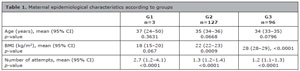
Table 1. Maternal epidemiological characteristics according to groups
G1, group 1 (underweight); G2, group 2 (eutrophic); G3, group 3 (overweight); n, number of cycles; CI, confidence interval; BMI, body mass index.
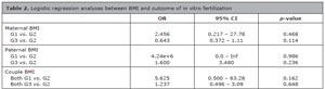
Table 2. Logistic regression analyses between BMI and outcome of in vitro fertilization
G1, group 1 (underweight); G2, group 2 (eutrophic); G3, group 3 (overweight); OR, odds ratio; CI, confidence interval; BMI, body mass index; Inf, infinity.
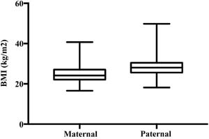
Figure 2. Boxplot showing mean maternal BMI and mean paternal BMI by genderBMI, body mass index
The binomial logistic regression analysis did not show a statistically significant association between maternal BMI and success of IVF cycles, either when comparing the underweight group with the eutrophic group or when comparing the overweight group with the eutrophic group. Paternal BMI also did not demonstrate a significant association with the outcome, and the same pattern was observed when the combined BMI of the couple was evaluated. These findings indicate that, in the sample analyzed, BMI alone did not influence IVF success rates (Table 2).
Considering possible perinatal changes, multinomial modeling did not show a statistically significant association between maternal BMI and newborn characteristics at birth. Gestational age did not differ significantly when comparing prematurity, abortion, or post-term birth with term birth, and no significant association was observed when comparing low birth weight or macrosomia with normal birth weight. Significant differences were identified only in the intercepts for abortion, preterm birth, low birth weight, and macrosomia, reflecting the higher frequency of term births and the higher frequency of newborns with normal birth weight in the sample. These findings were not related to maternal BMI (Table 3).
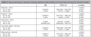
Table 3. Multinomial logistic regression analyses between maternal BMI and miscarriage, prematurity, and birth weight
G1, group 1 (underweight); G2, group 2 (eutrophic); G3, group 3 (overweight); OR, odds ratio; CI, confidence interval; BMI, body mass index.Source: Authors, 2025.
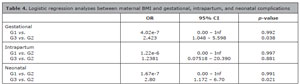
Table 4. Logistic regression analyses between maternal BMI and gestational, intrapartum, and neonatal complications
G1, group 1 (underweight); G2, group 2 (eutrophic); G3, group 3 (overweight); OR, odds ratio; CI, confidence interval; BMI, body mass index; Inf, infinity.Source: Authors, 2025.
DISCUSSION
Female obesity has been consistently associated with reduced fertility through multiple alterations in the neuroendocrine axis, including increased insulin resistance, hyperandrogenism, and systemic inflammation. These changes affect processes ranging from oogenesis and oocyte quality to endometrial receptivity (Marinelli et al., 2022). Although the relationship between metabolic dysfunction and female infertility has been well established in numerous studies, the results of this study did not identify a significant association between maternal BMI and IVF success. These findings align with those of Devranoğlu et al. (2024), who also reported that BMI alone was not a reliable predictor of obesity-related reproductive outcomes.
Debate continues regarding appropriate parameters for assessing obesity, which forms part of a broader cluster of metabolic alterations known as metabolic syndrome. This condition is evaluated according to criteria from the National Cholesterol Education Program Adult Treatment Panel III (NCEP, 2001), which include abdominal circumference, blood pressure, fasting blood glucose, triglycerides, and high-density lipoprotein. In contrast to this comprehensive approach, BMI is an indirect and often inaccurate measure that may overestimate or underestimate adiposity in individuals with atypical body composition (Farhangiyan et al., 2019).
The low prevalence of metabolic comorbidities in the analyzed medical records suggested underreporting, considering that 42.5% of the female participants were overweight. The absence of a systematic evaluation of laboratory markers may result in a characterization that does not accurately reflect the true systemic complexity and metabolic risk, as the underlying pathophysiological processes may not have been adequately identified. Furthermore, the literature indicates that an altered metabolic profile can compromise assisted reproductive outcomes even before manifesting as a clinically evident pathology, which may further obscure relevant risk conditions when combined with the underreporting of metabolic comorbidities (Silvestris et al., 2019).
Although BMI is a practical and widely used tool for population screening, it has important limitations as it does not differentiate between adipose tissue and lean mass, nor does it identify body fat distribution, particularly visceral fat, which has a greater metabolic impact. In addition, BMI is influenced by factors such as age, sex, and functional status, potentially leading to both underdiagnosis of obesity in individuals with high adiposity and normal BMI, and overdiagnosis in individuals with greater muscle mass. Therefore, BMI should be interpreted as an initial indicator and complemented by clinical assessment and other anthropometric or body composition measures for a more accurate diagnosis of obesity (Sweatt et al., 2024). From a pathophysiological perspective, obesity is characterized not only by excess body weight but also by adipose tissue dysfunction, which is associated with chronic low-grade inflammation, insulin resistance, alterations in the hypothalamic-pituitary-gonadal axis, and dysregulation of adipokine secretion, including leptin and adiponectin, none of which are captured by BMI. Therefore, the absence of an association between maternal BMI and IVF outcomes may reflect the limitations of this metric when used as the sole marker of obesity in fertility research.
A temporal analysis in Brazil between 1975 and 2019 revealed a reduction in the prevalence of underweight among women from 12.2% to 3.4%, reflecting a characteristic pattern of nutritional transition in which subclinical undernutrition is replaced by overweight and obesity (Conde et al., 2022). In the present study, only three participants were classified as underweight, a finding that may reflect the currently low prevalence of underweight status among adult women and by the profile of our sample, which consisted of women of reproductive age seeking fertility treatment. This population generally has greater access to healthcare services and a lower prevalence of food insecurity. In addition, selection bias, may have contributed, as underweight women with ovulatory dysfunctions are less likely to be referred for IVF.
Although less explored in the literature, paternal obesity can also directly affect fertility by altering spermatogenesis, reducing sperm motility, and inducing epigenetic changes in sperm DNA-effects that are more pronounced in men with pre-existing alterations in seminal parameters (Barbouni et al., 2025). Nikolic et al. (2024) emphasize that poor semen quality represents the leading cause of male infertility, reinforcing the need for continued investigation in this field. However, the findings of this study did not identify an association between paternal BMI and IVF success, which, as seen with maternal results, may reflect the limitations of BMI when used as a single indicator of metabolic status.
The findings of McPherson et al. (2021) indicate that maternal overweight and obesity show a stronger association with increased birth weight than paternal factors. This pattern is attributed to genetic and epigenetic mechanisms mediated by metabolic dysfunction related to obesity, with potential effects on both oocytes and sperm that may influence fetal development. In the present study, this association could not be demonstrated, with only two cases of macrosomia (1.5%) and nine cases of low birth weight (6.7%) identified among the 134 positive IVF cycles.
From a pathophysiological perspective, excessive accumulation of adipose tissue can induce a chronic inflammatory state with negative effects on reproductive tissues, including changes in the endometrium, which have been linked to higher rates of miscarriage (Broughton & Moley, 2017). In addition, Liu et al. (2022) conducted a prospective cohort study that evaluated 43,056 pregnancies between 2017 and 2019 and identified maternal obesity as a predictor of spontaneous preterm birth, preterm rupture of membranes, and medically indicated preterm birth. However, data in the present study did not identify a significant relationship between maternal BMI and prematurity.
During pregnancy, excessive weight gain-especially when above 9kg or occurring in the context of pre-pregnancy overweight or obesity-is consistently associated with increased risk of adverse outcomes, including cesarean delivery, gestational diabetes mellitus, hypertensive syndromes of pregnancy, and large-for-gestational-age newborns. Maternal obesity has also been linked to increased risk of spontaneous abortion, attributed to both genetic alterations and functional impairments of reproductive tissues (Schon et al., 2024).
In line with current literature, data from the present study showed that pregnant women with BMI ≥25kg/m2 had a 2.4-fold higher likelihood of developing complications during pregnancy and a 2.8-fold higher likelihood of neonatal complications compared with eutrophic women. Labor-related complications also merit attention, particularly postpartum hemorrhage, which is currently the second leading cause of maternal mortality in Brazil. However, consistent with the findings of Butwick et al. (2018), the analysis did not identify a statistically significant association between obesity and postpartum hemorrhage.
These gestational and neonatal alterations are directly associated with lipotoxicity, which is driven primarily by oxidative stress, systemic inflammation, and immune dysregulation. Chronic and excessive lipid consumption promotes increased oxidative stress, resulting in an imbalance in antioxidant enzymes and intensification of the systemic inflammatory response. In this context, there is an increase in immune responses mediated by Th1, Th17, and CD8⁺ cytotoxic T lymphocytes, concomitant with a reduction in Th2 and regulatory T cell (Treg) responses. Elevated levels of these inflammatory markers are associated with implantation failure, impaired placental development, and destruction of the syncytiotrophoblast, whereas Th2 and Treg responses play fundamental roles in fetal protection (Sheikhansari et al., 2019).
Another prominent mechanism is related to increased insulin resistance resulting from an inadequate response to insulin, which requires greater hormonal production to maintain glycemic homeostasis. Hyperinsulinemia leads to hyperstimulation of the adrenal glands and ovaries, increasing the production of androgens, which are subsequently aromatized in the ovaries and converted into estrogens (Silvestris et al., 2019). In addition, excess insulin exerts a hepatic effect by reducing the synthesis of sex hormone-binding globulin (SHBG), thereby increasing the free androgen fraction and perpetuating a reproductively unfavorable environment (Aghaei et al., 2023).
Subsequently, hyperestrogenism exerts negative feedback on the HPO axis, interfering with the pulsatility of gonadotropin-releasing hormone (GnRH) and favoring LH synthesis. As a result, there is an increased conversion of cholesterol into androgens by the theca cells mediated by LH. In parallel, reduced production of FSH is observed along with decreased secretion of inhibin B from nondominant follicles, further impairing FSH stimulation. Consequently, folliculogenesis may be disrupted, leading to menstrual cycle irregularities, anovulation, and infertility (Kasum et al., 2018).
Finally, regarding the associated endometrial alterations, several notable changes have been described. These include impaired decidualization due to dysregulation of the HPO axis and reduced progesterone synthesis, reflecting hormone-dependent nature of the endometrium, along with inadequate endometrial development related to increased insulin resistance. In addition, a systemic pro-inflammatory state may promote thrombus formation that compromises placental progression, and alterations in lipid metabolism can induce endothelial injury and thereby impair placental perfusion (Aghaei et al., 2023).
Among the limitations of the present study is its retrospective design, which may introduce biases inherent to this type of research research because the data were obtained from a single clinical encounter. Another relevant limitation is the exclusive use of BMI as an indicator of obesity. Although widely used, BMI is a global measure of body mass that does not distinguish between lean mass and adipose tissue, does not identify fat distribution (including visceral adiposity), and does not account for more specific metabolic markers, which may result in inaccurate classifications. Furthermore, the study population was derived exclusively from the private healthcare system, which limits the extrapolation of the results to users of the Brazilian Unified Health System and other population contexts. This context highlights the need for optimized protocols that address the challenges to reproductive health posed by male and female obesity.
CONCLUSION
In this study, maternal, paternal, and couple BMI did not show a significant association with the success of IVF cycles. However, pregnant women with high BMI had a higher risk of gestational and neonatal complications compared with the eutrophic group. No significant associations were identified for intrapartum complications, perinatal outcomes, or presence of gestational comorbidities. Given the relevance of this subject, further research is needed to better clarify the real impact of obesity, incorporating additional clinical and anthropometric parameters to support the development of preventive strategies.
Acknowledgments:
The authors thank all individuals who contributed to this study.
REFERENCES
Aghaei B, Moradi F, Soleimani D, Moradinazar M, Khosravy T, Samadi M. Glycemic index, glycemic load, dietary inflammatory index, and risk of infertility in women. Food Sci Nutr. 2023;11:6413-24. PMID: 37823091 DOI: 10.1002/fsn3.3584
Medline
Barbouni K, Jotautis V, Metallinou D, Diamanti A, Orovou E, Liepinaitienė A, Nikolaidis P, Karampas G, Sarantaki A. When Weight Matters: How Obesity Impacts Reproductive Health and Pregnancy-A Systematic Review. Curr Obes Rep. 2025;14:37. PMID: 40238039 DOI: 10.1007/s13679-025-00629-9
Medline
Blundell JE, Dulloo AG, Salvador J, Frühbeck G; EASO SAB Working Group on BMI. Beyond BMI--phenotyping the obesities. Obes Facts. 2014;7:322-8. PMID: 25485991 DOI: 10.1159/000368783
Medline
Broughton DE, Moley KH. Obesity and female infertility: potential mediators of obesity’s impact. Fertil Steril. 2017;107:840-7. PMID: 28292619 DOI: 10.1016/j.fertnstert.2017.01.017
Medline
Butwick AJ, Abreo A, Bateman BT, Lee HC, El-Sayed YY, Stephansson O, Flood P. Effect of maternal body mass index on postpartum hemorrhage. Anesthesiology. 2018;128:774-83. PMID: 29346134 DOI: 10.1097/ ALN.0000000000002082
Medline
Conde WL, Silva IVD, Ferraz FR. Undernutrition and obesity trends in Brazilian adults from 1975 to 2019 and its associated factors. Cad Saude Publica. 2022;38:e00149721. PMID: 35613255 DOI: 10.1590/0102-311XEN149721
Medline
Devranoğlu B, Yilmaz MB, Peker G, Emekçi Özay Ö, Özay AC, Güzel Aİ. Effects of obesity on clinical outcomes in diminished ovarian reserve patients undergoing intracytoplasmic sperm injection cycles. Medicine (Baltimore). 2024;103:e38942. PMID: 38996138 DOI: 10.1097/MD.0000000000038942
Medline
Expert Panel on Detection, Evaluation, and Treatment of High Blood Cholesterol in Adults. Executive Summary of The Third Report of The National Cholesterol Education Program (NCEP) Expert Panel on Detection, Evaluation, And Treatment of High Blood Cholesterol In Adults (Adult Treatment Panel III). JAMA. 2001;285:2486-97. PMID: 11368702 DOI: 10.1001/jama.285.19.2486
Medline
Farhangiyan Z, Latifi SM, Rashidi H, Shahbazian H. The most appropriate cut-off point of anthropometric indices in predicting the incidence of metabolic syndrome and its components. Diabetes Metab Syndr. 2019;13:2739-45. PMID: 31405702 DOI: 10.1016/j.dsx.2019.07.009
Medline
Kasum M, Orešković S, Čehić E, Lila A, Ejubović E, Soldo D. The role of female obesity on in vitro fertilization outcomes. Gynecol Endocrinol. 2018;34:184-8. PMID: 29037105 DOI: 10.1080/09513590.2017.1391209
Medline
Liu K, Chen Y, Tong J, Yin A, Wu L, Niu J. Association of maternal obesity with preterm birth phenotype and mediation effects of gestational diabetes mellitus and preeclampsia: a prospective cohort study. BMC Pregnancy Childbirth. 2022;22:459. PMID: 35650604 DOI: 10.1186/ s12884-022-04780-2
Medline
Marinelli S, Napoletano G, Straccamore M, Basile G. Female obesity and infertility: outcomes and regulatory guidance. Acta Biomed. 2022;93:e2022278. PMID: 36043953 DOI: 10.23750/abm.v93i4.13466
Medline
McPherson NO, Vincent AD, Zander-Fox D, Grieger JA. Birthweight associations with parental obesity: retrospective analysis of 1,778 singleton term births following assisted reproductive treatment. F S Rep. 2021;2:405-12. PMID: 34934980 DOI: 10.1016/j.xfre.2021.04.011
Medline
Nikolic AZ, Dragojevic-Dikic S, Kocic J, Babic U, Joksimovic A, Radakovic-Cosic J, Gerginic V, Spasic D, Dugalic S, Petrovic A, Mandic-Rajcevic S, Perovic M. Influence of male body mass index on semen analysis parameters and in vitro fertilization outcomes. Medicine (Baltimore). 2024;103:e38949. PMID: 39093753 DOI: 10.1097/ MD.0000000000038949
Medline
Schon SB, Cabre HE, Redman LM. The impact of obesity on reproductive health and metabolism in reproductive-age females. Fertil Steril. 2024;122:194-203. PMID: 38704081 DOI: 10.1016/j.fertnstert.2024.04.036
Medline
Sheikhansari G, Soltani-Zangbar MS, Pourmoghadam Z, Kamrani A, Azizi R, Aghebati-Maleki L, Danaii S, Koushaeian L, Hojat-Farsangi M, Yousefi M. Oxidative stress, inflammatory settings, and microRNA regulation in the recurrent implantation failure patients with metabolic syndrome. Am J Reprod Immunol. 2019;82:e13170. PMID: 31310689 DOI: 10.1111/aji.13170
Medline
Silvestris E, Lovero D, Palmirotta R. Nutrition and Female Fertility: An Interdependent Correlation. Front Endocrinol (Lausanne). 2019;10:346. PMID: 31231310 DOI: 10.3389/fendo.2019.00346
Medline
Sweatt K, Garvey WT, Martins C. Strengths and Limitations of BMI in the Diagnosis of Obesity: What is the Path Forward? Curr Obes Rep. 2024;13:584-95. PMID: 38958869 DOI: 10.1007/s13679-024-00580-1. Erratum in: Curr Obes Rep. 2024;13:831. PMID: 38958869 DOI: 10.1007/s13679-024-00584-x
Medline
Tomiyama AJ, Hunger JM, Nguyen-Cuu J, Wells C. Misclassification of cardiometabolic health when using body mass index categories in NHANES 2005-2012. Int J Obes (Lond). 2016;40:883-6. PMID: 26841729 DOI: 10.1038/ijo.2016.17
Medline
Zheng L, Yang L, Guo Z, Yao N, Zhang S, Pu P. Obesity and its impact on female reproductive health: unraveling the connections. Front Endocrinol (Lausanne). 2024;14:1326546. PMID: 38264286 DOI: 10.3389/ fendo.2023.1326546
Medline