JBRA Assist. Reprod. 2026 2026;00(0):00-00
REVIEW
doi: 10.5935/1518-0557.20260084
Platelet-Rich Plasma in Human Reproduction: A Review of the Evidence in Endometrium, Ovary, and Male Infertility
1Federal Rural University of Pernambuco - UFRPE, Recife, PE, Brazil
2Catholic University of Pernambuco - UNICAP, Recife, PE, Brazil
CONFLICT OF INTEREST
The authors declare no conflicts of interest related to this study.
ABSTRACT
Although platelet-rich plasma (PRP) is an experimental therapeutic approach under investigation in different areas of medicine, several questions remain unresolved, and its use in human reproduction is still considered experimental. This limitation stems mainly from the scarcity of robust clinical studies and the heterogeneity of PRP preparation and application methods, factors that hinder comparisons among studies and contribute to inconsistent results. This narrative review aimed to evaluate the different applica-tions of PRP in reproductive medicine and its effects on patients with infertility. The findings suggest that PRP may be associated with improved pregnancy rates in some studies compared with control groups and may also favor outcomes in assisted reproduction treatments in humans.
Keywords: regenerative medicine, assisted reproductive techniques, growth factors, fertility, biological therapies
INTRODUCTION
The use of platelet-rich plasma (PRP) began in the 1970s, when hematology described it as plasma with a supraphysiological platelet count compared with peripheral blood, initially for the treatment of hemorrhage in thrombocytopenic patients (Murphy & Gardner, 1971; Alves & Grimalt, 2018). PRP has been shown to induce the differentiation of several cell types, presumably due to the action of cytokines (Marx, 2001; Amable et al., 2013). Since then, other areas of human medicine, such as orthopedics (Floryan & Berghoff, 2004), plastic surgery (Cervelli et al., 2009), and dermatology (Chi et al., 2023), as well as veterinary medicine (Dart et al., 2005) and dentistry (Foitzik & Staus, 2003), have begun to evaluate its potential therapeutic benefits. From the 1990s onward, reproductive medicine expanded this research, aiming to improve fertility in women and men as an alternative therapeutic approach, either alone or in combination with other therapies (Atkinson et al., 2021). Early studies investigating the use of PRP in assisted reproduction described PRP as a potential adjuvant strategy, as reported by Frantz et al. (2020). However, these initial publications emphasized the experimental nature of the intervention and the lack of robust randomized clinical evidence at that time. Despite its therapeutic potential, there is still no established clinical consensus, which justifies this critical narrative review.
PRP is a biological product prepared from autologous venous blood to obtain a high platelet concentration. Platelets carry cytokines and may be associated with macrophages and neutrophils; they also contain components such as serotonin, dopamine, histamine, adenosine, and calcium (Pixley et al., 2023). In addition, platelets contain numerous growth factors (GFs), the most relevant being platelet-derived growth factor (PDGF), transforming growth factor β (TGF-β), vascular endothelial growth factor (VEGF), insulin-like growth factor 1 (IGF-1), epidermal growth factor (EGF), hepatocyte growth factor (HGF), and basic fibroblast growth factor (bFGF). These growth factors are released from the alpha granules of activated platelets and play an important role in tissue repair because of their mitogenic, chemotactic, neovascular, and anti-inflammatory effects (Foster et al., 2009). Figure 1 shows the composition of platelets and the main bioactive compounds of PRP.
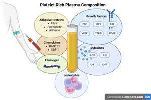
Figure 1. Biochemical composition: PRP is an autologous concentrate derived from peripheral blood, rich in platelets and composed of growth factors (PDGF, TGF-β, VEGF, IGF-1, EGF, FGF), cytokines (IL-1β, IL-6, IL-8, IL-10), chemokines (RANTES, SDF-1), adhesive proteins (fibrin, fibronectin, adhesin), small amounts of leukocytes, and plasma proteins such as fibrinogen and albumin. These elements act synergistically in tissue regeneration, inflammatory modulation, and angiogenesis induction, which are important for clinical applications in assisted reproduction.
Given these properties, PRP has been used in female reproduction to promote endometrial growth and repair, as well as follicular development. It has also been investigated as a potential strategy for improving ovarian reserve parameters in poor ovarian responders (PORs), with promising results (Pantos et al., 2016). Other increasingly investigated applications include its use in recurrent unexplained implantation failure in in vitro fertilization (IVF) and in recurrent pregnancy loss (RPL), as reported by Li et al. (2022). PRP may also be used as a first-line treatment for persistent chronic endometritis in women who do not respond to conventional antimicrobial therapy (Sfakianoudis et al., 2019).
In male infertility, studies with PRP are still scarce and mainly involve intracavernous PRP injections for the treatment of erectile dysfunction (ED), an uncommon cause of infertility in men (Anastasiadis et al., 2022). Additional studies have evaluated PRP as a supplement to conventional cryopreservation media for semen cryopreservation (Nabavinia et al., 2023). Khadivi et al. (2020) investigated the effect of adding PRP to two-dimensional (2D) and three-dimensional (3D) spermatogonial stem cell (SSC) culture systems for in vitro spermatogenesis. More recently, although still preliminary, studies on the use of PRP in testicular tissue have intensified, focusing on spermatogenesis and the improvement of semen parameters (Alonso-Frías & Esbert, 2024).
Despite evidence regarding the biological mechanisms that support the clinical efficacy of platelets, the technical aspects of PRP preparation are closely related to its clinical effects (Ferrari et al., 2021). The lack of standardization in preparation techniques and qualitative sample evaluation remains a common problem in studies in this field and may explain the lack of efficacy reported by some authors. Therefore, this study aimed to conduct a narrative literature review to evaluate the effects of PRP and its various applications in human reproduction.
MATERIALS AND METHODS
The literature search was conducted using the PubMed/ MEDLINE, Scopus, Web of Science, and SciELO databases between 2003 and 2026. The following terms in English were used: “platelet-rich plasma”, “thin endometrium”, “chronic endometritis”, “recurrent implantation failure”, “poor ovarian response”, “male infertility”, and “erectile dysfunction”. The references included randomized controlled clinical trials, literature reviews, retrospective and prospective cohort studies, original articles, systematic reviews, clinical studies, pilot studies, case series, and case reports. Duplicate articles and opinion pieces were excluded. The articles were divided into five categories: PRP and thin endometrium, PRP and chronic endometritis, PRP and recurrent implantation failure, PRP and ovarian insufficiency, and PRP and male infertility. Thin endometrium was defined as <7 mm despite hormone replacement therapy (Kasius et al., 2014). Ovarian insufficiency was assessed using the POSEIDON Group criteria (Alviggi et al., 2016). Recurrent implantation failure was defined as the failure to achieve clinical pregnancy after at least three in vitro fertilization cycles using high-quality embryos (Coughlan et al., 2014).
PRP preparation
PRP preparation is not standardized, and its composition is closely related to processing time, the number of active platelets, the preservation method and temperature (Sobral et al., 2023; Kim et al., 2020), centrifugation, final pH, and the use of activators (Zhuang et al., 2018). Factors such as sex, age, hematocrit, associated clinical conditions, and some medications can influence the quality of the compound and its clinical efficacy.To prepare PRP, whole blood samples (10-25 mL) are collected by peripheral venipuncture using vacuum needles. The samples are placed in polypropylene tubes containing anticoagulant, usually sodium citrate or acid citrate dextrose. The blood is centrifuged using a single-or double-spin protocol, depending on the study, with the latter being the most frequent. At the end of processing, the tubes contain three layers: the red blood cell layer at the bottom; the intermediate leukocyte layer, which corresponds to the PRP; and the upper layer, where platelet-poor plasma (PPP) is located. The leukocyte layer, together with the PPP, is pipetted into another tube and, depending on the protocol, undergoes a second centrifugation. After platelet deposition at the bottom of the tube, PRP is obtained, with an average platelet concentration 3-5 times higher than that of whole blood. The average volume of PRP obtained ranges from 1 to 4.5 mL. For intraovarian injection, an average of 0.5 mL is used in each ovary. For intrauterine infusion, the amount of PRP ranges from 0.5 to 1 mL. In men, an average of 0.5 mL is injected into each corpus cavernosum (Poulios et al., 2021) and 1.5 mL into each testicle (Fazli et al., 2024). Platelets may or may not be activated before PRP application, although there is no consensus on the advantages of pre-application activation (Singh & Singh, 2023) or on the best activating substance (Bolton, 2021). The preparation steps described above are summarized in Figure 2.
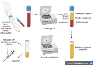
Figure 2. Schematic representation of PRP preparation: collection of autologous venous blood with anticoagulant, centrifugation in one or two cycles, separation of the platelet-rich fraction, and clinical application. PRP activation may or may not be performed before infusion, depending on the protocol and application site (endometrium, ovary, testicle, or penis).
PRP and thin endometrium
Thin endometrium is most often defined by ultrasound as an endometrial thickness of <7 mm despite hormone replacement therapy, which is the cutoff most frequently cited in the literature (Kasius et al., 2014). It is one cause of failure in spontaneous conception and assisted reproduction treatments and is associated with a higher incidence of implantation failure, ectopic pregnancy, spontaneous abortion, and abnormal placentation (Yuan et al., 2016). In addition, it can lead to embryo transfer cycle cancellation, negative psychological and financial effects, and, in some cases, the inability to conceive, leaving surrogacy as the only option.There is no robust evidence to date supporting the use of any adjuvant to treat refractory thin endometrium. Granulocyte colony-stimulating factor (G-CSF), sildenafil citrate, aspirin, pentoxifylline, tocopherol, and pelvic floor electrostimulation with biofeedback have shown limited or conflicting results. However, strategies such as a GnRH agonist in the luteal phase and intrauterine PRP on the day of the trigger appear to have positive potential, although they need to be confirmed by large randomized controlled trials (Ranisavljevic et al., 2019).The pioneering study on autologous human endometrial PRP was published by Chang et al. (2015) and reported endometrial thickening and pregnancies in a small pilot cohort (n=5) after PRP infusion. Thus, intrauterine PRP application emerged as a new adjuvant method for treating thin endometrium poorly responsive to hormone replacement therapy, encouraging further studies.In light of this, Kim et al. (2019) conducted a pilot study including 24 women with persistent thin endometrium (<7 mm) who were treated with intrauterine infusion of autologous PRP for frozen embryo transfer (FET). On average, endometrial thickness increased by 0.6 mm, but without a statistically significant difference. Paradoxically, four of the six clinical pregnancies occurred in women with smaller endometrial measurements after PRP. Therefore, the results suggested that intrauterine PRP administration improved endometrial receptivity without necessarily increasing endometrial thickness. This indicates that PRP may also act through mechanisms other than endometrial thickening to increase implantation and pregnancy rates, contradicting previous findings on endometrial metrics and receptivity. In line with these findings, Frantz et al. (2020), in a prospective clinical study, demonstrated that intrauterine PRP infusion improved pregnancy outcomes in women with impaired endometrial receptivity, even though endometrial thickness was not reported as a primary outcome. These data support the concept that PRP enhances the functional quality of the endometrium rather than acting exclusively through morphological thickening.The improvement in endometrial receptivity, independent of increased endometrial thickness, may occur through cellular and molecular mechanisms. PRP has also been reported to stimulate local angiogenesis, increase endometrial perfusion and oxygenation, and activate resident stem cells in experimental models (Chen et al., 2024; Chen et al., 2025a), contributing to functional tissue renewal. Evidence also indicates that PRP regulates the expression of genes and hormone receptors associated with implantation, such as LIF, HOXA10, ITGB3, and VEGFA, in addition to reducing oxidative stress and cell apoptosis, thereby optimizing the uterine microenvironment (Artimani et al., 2024; Chen et al., 2024). Thus, the beneficial effect of PRP on female fertility appears to stem from qualitative improvement in the endometrium, related to its functionality and capacity for embryo receptivity, rather than from morphological changes in thickness.Chang et al. (2019) recruited 64 patients with thin endometrium, and after PRP infusion, endometrial thickness was significantly greater in the PRP group than in the control group. The group also showed significantly higher amounts of growth factors in PRP than in peripheral blood (p<0.01), which could explain the higher implantation rate and clinical pregnancy rate (27.94% vs. 11.67%, p<0.05; 44.12% vs. 20%, p<0.05, respectively).Similarly, a prospective randomized controlled trial selected 120 women with thin endometrium who underwent ovarian stimulation and intrauterine insemination. The authors observed a significant increase in endometrial thickness and vascularization compared with the control group after three cycles of intrauterine PRP infusion. Considering that all pregnancies occurred after the third infusion cycle, the results indicated that PRP has a cumulative trophic effect on the thin endometrium (Pandey et al., 2023).Recent studies reinforce the previously discussed data on the use of PRP in women with thin endometrium. In a prospective cohort study, Shin et al. (2024) observed that intrauterine PRP infusion was associated with higher implantation rates compared with the previous cycle without PRP (16.4% vs. 3.1%, p<0.001). The intervention was also associated with a statistically significant increase in endometrial thickness, with a mean post-treatment thickness of 6.1 mm and a mean increase of 0.8 mm (p<0.001). However, confidence intervals were not reported; therefore, the magnitude of the effect should be interpreted cautiously. For the first time, endometrial transcriptomic analysis conducted by Zeng et al. (2025) demonstrated that PRP therapy not only increased endometrial thickness in all 10 patients with thin endometrium refractory to conventional hormonal therapy but also activated regenerative pathways. The authors observed that PRP increased the potential of stromal and epithelial stem cells and the expression of genes associated with cell proliferation and tissue repair, while also stimulating mesenchymal-epithelial transition and inducing a macrophage-to-progenitor cell transition phenotype, suggesting a role in molecular plasticity through modulation of gene expression.More recent evidence indicates that PRP combined with hormone replacement therapy has been associated with improvements in menstrual parameters and pregnancy outcomes in patients with thin endometrium undergoing frozen embryo transfer cycles, reinforcing the hypothesis that PRP efficacy may depend on the endometrial context and endocrine support (Zhu et al., 2026).Therefore, PRP may represent a potential candidate therapy, with promising results and ease of application, in infertile women with thin endometrium who do not respond to conventional treatment, although methodological standardization and robust clinical validation are still lacking. Table 1 summarizes the main publications on the use of PRP in thin endometrium and highlights the observed outcomes.
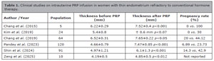
Table 1. Clinical studies on intrauterine PRP infusion in women with thin endometrium refractory to conventional hormone therapy.
PRP and chronic endometritis
Chronic endometritis (CE) is a persistent inflammatory condition of the inner lining of the uterine cavity. Although it causes few or no symptoms, CE is associated with reduced endometrial receptivity and poorer reproductive outcomes (Li et al., 2023). Therefore, CE is usually diagnosed by hysteroscopy, which is considered the standard procedure for evaluating the uterine cavity in patients with endometrial infertility conditions (Cicinelli et al., 2019). Growing evidence points to an association between CE and infertility of unknown etiology, unexplained repeated pregnancy loss, and embryo implantation failure after in vitro fertilization (IVF) in women (Kitaya et al., 2017). Oral antibiotic regimens are considered the gold standard for treating chronic endometritis (Cicinelli et al., 2019). However, some cases of CE do not respond satisfactorily to these traditional treatments. This problem is compounded by the increasing incidence of resistant microorganisms.Given this scenario, PRP has been investigated as a possible adjunct therapy capable of modulating the endometrial inflammatory response and may be effective in treating patients with chronic endometritis. Sfakianoudis et al. (2019) reported clinical remission and reproductive success in a woman diagnosed with persistent chronic endometritis due to Candida albicans who received PRP as monotherapy after two failed courses of antifungal treatment.Li et al. (2023) selected 117 of 327 patients with recurrent implantation failure who had positive MUM-1+/CD138+ immunostaining for chronic endometritis. All patients received antibiotic therapy with doxycycline 100 mg and metronidazole 500 mg every 12 hours for 14 days, in addition to endometrial PRP infusion, and underwent endometrial biopsy in the following luteal phase, with repeat immunohistochemical analysis. Of the 117 patients, 83 (70.94%) became negative after PRP administration combined with antibiotic therapy. Although this combination achieved statistical significance for clinical pregnancy and implantation rates (p<0.01) and live birth rate (p<0.05), studies with larger sample sizes and prospective designs are needed to confirm the true clinical benefit in refractory patients.In line with findings in humans, animal models have demonstrated failures of conventional treatments for endometritis, and the intrauterine use of PRP as adjuvant therapy has proven effective in horses (Carneiro et al., 2020) and cattle (Marini et al., 2016). However, results from animal models cannot be directly extrapolated to humans.Recently, a review published by Alonso-Frías et al. (2025) comprehensively addressed the efficacy and safety of PRP in endometrial disorders, including endometritis. The study highlighted that, in addition to the anti-inflammatory and regenerative effects of PRP on the endometrium, modulation of the endometrial microbial population may restore endometrial receptivity compromised by persistent infection and local inflammation. This review corroborates the findings of Sfakianoudis et al. (2019), strengthening the proposal of PRP as a promising alternative for patients with chronic endometritis resistant to conventional antibiotic therapy.In a study published by Chen et al. (2025b), endometrial transcriptome analysis suggested that PRP treatment effectively improved the endometrial microenvironment in patients with persistent endometritis. The therapy promoted conversion of the endometrial inflammatory profile, reducing the expression of pro-inflammatory cytokines (such as IL-6, TNF-α, and IFN-γ) and increasing the expression of anti-inflammatory and regulatory mediators (such as IL-10 and TGF-β). There was also a transition from the M1 to M2 macrophage phenotype, favoring a microenvironment receptive to implantation and reinforcing the potential of PRP as an adjuvant therapeutic strategy in cases of infertility associated with chronic endometritis. The main studies on the use of PRP in chronic endometritis are presented in Table 2, which shows significant rates of clinical remission and improvement in reproductive outcomes. Because PRP was combined with antibiotics, its isolated effect cannot be determined.
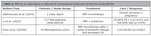
Table 2. PRP as an alternative or adjunct to conventional treatment of chronic endometritis
PRP and recurrent implantation failure
Recurrent implantation failure (RIF) is defined as the failure to achieve clinical pregnancy after at least three cycles of in vitro fertilization using high-quality embryos (Coughlan et al., 2014). This definition is not exact and is still debated by some scholars, considering the number and type of embryos transferred and the distinction between biochemical and clinical pregnancy (Cakiroglu & Tiras, 2020). Because of the complexity of the subject, it has also been conceptually accepted that RIF involves at least two embryo transfer failures (Zargar et al., 2021).RIF is a challenging infertility disorder. The mechanisms at the embryo-endometrium interface that lead to a successful pregnancy remain unclear. Although assisted reproductive technology has developed rapidly in recent years, most RIF treatment options lack sufficient high-quality evidence to support formal clinical recommendation. Several endometrial-targeted strategies have been investigated to improve receptivity, including endometrial scratching, topical immunomodulators, endometrial receptivity array (ERA) testing, and PRP. Embryonic interventions, such as preimplantation genetic testing for aneuploidy (PGT-A) and assisted hatching, have also been explored (Makrigiannakis et al., 2021). Clinical studies have suggested that autologous intrauterine PRP may improve reproductive outcomes in women with recurrent implantation failure, particularly in cases associated with impaired endometrial receptivity (Aghajanzadeh et al., 2020). However, not all studies have demonstrated consistent benefit. In women with a history of recurrent implantation failure but without thin endometrium, PRP infusion did not significantly improve pregnancy outcomes, suggesting that its potential effect may be restricted to specific endometrial phenotypes (Tehraninejad et al., 2021).At the molecular level, impaired signaling during the implantation window may be associated with alterations in protein synthesis and the release of cytokines, growth factors, prostaglandins, and adhesion molecules (Simon & Laufer, 2012). In this context, the biological properties of activated platelets in PRP have been proposed as a potential mechanism by which PRP could influence implantation, including modulation of growth factor signaling, increased expression of IL1 and IL8, increased progesterone receptor expression, and promotion of endometrial cell proliferation (Artimani et al., 2024). In a prospective double-blind clinical trial including 100 women with at least two prior implantation failures, Bakhsh et al. (2022) reported higher pregnancy rates in the PRP group than in the intrauterine catheterization-alone group (20% vs. 13.33%), and the difference was reported as statistically significant by the authors.Li et al. (2022) subsequently published the first systematic review and meta-analysis evaluating intrauterine PRP infusion in patients with unexplained recurrent implantation failure, including 1,629 participants (795 cases and 834 controls) from 10 studies. The pooled analysis demonstrated significantly higher clinical pregnancy rates in the PRP group than in controls (Nazari et al., 2022; Safdarian et al., 2022), whereas miscarriage rates did not differ significantly, consistent with previous reports (Safdarian et al., 2022; Zamaniyan et al., 2021). Subgroup analysis suggested a dose-dependent effect, with PRP volumes between 0.5 and 1 mL associated with more favorable outcomes than doses ≥1 mL. However, heterogeneity across included studies ranged from moderate to high, and substantial methodological variability was observed in PRP preparation protocols and study design. Therefore, although pooled estimates suggest potential benefit, these findings should be interpreted cautiously given the heterogeneity and risk of bias inherent in the available evidence.Similarly, another meta-analysis including 1,555 patients (775 cases and 780 controls) from six randomized clinical trials and four cohort studies reported that intrauterine autologous PRP infusion was associated with significant improvements in clinical pregnancy, chemical pregnancy, implantation, and live birth rates (Deng et al., 2023). Interpretation of these findings should consider variability in study design, PRP preparation protocols, and dosing strategies across included trials.More recently, a multicenter study by Yahyaei et al. (2024) evaluated intrauterine PRP infusion in 80 patients with RIF and reported significantly higher clinical pregnancy rates (55.8% vs. 20%; p<0.0001) and live birth rates (52.9% vs. 8.6%; p<0.0001) compared with controls when PRP was administered 48 hours before embryo transfer. These results support the hypothesis that PRP may modulate the endometrial implantation window and improve outcomes in patients with compromised receptivity, especially in IVF cycles using good-quality embryos. Although PRP has been increasingly proposed as an adjunct therapy in assisted reproduction, recent analyses have questioned whether intrauterine infusion should be routinely offered to women with recurrent implantation failure, highlighting the need for cautious interpretation of available clinical data and the importance of further high-quality trials (Katsika et al., 2025). The main studies investigating PRP use in RIF are summarized in Table 3.
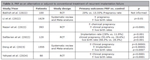
Table 3. PRP as an alternative or adjunct to conventional treatment of recurrent implantation failure
PRP and ovarian insufficiency
Ovarian insufficiency is a major reproductive obstacle for women, encompassing conditions such as premature ovarian insufficiency (POI), poor ovarian response (POR), and advanced maternal age (AMA) (Pantos et al., 2021). It is also an important focus of regenerative medicine, which seeks to restore female fertility impaired by the deleterious effects of associated pathophysiological conditions (Sfakianoudis et al., 2020). In this context, publications have increased in the last five years, suggesting that ovarian reactivation can be achieved using factors with recognized regenerative potential, such as PRP (Pantos et al., 2019).Danforth et al. (2003) observed increased follicular development and decreased apoptotic events in rats after intraovarian infusion of growth factors. Based on these findings, PRP injection into ovaries was subsequently introduced in humans, performed under anesthetic sedation and guided by transvaginal ultrasound. Two pioneering case series reported ovarian rejuvenation after intraovarian PRP infusion in patients characterized as perimenopausal (Pantos et al., 2016) or with premature ovarian failure (Sfakianoudis et al., 2018). Randomized controlled data have also emerged regarding intraovarian PRP administration. Recent trials evaluating its use in women with poor ovarian response have investigated potential improvements in IVF outcomes following intraovarian injection (Herlihy et al., 2024).In a systematic review, PRP intervention was associated with beneficial improvements in ovarian reserve parameters in women, including increased anti-Müllerian hormone (AMH), increased antral follicle count (AFC), and decreased follicle-stimulating hormone (FSH). Furthermore, it improved the performance of intracytoplasmic sperm injection (ICSI) cycles in terms of the total number of oocytes retrieved, the number of two-pronucleated embryos, the fertilization rate, and the number of cleavage-stage embryos (Panda et al., 2020). These findings were supported by a large cohort of 510 women, with a mean age of 40.3 years and poor ovarian reserve (POR), who were candidates for in vitro fertilization and treated with intraovarian PRP injection. The authors observed that this intervention resulted in a 20.5% pregnancy rate and a 12.9% live birth rate (Cakiroglu et al., 2022). Additionally, Éliás et al. (2024), in a systematic review and meta-analysis with 2,256 eligible women, confirmed the reproductive benefits of PRP, especially in the recovery of ovarian function and the response to hormonal stimulation in women with ovarian insufficiency (Figure 3).
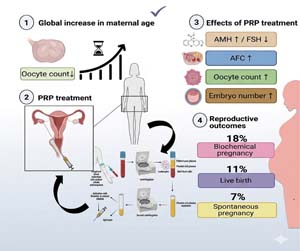
Figure 3. Overview of platelet-rich plasma (PRP) therapy in ovarian regeneration and reproductive outcomes. Adapted from Éliás et al. (2024).
Among current publications, a recent study conducted by Yu et al. (2025) evaluated the efficacy of intraovarian PRP injection in 74 women with poor ovarian response, including 30 controls. The results showed significant increases in oocyte maturation rate, fertilization, and blastocyst development, as well as improved clinical pregnancy rates. Another relevant study is the systematic review by Zamaniyan et al. (2025), which evaluated a total of 1,563 women with POR and premature ovarian insufficiency undergoing IVF after ovarian PRP treatment. This study demonstrated that PRP application in women with POR can improve oocyte quality and implantation rate, showing significant differences compared with control groups.The authors suggest that PRP may act through growth factor-mediated mechanisms, promoting cellular regeneration and supporting mitochondrial function, resulting in healthier oocytes and higher-quality embryos, which could favor implantation and reduce the risk of early miscarriage. Although promising, the overall pregnancy rate remains low, reinforcing the need for larger, standardized clinical studies to confirm these findings and better understand variations in response according to individual patient characteristics (Table 4).
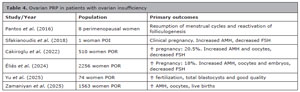
Table 4. Ovarian PRP in patients with ovarian insufficiency
PRP and male infertility
Erectile dysfunction (ED) is a disorder that affects 12%-19% of men of reproductive age. Among infertile men and couples, the prevalence is even higher, reaching 48%-58% (Starc et al., 2019). Despite this, ED is considered a rare cause of male infertility, accounting for 5% of cases (Goldstein et al., 2019). Current therapies do not affect the degenerative progression of the disease and function as temporary symptomatic palliation. In contrast, intracavernous PRP may potentially reverse or halt disease progression (Anastasiadis et al., 2022).Poulios et al. (2021) demonstrated a significant improvement in erectile function in a group of men, suggesting that intracavernous PRP injection may represent a new treatment strategy for ED because of its regenerative potential.Khadivi et al. (2020) evaluated the in vitro proliferative capacity of testicular tissue from prepubertal males when PRP was added to two-dimensional (2D) and three-dimensional (3D) culture systems. These systems are used in many species to promote in vitro spermatogenesis of spermatogonial stem cells (SSCs) (Ibtisham et al., 2021; Rahbar et al., 2022). These cells are highly sensitive to chemotherapy and radiotherapy; therefore, their improved preservation is of reproductive interest for male fertility preservation. The group concluded that optimizing these culture media with PRP improved testicular cell function and increased the number of human SSC colonies, making it a valid tool for fertility preservation in prepubertal males. PRP was associated with a sperm-protective effect and improved progressive sperm motility, viability, and membrane integrity after thawing (p<0.05) in semen cryopreservation studies in humans (Yan et al., 2021;Tamburrino et al., 2023) and animals (El-Sherbiny et al., 2022).Nabavinia et al. (2023) collected normal semen samples from healthy men and treated them with different concentrations of PRP as a cryoprotectant supplement, analyzing sperm before and after freezing. They concluded that PRP did not have a significant effect on sperm count, but it increased progressive motility and sperm viability significantly more than in the control group. In addition, the rate of immotile sperm in these samples was significantly lower than in controls, as was the rate of sperm DNA fragmentation.Within the same context of male infertility, additional studies have demonstrated promising applications of PRP. Fazli et al. (2024), in a clinical trial with 88 participants, administered bilateral PRP injections directly into the testes of 44 men with oligoasthenoteratozoospermia (OAT), observing significant improvements in concentration, progressive motility, and sperm DNA integrity. Similar results were found by Alonso-Frías & Esbert (2024) in a systematic review on the use of PRP in various male infertility conditions, including semen cryopreservation, oxidative stress, SSC culture, and non-obstructive azoospermia. According to the literature, supplementing cryopreservation medium with PRP significantly improved sperm parameters and preserved cellular integrity during freezing and thawing. Samples incubated with PRP showed a significant increase in antioxidant capacity, suggesting a protective effect against oxidative stress induced by the cryogenic process. Furthermore, PRP has been shown to stimulate the proliferation and vitality of spermatogonial stem cells, favoring sperm recovery in cases of non-obstructive azoospermia.Recently published evidence supports these conclusions. Hosseini et al. (2025) evaluated the addition of PRP to swim-up and semen incubation protocols, reporting statistically significant increases in concentration and total, progressive, and non-progressive motility in samples from asthenoteratozoospermic and normozoospermic men, but without a significant increase in sperm morphology. These data were corroborated by Moradian et al. (2025), who, in a systematic review, highlighted the potential of PRP to improve spermatogenesis and hormonal profiles through its antioxidant, anti-inflammatory, and tissue-repair properties, as well as sperm recovery in cases of non-obstructive azoospermia. Collectively, these findings indicate that PRP may play an adjuvant role in modulating the testicular microenvironment and improving sperm quality (Table 5). The various therapeutic applications of PRP in male reproductive function are illustrated in Figure 4.
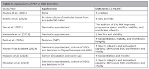
Table 5. Applications of PRP in Male InfertilityStudy/Year Application Outcomes (p<0.05) Poulios et al. (2021)Penis ↑ erection Khadivi et al. (2020)In vitro culture of testicular tissue from pre-pubertal males ↑ SSC colonies Yan et al. (2021)Seminal cryoprotectant The addition of 5% PRP improved progressive sperm motility, viability, and membrane integrity Nabavinia et al. (2023)Seminal cryoprotectant ↑ Motility and viability Fazli et al. (2024)Testicles (OAT) ↑ Concentration, motility, and membrane integrity Alonso-Frías & Esbert (2024)Seminal cryoprotectant, culture of SSCs and testicles in Oligoasthenospermia (OA) ↑ Sperm integrity and antioxidant capacity. Stimulates SSC proliferation and vitality Hosseini et al. (2025)Semen (incubation and swim-up) ↑ Concentration and motility Moradian et al. (2025)Seminal cryoprotectant, culture of SSCs and testicles in OA ↑ Sperm integrity and antioxidant capacity. Stimulates SSC proliferation and vitality
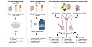
Figure 4. Therapeutic applications of platelet-rich plasma (PRP) in male reproductive function. Adapted from Moradian et al. (2025).
Although the available findings are promising, the current evidence still lacks robustness. The wide heterogeneity in PRP preparation protocols, the scarcity of data on long-term effects, and interspecies differences limit the extrapolation of results. Thus, future studies with better-structured methods, prolonged evaluation of clinical outcomes, and in-depth investigation of molecular mechanisms are needed to validate the true therapeutic potential of PRP and ensure its safe and reproducible clinical application.
CONCLUSION
Current evidence suggests that PRP may have potential as an adjuvant therapeutic approach in the management of infertility in both women and men. Interest in its clinical application has increased in recent years because of its immunomodulatory and anti-inflammatory properties; however, the available evidence remains inconclusive. As an autologous biological product, PRP is considered safe and easily obtainable and has low production costs. Some studies have reported that PRP can increase clinical pregnancy and live birth rates in cases of recurrent unexplained implantation failure in IVF, thin endometrium refractory to hormone therapy, chronic endometritis, and low ovarian reserve. PRP has also shown potential in male infertility, including erectile dysfunction and the protection of human semen quality after cryopreservation. Variability in processing protocols, application methods, and the absence of standardized qualitative assessment of PRP preparations may contribute to differences in efficacy reported across studies. Further well-designed studies are required to confirm these findings. Future research should expand the investigation of PRP in reproductive medicine, particularly in male infertility, supported by data from preclinical and translational models.
Acknowledgments:
The authors thank all contributors who provided academic support, technical assistance, and critical input during the preparation of this manuscript.
Funding:
REFERENCES
Aghajanzadeh F, Esmaeilzadeh S, Basirat Z, Mahouti T, Heidari FN, Golsorkhtabaramiri M. Using autologous intrauterine platelet-rich plasma to improve the reproductive outcomes of women with recurrent implantation failure. JBRA Assist Reprod. 2020;24:30-3. PMID: 31689042 DOI: 10.5935/1518-0557.20190055
Medline
Alonso-Frías P, Esbert M. Therapeutic use of platelet-rich plasma for the treatment of male infertility. Rev Int Androl. 2024;22:16-23. PMID: 39394745 DOI: 10.22514/j.androl.2024.018
Medline
Alonso-Frías P, Francés-Herrero E, Bueno-Fernandez C, Gómez-Álvarez M, Agustina-Hernández M, Cervelló I, Cozzolino M. Beneficial effects of infiltration of platelet-rich plasma in the endometrium. Biology (Basel). 2025;14:319. PMID: 40282184 DOI: 10.3390/biology14040319
Medline
Alves R, Grimalt R. A review of platelet-rich plasma: history, biology, mechanism of action, and classification. Skin Appendage Disord. 2018;4:18-24. PMID: 29457008 DOI: 10.1159/000477353
Medline
Amable PR, Carias RB, Teixeira MV, da Cruz Pacheco I, Corrêa do Amaral RJ, Granjeiro JM, Borojevic R. Platelet-rich plasma preparation for regenerative medicine: optimization and quantification of cytokines and growth factors. Stem Cell Res Ther. 2013;4:67. PMID: 23759113 DOI: 10.1186/scrt218
Medline
Anastasiadis E, Ahmed R, Khoja AK, Yap T. Erectile dysfunction: Is platelet-rich plasma the new frontier? A review of the existing evidence. Front Reprod Health. 2022;4:944765. PMID: 36303622 DOI: 10.3389/ frph.2022.944765
Medline
Artimani T, Asl SS, Amiri I, Pilehvari S, Yavangi M, Mohammadpour N, Mehdizadeh M. Effect of different concentrations of PRP on the expression of factors involved in endometrial receptivity in human endometrial cells from RIF patients compared to controls. Reprod Sci. 2024;31:3870-9. PMID: 39496922 DOI: 10.1007/s43032-024-01744-z
Medline
Atkinson L, Martin F, Sturmey RG. Intraovarian injection of platelet-rich plasma in assisted reproduction: too much too soon? Hum Reprod. 2021;36:1737-50. PMID: 33963408 DOI: 10.1093/humrep/deab106
Medline
Bakhsh AS, Maleki N, Sadeghi MR, SadeghiTabar A, Tavakoli M, Zafardoust S, Ghaemmaghami P. Effects of autologous platelet-rich plasma in women with repeated implantation failure undergoing assisted reproduction. JBRA Assist Reprod. 2022;26:84-7. PMID: 34514770 DOI: 10.5935/1518-0557.20210046
Medline
Bolton L. Platelet-rich plasma: optimal use in surgical wounds. Wounds. 2021;33:219-21. PMID: 34357880 DOI: 10.25270/wnds/2021.219221
Medline
Cakiroglu Y, Tiras B. Determining diagnostic criteria and cause of recurrent implantation failure. Curr Opin Obstet Gynecol. 2020;32:198-204. PMID: 32251092 DOI: 10.1097/GCO.0000000000000620
Medline
Cakiroglu Y, Yuceturk A, Karaosmanoglu O, Kopuk SY, Korun ZEU, Herlihy N, Scott RT, Tiras B, Seli E. Ovarian reserve parameters and IVF outcomes in 510 women with poor ovarian response (POR) treated with intraovarian injection of autologous platelet rich plasma (PRP). Aging (Albany NY). 2022;14:2513-23. PMID: 35320118 DOI: 10.18632/aging.203972
Medline
Cervelli V, Gentile P, Scioli MG, Grimaldi M, Casciani CU, Spagnoli LG, Orlandi A. Application of platelet-rich plasma in plastic surgery: clinical and in vitro evaluation. Tissue Eng Part C Methods. 2009;15:625-34. PMID: 19231923 DOI: 10.1089/ten.TEC.2008.0518
Medline
Chang Y, Li J, Chen Y, Wei L, Yang X, Shi Y, Liang X. Autologous platelet-rich plasma promotes endometrial growth and improves pregnancy outcome during IVF. Int J Clin Exp Med. 2015;8:1286-90. PMID: 25785127
Medline
Chang Y, Li J, Wei LN, Pang J, Chen J, Liang X. Autologous infusion of platelet-rich plasma improves clinical pregnancy rate in frozen embryo transfer cycles for women with thin endometrium. Medicine (Baltimore). 2019;98:e14062. PMID: 30653117 DOI: 10.1097/MD.0000000000014062
Medline
Chen K, Chen YC, Yu TY, Lin LP, Hsu HC, Liao YC, Su Pang JH, Tsai WC. Platelet-rich plasma promotes cell migration of adipose-derived stem cells by upregulation of focal adhesion kinase and paxillin. Regen Ther. 2025a;30:9-19. PMID: 40487227 DOI: 10.1016/j.reth.2025.05.004
Medline
Chen PF, Liang YL, Chuang YJ, Wu MH. Autologous PRP therapy for thin endometrium: a self-controlled case series study across menstrual cycles. Eur J Obstet Gynecol Reprod Biol. 2024;299:12-7. PMID: 38820688 DOI: 10.1016/j.ejogrb.2024.05.032
Medline
Chen X, Chen M, Liu M, Qi L, Liu Z, Chen C, Liang B, Yang X, Zhang T, Li Y, Lian R. Intrauterine infusion of autologous platelet-rich plasma modulates endometrial immune status and improves pregnancy outcomes in patients with persistent chronic endometritis. Front Immunol. 2025b;16:1528522. PMID: 40443660 DOI: 10.3389/ fimmu.2025.1528522
Medline
Chi L, Wang Q, Zhong W, Pan X, Li Y. Usefulness of platelet-rich plasma to manage skin wounds: a meta-analysis. Int Wound J. 2023;20:3123-30. PMID: 37194622 DOI: 10.1111/iwj.14179
Medline
Cicinelli E, Vitagliano A, Kumar A, Lasmar RB, Bettocchi S, Haimovich S; International Working Group for Standardization of Chronic Endometritis Diagnosis. Unified diagnostic criteria for chronic endometritis at fluid hysteroscopy: proposal and reliability evaluation through an international randomized-controlled observer study. Fertil Steril. 2019;112:162-73.e2. PMID: 31200985 DOI: 10.1016/j.fertnstert.2019.03.004
Medline
Coughlan C, Ledger W, Wang Q, Liu F, Demirol A, Gurgan T, Cutting R, Ong K, Sallam H, Li TC. Recurrent implantation failure: definition and management. Reprod Biomed Online. 2014;28:14-38. PMID: 24269084 DOI: 10.1016/j. rbmo.2013.08.011
Medline
Danforth DR, Arbogast LK, Ghosh S, Dickerman A, Rofagha R, Friedman CI. Vascular endothelial growth factor stimulates preantral follicle growth in the rat ovary. Biol Reprod. 2003;68:1736-41. PMID: 12606455 DOI: 10.1095/biolreprod.102.011460
Medline
Dart AJ, Dowling BA, Smith CL. Topical treatments in equine wound management. Vet Clin North Am Equine Pract. 2005;21:77-89. PMID: 15691601 DOI: 10.1016/j. cveq.2004.11.003
Medline
Deng H, Wang S, Li Z, Xiao L, Ma L. Effect of intrauterine infusion of platelet-rich plasma for women with recurrent implantation failure: a systematic review and meta-analysis. J Obstet Gynaecol. 2023;43:2144177. PMID: 36397660 DOI: 10.1080/01443615.2022.2144177
Medline
Éliás M, Kónya M, Kekk Z, Turan C, das Virgens IPA, Tóth R, Keszthelyi M, Hegyi P, Várbíró S, Sipos M. Platelet-rich plasma (PRP) treatment of the ovaries significantly improves fertility parameters and reproductive outcomes in diminished ovarian reserve patients: a systematic review and meta-analysis. J Ovarian Res. 2024;17:104. PMID: 38760869 DOI: 10.1186/s13048-024-01423-2
Medline
El-Sherbiny HR, Abdelnaby EA, Samir H, Fathi M. Addition of autologous platelet-rich plasma to semen extender enhances cryotolerance and fertilizing capacity of buffalo bull spermatozoa. Theriogenology. 2022;194:104-9. PMID: 36219886 DOI: 10.1016/j.theriogenology.2022.09.029
Medline
Fazli F, Torkashvand H, Soltanian AR, Babalhavaeji A, Olomi H, Pilehvari S. Effects of Testicular Platelet-Rich Plasma (PRP) Injection on Sperm Parameters in Men with Severe Oligoasthenoteratozoospermia (OAT): A Clinical Evaluation. Int J Fertil Steril. 2024;18:71-6. PMID: 39033373 DOI: 10.22074/IJFS.2024.2011066.1535
Medline
Ferrari AR, Cortrezzi S, Borges E Junior, Braga D, Souza MDCB, Antunes RA. Evaluation of the Effects of Platelet-Rich Plasma on Follicular and Endometrial Growth: A Literature Review. JBRA Assist Reprod. 2021;25:601-7. PMID: 34415119 DOI: 10.5935/1518-0557.20210036
Medline
Floryan KM, Berghoff WJ. Intraoperative use of autologous platelet-rich and platelet-poor plasma for orthopedic surgery patients. AORN J. 2004;80:668-74. PMID: 15526700 DOI: 10.1016/S0001-2092(06)61320-3
Medline
Foitzik C, Staus H. Le Fort I osteotomy in atrophied maxilla and bone regeneration with beta-tricalcium phosphate and PRP. Implant Dent. 2003;12:132-9. PMID: 12861880 DOI: 10.1097/01.id.0000061084.09518.3e
Medline
Foster TE, Puskas BL, Mandelbaum BR, Gerhardt MB, Rodeo SA. Platelet-rich plasma: from basic science to clinical applications. Am J Sports Med. 2009;37:2259-72. PMID: 19875361 DOI: 10.1177/0363546509349921
Medline
Frantz N, Ferreira M, Kulmann MI, Frantz G, Bos-Mikich A, Oliveira R. Platelet-rich plasma as an effective alternative approach for improving endometrial receptivity: a clinical retrospective study. JBRA Assist Reprod. 2020;24:442-6. PMID: 32558323 DOI: 10.5935/1518-0557.20200026
Medline
Goldstein I, Goren A, Li VW, Maculaitis MC, Tang WY, Hassan TA. Erectile dysfunction and productivity: global association. Int J Clin Pract. 2019;73:e13384. PMID: 31389146 DOI: 10.1111/ijcp.13384
Medline
Herlihy NS, Cakiroglu Y, Whitehead C, Reig A, Tiras B, Scott RT Jr, Seli E. Effect of intraovarian platelet-rich plasma injection on IVF outcomes in women with poor ovarian response: the PROVA randomized controlled trial. Hum Reprod. 2024;39:1215-25. PMID: 38721967 DOI: 10.1093/humrep/deae093
Medline
Hosseini S, Kazemi M, Salehpour S, Saharkhiz N, Majdi L. Evaluation of the effect of platelet-rich plasma (PRP) on the sperm parameters. Taiwan J Obstet Gynecol. 2025;64:313-8. PMID: 40049817 DOI: 10.1016/j.tjog.2024.12.010
Medline
Ibtisham F, Zhao Y, Nawab A, Wu J, Mei X, Honaramooz A, An L. In vitro production of haploid germ cells from murine spermatogonial stem cells using a two-dimensional cell culture system. Theriogenology. 2021;162:84-94. PMID: 33450717 DOI: 10.1016/j.theriogenology.2020.12.024
Medline
Kasius A, Smit JG, Torrance HL, Eijkemans MJC, Mol BW, Opmeer BC, Broekmans FJ, Torrance JM. Endometrial thickness and pregnancy rates after IVF: a systematic review and meta-analysis. Hum Reprod Update. 2014;20:530-41. PMID: 24664156 DOI: 10.1093/humupd/dmu011
Medline
Katsika ET, Venetis CA, Bosdou JK, Kolibianakis EM. Is it justified to offer intrauterine infusion of autologous PRP in women with repeated implantation failure? Hum Reprod. 2025;40:771-84. PMID: 40052514 DOI: 10.1093/humrep/deaf039
Medline
Khadivi F, Koruji M, Akbari M, Jabari A, Talebi A, Ashouri Movassagh S, Panahi Boroujeni A, Feizollahi N, Nikmahzar A, Pourahmadi M, Abbasi M. Application of platelet-rich plasma (PRP) improves self-renewal of human spermatogonial stem cells in two-dimensional and three-dimensional culture systems. Acta Histochem. 2020;122:151627. PMID: 33002788 DOI: 10.1016/j.acthis.2020.151627
Medline
Kim H, Shin JE, Koo HS, Kwon H, Choi DH, Kim JH. Effect of Autologous Platelet-Rich Plasma Treatment on Refractory Thin Endometrium During the Frozen Embryo Transfer Cycle: A Pilot Study. Front Endocrinol (Lausanne). 2019;10:61. PMID: 30837945 DOI: 10.3389/fendo.2019.00061
Medline
Kim JI, Bae HC, Park HJ, Lee MC, Han HS. Effect of storage and activation on growth factor concentration in platelet-rich plasma. J Orthop Res. 2020;38:777-84. PMID: 31709604 DOI: 10.1002/jor.24520
Medline
Kitaya K, Matsubayashi H, Takaya Y, Nishiyama R, Yamaguchi K, Takeuchi T, Ishikawa T. Live birth rate following oral antibiotic treatment for chronic endometritis in infertile women with repeated implantation failure. Am J Reprod Immunol. 2017;78:e12719. PMID: 28608596 DOI: 10.1111/aji.12719
Medline
Li J, Li X, Ding J, Zhao J, Chen J, Guan F, Deng H, Zhou M, Han Y, Xiao Z, Yang J. Analysis of pregnancy outcomes in recurrent implantation failure with chronic endometritis. Front Cell Dev Biol. 2023;11:1088586. PMID: 36861040 DOI: 10.3389/fcell.2023.1088586
Medline
Li M, Kang Y, Wang Q, Yan L. Efficacy of Autologous Intrauterine Infusion of Platelet-Rich Plasma in Patients with Unexplained Repeated Implantation Failures in Embryo Transfer: A Systematic Review and Meta-Analysis. J Clin Med. 2022;11:6753. PMID: 36431229 DOI: 10.3390/ jcm11226753
Medline
Makrigiannakis A, Makrygiannakis F, Vrekoussis T. Approaches to improve endometrial receptivity in case of repeated implantation failures. Front Cell Dev Biol. 2021;9:613277. PMID: 33796523 DOI: 10.3389/ fcell.2021.613277
Medline
Marini MG, Perrini C, Esposti P, Corradetti B, Bizzaro D, Riccaboni P, Fantinato E, Urbani G, Gelati G, Cremonesi F, Lange-Consiglio A. Effects of platelet-rich plasma in bovine endometrial inflammation in vitro. Reprod Biol Endocrinol. 2016;14:58. PMID: 27619959 DOI: 10.1186/s12958-016-0195-4
Medline
Marx RE. Platelet-rich plasma (PRP): what is PRP and what is not PRP? Implant Dent. 2001;10:225-8. PMID: 11813662 DOI: 10.1097/00008505-200110000-00002
Medline
Moradian SA, Amirkhani Z, Movahedin M, Gharib SN, Varghaiyan Y, Niknafs B, Hamdi K, Hajipour H, Fattahi A. Platelet-rich plasma (PRP) and the future of male fertility: a path forward for personalized and regenerative therapies. Stem Cell Res Ther. 2025;16:486. PMID: 40898310 DOI: 10.1186/s13287-025-04602-0
Medline
Murphy S, Gardner FH. Platelet storage at 22 degrees C; metabolic, morphologic, and functional studies. J Clin Invest. 1971;50:370-7. PMID: 5540174 DOI: 10.1172/ JCI106504
Medline
Nabavinia MS, Yari A, Ghasemi-Esmailabad S, Gholoobi A, Gholizadeh L, Nabi A, Lotfi M, Khalili MA. Improvement of human sperm properties with platelet-rich plasma as a cryoprotectant supplement. Cell Tissue Bank. 2023;24:307-15. PMID: 36074213 DOI: 10.1007/s10561-022-10032-6
Medline
Nazari L, Salehpour S, Hosseini S, Sheibani S, Hosseinirad H. The Effects of Autologous Platelet-Rich Plasma on Pregnancy Outcomes in Repeated Implantation Failure Patients Undergoing Frozen Embryo Transfer: A Randomized Controlled Trial. Reprod Sci. 2022;29:993-1000. PMID: 34651260 DOI: 10.1007/s43032-021-00669-1
Medline
Panda SR, Sachan S, Hota S. A Systematic Review Evaluating the Efficacy of Intra-Ovarian Infusion of Autologous Platelet-Rich Plasma in Patients With Poor Ovarian Reserve or Ovarian Insufficiency. Cureus. 2020;12:e12037. PMID: 33457137 DOI: 10.7759/cureus.12037
Medline
Pandey D, Bajaj B, Kapoor G, Bharti R. Intrauterine platelet-rich plasma in infertile females with thin endometrium undergoing intrauterine insemination: randomized trial. AJOG Glob Rep. 2023;3:100172. PMID: 36960130 DOI: 10.1016/j.xagr.2023.100172
Medline
Pantos K, Simopoulou M, Pantou A, Rapani A, Tsioulou P, Nitsos N, Syrkos S, Pappas A, Koutsilieris M, Sfakianoudis K. A case series on natural conceptions resulting in ongoing pregnancies in menopausal and prematurely menopausal women following platelet-rich plasma treatment. Cell Transplant. 2019;28:1333-40. PMID: 31271054 DOI: 10.1177/0963689719859539
Medline
Pixley JN, Cook MK, Singh R, Larrondo J, McMichael AJ. Platelet-rich plasma for dermatologic disorders: comprehensive review. J Dermatolog Treat. 2023;34:2142035. PMID: 36318219 DOI: 10.1080/09546634.2022.2142035
Medline
Poseidon Group (Patient-Oriented Strategies Encompassing IndividualizeD Oocyte Number); Alviggi C, Andersen CY, Buehler K, Conforti A, De Placido G, Esteves SC, Fischer R, Galliano D, Polyzos NP, Sunkara SK, Ubaldi FM, Humaidan P. A new more detailed stratification of low responders to ovarian stimulation: from a poor ovarian response to a low prognosis concept. Fertil Steril. 2016;105:1452-3. PMID: 26921622 DOI: 10.1016/j.fertnstert.2016.02.005
Medline
Poulios E, Mykoniatis I, Pyrgidis N, Zilotis F, Kapoteli P, Kotsiris D, Kalyvianakis D, Hatzichristou D. Platelet-Rich Plasma (PRP) Improves Erectile Function: A Double-Blind, Randomized, Placebo-Controlled Clinical Trial. J Sex Med. 2021;18:926-35. PMID: 33906807 DOI: 10.1016/j. jsxm.2021.03.008
Medline
Rahbar M, Asadpour R, Azami M, Mazaheri Z, Hamali H. Improving the process of spermatogenesis in azoospermic mice using spermatogonial stem cells co-cultured with epididymosomes in three-dimensional culture system. Life Sci. 2022;310:121057. PMID: 36220369 DOI: 10.1016/j. lfs.2022.121057
Medline
Ranisavljevic N, Raad J, Anahory T, Grynberg M, Sonigo C. Embryo transfer strategy and therapeutic options in infertile patients with thin endometrium: a systematic review. J Assist Reprod Genet. 2019;36:2217-31. PMID: 31502111 DOI: 10.1007/s10815-019-01576-w
Medline
Sfakianoudis K, Simopoulou M, Nitsos N, Rapani A, Pappas A, Pantou A, Chronopoulou M, Deligeoroglou E, Koutsilieris M, Pantos K. Autologous platelet-rich plasma treatment enables pregnancy for a woman in premature menopause. J Clin Med. 2018;8:1. PMID: 30577435 DOI: 10.3390/ jcm8010001
Medline
Sfakianoudis K, Simopoulou M, Nitsos N, Lazaros L, Rapani A, Pantou A, Koutsilieris M, Nikas Y, Pantos K. Successful live birth after platelet-rich plasma treatment in a patient with recurrent implantation failure and chronic endometritis. In Vivo. 2019;33:515-21. PMID: 30804135 DOI: 10.21873/invivo.11504
Medline
Sfakianoudis K, Rapani A, Grigoriadis S, Retsina D, Maziotis E, Tsioulou P, Giannelou P, Pantos K, Koutsilieris M, Vlahos N, Mastorakos G, Simopoulou M. Novel Approaches in Addressing Ovarian Insufficiency in 2019: Are We There Yet? Cell Transplant. 2020;29:963689720926154. PMID: 32686983 DOI: 10.1177/0963689720926154
Medline
Shin SY, Chung N, Shin JE, Kim JH, Park C, Kwon H, Choi DH, Lee JR, Kim JH. Angiogenic factor-driven improvement of refractory thin endometrium with autologous platelet-rich plasma intrauterine infusion in frozen embryo transfer cycles. Front Endocrinol (Lausanne). 2024;15:1431453. PMID: 39290323 DOI: 10.3389/fendo.2024.1431453
Medline
Simon A, Laufer N. Repeated implantation failure: clinical approach. Fertil Steril. 2012;97:1039-43. PMID: 22464086 DOI: 10.1016/j.fertnstert.2012.03.010
Medline
Singh SK, Singh S. Effect of platelet counts and activator in platelet-rich plasma on the treatment of androgenetic alopecia, split-head comparison: A randomised, double-blind study. Indian J Dermatol Venereol Leprol. 2023;89:647-55. PMID: 37067143 DOI: 10.25259/ IJDVL_1254_20
Medline
Starc A, Trampuš M, Pavan Jukić D, Rotim C, Jukić T, Mivšek PA. Infertility and sexual dysfunctions: systematic review. Acta Clin Croat. 2019;58:508-15. PMID: 31969764 DOI: 10.20471/acc.2019.58.03.15
Medline
Tamburrino L, Traini G, Marcellini A, Vignozzi L, Baldi E, Marchiani S. Cryopreservation of human spermatozoa: functional, molecular and clinical aspects. Int J Mol Sci. 2023;24:4656. PMID: 36902084 DOI: 10.3390/ijms24054656
Medline
Tehraninejad ES, Kashani NG, Hosseini A, Tarafdari A. Autologous platelet-rich plasma infusion does not improve pregnancy outcomes in frozen embryo transfer cycles in women with history of repeated implantation failure without thin endometrium. J Obstet Gynaecol Res. 2021;47:147-51. PMID: 32840031 DOI: 10.1111/jog.14445
Medline
Yahyaei A, Madani T, Vesali S, Mashayekhi M. Intrauterine infusion of autologous platelet-rich plasma in unexplained recurrent implantation failure. Sci Rep. 2024;14:26009. PMID: 39472511 DOI: 10.1038/s41598-024-77578-1
Medline
Yan B, Zhang Y, Tian S, Hu R, Wu B. Effect of autologous platelet-rich plasma on human sperm quality during cryopreservation. Cryobiology. 2021;98:12-6. PMID: 33476642 DOI: 10.1016/j.cryobiol.2021.01.009
Medline
Yu TN, Chen MJ, Lee TH, Chen YC, Cheng EH, Huang CC, Chen CI, Lee CI, Lee MS, Lin PY. Intraovarian platelet-rich plasma injection significantly improves blastocyst yield and quality in IVF patients. Sci Rep. 2025;15:1301. PMID: 39779754 DOI: 10.1038/s41598-024-82630-1
Medline
Yuan X, Saravelos SH, Wang Q, Xu Y, Li TC, Zhou C. Endometrial thickness and pregnancy outcomes in 30,495 IVF-ICSI cycles. Reprod Biomed Online. 2016;33:197-205. PMID: 27238372 DOI: 10.1016/j.rbmo.2016.05.002
Medline
Zamaniyan M, Peyvandi S, Heidaryan Gorji H, Moradi S, Jamal J, Yahya Poor Aghmashhadi F, Hossein Mohammadi M. Effect of platelet-rich plasma on pregnancy outcomes in infertile women with recurrent implantation failure: a randomized controlled trial. Gynecol Endocrinol. 2021;37:141-5. PMID: 32363968 DOI: 10.1080/09513590.2020.1756247
Medline
Zamaniyan M, Peivandi S, Pashazadeh M, Safarloo F. Efficacy of intraovarian injection of autologous platelet-rich plasma in women with poor ovarian response: systematic review. Int J Reprod Biomed. 2025;23:459-74. PMID: 40959472 DOI: 10.18502/ijrm.v23i6.19397
Medline
Zeng J, Quan J, Liu H, Geng W, Qiu F, Liu J. Single-cell transcriptome analysis of platelet-rich plasma therapy on human thin endometrium. Sci Rep. 2025;15:14637. PMID: 40287476 DOI: 10.1038/s41598-025-99468-w
Medline
Zhu X, Deng X, Li L, Zhao X, Guo Y. Intrauterine platelet-rich plasma infusion with hormone replacement therapy improves menstrual and pregnancy outcomes in thin endometrium patients undergoing frozen-thawed embryo transfer. Gynecol Endocrinol. 2026;42:2610084. PMID: 41493055 DOI: 10.1080/09513590.2025.2610084
Medline
Zhuang YW, Zeng YM, Chen YF, Zhang HP, Chen XY, Yang DY, Wu WJ. The effects of different activators on the release curve of human platelet-rich plasma. Zhonghua Jie He He Hu Xi Za Zhi. 2018;41:868-72. PMID: 30423630 DOI: 10.3760/cma.j.issn.1001-0939.2018.11.008
Medline