JBRA Assist. Reprod. 2026 2026;00(0):00-00
ORIGINAL ARTICLE
doi: 10.5935/1518-0557.20260093
Correlation between Sperm DNA Fragmentation and Motility Pa-rameters: A Comprehensive Study
1Urology Department of Centro Universitário FMABC, Santo André, São Paulo, Brazil
2Instituto Ideia Fértil de Saúde Reprodutiva, Santo André, São Paulo, Brazil
3Centro Universitário FMABC, Department of Collective Health, Discipline of Sexual and Reproductive Health and Population Genetics, Santo André, São Paulo, Brazil
CONFLICTS OF INTEREST
None.
ABSTRACT
Objective: To investigate the relationship between sperm DNA fragmentation (SDF) and sperm motility parameters in men undergoing fertility evaluation.
Methods: This retrospective observational study in-cluded 589 men aged 18-50 years evaluated at a sin-gle Brazilian andrology center between August 2021 and February 2024. Only the first semen sample per patient was analyzed. SDF was assessed using the sperm chromatin dispersion (SCD) test, and semen parameters were evaluated according to the sixth edition of the WHO Laboratory Manual. Data distribution was assessed using Shapiro-Wilk testing. As most variables were non-normally distributed, associations were analyzed using Spearman’s rank correlation, with Pearson’s test performed as sensitivity analysis. Comparisons be-tween two groups (<20% vs ≥20%) were performed us-ing the Mann-Whitney U test. Comparisons among three fragmentation categories (<20%, 20-30%, >30%) were conducted using the Kruskal-Wallis test followed by Dunn’s post-hoc analysis. A p-value <0.05 was considered statistically significant.
Results: The mean SDF was 23.5%. SDF demonstrat-ed a moderate inverse correlation with rapid progressive motility (rho=-0.289, 95% CI [-0.361, -0.213], p<0.001) and a moderate positive correlation with immotile sper-matozoa (rho=0.335, 95% CI [0.261, 0.405], p<0.001).Samples with SDF ≥20% (n=276) showed significant-ly lower rapid progressive motility and higher immotility compared to those with SDF <20% (n=313) (p<0.01). Total motile sperm count was also reduced in the higher fragmentation group (p<0.01). Similar patterns were observed across the three fragmentation categories.
Conclusion: Higher sperm DNA fragmentation levels were associated with impaired motility patterns, particularly reduced rapid progressive motility and increased immotility. These findings support the integration of SDF testing as a complementary parameter in the evaluation of male fertility.
Keywords: Sperm DNA fragmentation, Sperm motility, Male infertility
INTRODUCTION
Male infertility represents a substantial component of reproductive failure, contributing to approximately 40-50% of infertility cases worldwide (Agarwal et al., 2015). Although conventional semen analysis remains the cornerstone of male fertility assessment, increasing evidence suggests that traditional parameters alone may not fully capture sperm functional competence (Björndahl & Kirkman-Brown, 2022). Among the factors increasingly recognized as relevant to reproductive outcomes are sperm motility and sperm DNA integrity.
Progressive motility enables spermatozoa to migrate through the female reproductive tract and ultimately reach the oocyte, whereas intact DNA structure is fundamental for proper fertilization and embryonic development (Esteves et al., 2018; Simon et al., 2019).
Increasing attention has been directed toward sperm DNA fragmentation (SDF) as an additional marker of sperm quality, particularly in cases of unexplained infertility and adverse reproductive outcomes (Caliskan et al., 2022; Simon et al., 2019).
Previous investigations have suggested that elevated DNA fragmentation may be associated with impaired semen parameters, particularly reduced sperm motility (Belloc et al., 2014). However, available data remain heterogeneous, relatively few studies have evaluated this relationship in large real-world cohorts of patients undergoing andrological evaluation (Zhang et al., 2021). Clarifying this association may enhance interpretation of semen analysis and improve the clinical relevance of DNA integrity testing.
The present study aimed to investigate the relationship between sperm DNA fragmentation and sperm motility parameters in a large cohort of men undergoing fertility assessment in a specialized andrology laboratory.
MATERIAL AND METHODS
This retrospective observational study analyzed semen samples from men undergoing fertility evaluation at a single andrology center in Brazil between August 2021 and February 2024. The study included 589 semen analyses with complete data on sperm DNA fragmentation and conventional semen parameters.
Eligible participants were men aged between 18 and 50 years who underwent semen analysis as part of fertility investigation. Samples from individuals with incomplete laboratory data and known genetic disorders affecting fertility were excluded from the analysis. To ensure independence of observations and avoid pseudo-replication, only the first chronologically semen sample from each individual was analyzed.
Semen samples were collected by masturbation into sterile containers following a recommended abstinence period of 2-5 days according to routine laboratory practice. Conventional semen parameters, including volume, sperm concentration, and motility characteristics, were evaluated according to the World Health Organization (WHO) laboratory manual for the examination and processing of human semen (6th edition) (World Health Organization, 2021). Motility was categorized into rapid progressive motility (MPR), slow progressive motility (MPL), non-progressive motility (MNP), and immotile spermatozoa (IMOV).
Total motile sperm count (TMSC) was calculated using the standard formula: TMSC=semen volume × sperm concentration × total motility fraction where total motility corresponds to the sum of progressive and non-progressive motile spermatozoa.
Sperm DNA fragmentation was evaluated using the sperm chromatin dispersion (SCD) test (Halosperm G2 kit; Halotech DNA, Madrid, Spain), a method based on the differential dispersion of DNA loops following chromatin denaturation and protein removal. All evaluations were performed by trained laboratory personnel following standardized laboratory protocols. After semen processing, aliquots of the sample were mixed with low-melting-point agarose and placed on pretreated microscope slides. The slides were then exposed to an acid denaturation solution, followed by a lysis solution that removes nuclear proteins from sperm chromatin. Analysis was performed by two experienced observers blinded to the conventional semen parameters. Internal quality control was maintained through periodic inter-observer agreement assessments (Cohen’s kappa >0.80). Spermatozoa with intact DNA produce nucleoids with large halos of dispersed DNA loops surrounding a compact nuclear core. In contrast, spermatozoa with fragmented DNA display small halos or no halo due to the inability of DNA loops to expand following chromatin relaxation. Slides were stained and examined under bright-field microscopy. A minimum of 500 spermatozoa were evaluated per sample, and results were expressed as the percentage of spermatozoa exhibiting DNA fragmentation. This methodology follows previously validated protocols for SCD testing described in the literature (Feijó & Esteves, 2014; Zhang et al., 2010).
For descriptive subgroup analyses, samples were categorized according to three fragmentation ranges commonly reported in clinical practice: <20%, 20-30%, and >30%. These ranges have been commonly used in clinical andrology literature to stratify sperm DNA fragmentation severity in SCD-based assays. These thresholds are based on clinical studies using the SCD assay, where a DFI > 30% has been associated with significantly reduced pregnancy rates in both natural conception and assisted reproduction (Esteves et al., 2020; Evenson & Wixon, 2008; Feijó & Esteves, 2014).
Data distribution was assessed using Shapiro-Wilk testing and graphical inspection. As most variables showed non-normal distribution, and monotonic relationships were assumed, Spearman’s rank correlation coefficient was used to assess associations between SDF and semen parameters. Confidence intervals for correlation coefficients were calculated using Fisher’s z transformation.
Comparisons between two groups (<20% vs. ≥20%) were conducted using the Mann-Whitney U test. Comparisons across three groups (<20%, 20-30%, >30%) were performed using the Kruskal-Wallis test, followed by Dunn’s post-hoc test when appropriate. Continuous variables are presented as mean ± standard deviation and median with interquartile range when appropriate. Statistical significance was defined as p<0.05.
The study was approved by the institutional review board (approval number 80838924.0.00000082).
RESULTS
The mean sperm DNA fragmentation index in the study population was 23.5%. Baseline semen parameters for the entire cohort are presented in Table 1.
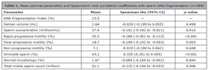
Table 1. Mean seminal parameters and Spearman’s rank correlation coefficients with sperm DNA fragmentation (n=589)
Correlation analysis performed using Spearman’s rank correlation coefficient.
No positive association was observed between SDF and total motile sperm. Instead, higher fragmentation levels were consistently associated with reduced progressive motility and increased immotility. Detailed correlation coefficients are presented in Table 1.
Samples were initially categorized into two groups according to sperm DNA fragmentation levels: 313 presented SDF<20% and 276 presented SDF ≥ 20%. The group with elevated DNA fragmentation showed lower values of rapid progressive motility and higher proportions of immotile spermatozoa (p<0.01). Total motile sperm count was also lower in this group. The complete comparison between groups is shown in Table 2.
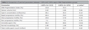
Table 2. Comparison of seminal parameters between <20% and ≥20% sperm DNA fragmentation groups
* Mann-Whitney U test.
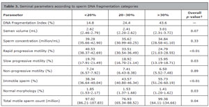
Table 3. Seminal parameters according to sperm DNA fragmentation categories
†Kruskal-Wallis test with Dunn’s post-hoc analysis.
DISCUSSION
This study evaluated the relationship between sperm DNA fragmentation and conventional semen parameters in a large cohort of men undergoing fertility evaluation in a specialized andrology laboratory.
The main finding was an association between higher levels of sperm DNA fragmentation and impaired sperm motility characteristics. Specifically, increased DNA fragmentation was correlated with reduced rapid progressive motility and increased proportions of immotile spermatozoa.
These findings are consistent with previous reports suggesting that sperm DNA integrity is closely related to sperm functional competence (Belloc et al., 2014; Ferrigno et al., 2021). Although conventional semen analysis evaluates motility and concentration, it does not directly assess chromatin integrity. The present results suggest that structural DNA damage may coexist with functional impairment of sperm movement.
The biological mechanisms underlying this association are likely multifactorial. Oxidative stress, apoptosis, and defective chromatin remodeling may simultaneously compromise DNA integrity and flagellar function, resulting in the combined phenotype of increased fragmentation and reduced motility (Faja et al., 2019; Simon et al., 2019).
An interesting observation in this study was the reduction in total motile sperm count among samples with higher DNA fragmentation levels. Although TMSC showed only a weak linear correlation with SDF in continuous analysis, group comparisons revealed significantly lower values in patients with elevated DNA fragmentation. This finding may reflect non-linear relationships between sperm DNA damage and composite semen parameters (Zhang et al., 2021).
From a clinical perspective, these results reinforce the concept that sperm DNA integrity testing may provide complementary information to conventional semen analysis (Esteves et al., 2018; Esteves et al., 2020; Kim, 2018). Evaluation of sperm DNA fragmentation may be useful in cases where standard semen parameters appear normal but reproductive outcomes remain suboptimal (Marinaro, 2023; McQueen et al., 2019; Nicopoullos et al., 2019; Rex et al., 2021; Tan et al., 2019).
It is important to acknowledge the limitations of this study. First, the retrospective and observational design does not allow causal inferences. Potential confounding factors such as age (Belloc et al., 2014), abstinence time, smoking status, varicocele, infections, and systemic inflammatory conditions were not controlled and may partially influence the associations observed. Furthermore, sperm DNA fragmentation was assessed using a single method. Although SCD is widely used due to its relative simplicity and reproducibility, different assays for sperm DNA damage measure distinct aspects of chromatin integrity and may yield different results (Evenson, 2013; Liffner et al., 2019). Additionally, the study was conducted in a single andrology center, which may limit the generalizability of the findings. Nevertheless, the consistency of the observed correlations and the relatively large sample size strengthen the robustness of the findings.
CONCLUSION
Higher levels of sperm DNA fragmentation were associated with reduced rapid progressive motility and increased immotility in men undergoing fertility evaluation. These findings support the integration of sperm DNA fragmentation assessment as a complementary tool in the comprehensive evaluation of male reproductive potential. Prospective studies incorporating reproductive outcomes and adjustment for relevant confounding variables are warranted to further clarify the clinical implications of this association.
§Senior Authors
* The content of this manuscript was previously presented in the form of a moderated poster at the Brazilian Congress of Assisted Reproduction, Florianópolis, Brazil, 2023.
REFERENCES
Agarwal A, Mulgund A, Hamada A, Chyatte MR. A unique view on male infertility around the globe. Reprod Biol Endocrinol. 2015;13:37. PMID: 25928197 DOI: 10.1186/ s12958-015-0032-1
Medline
Belloc S, Benkhalifa M, Cohen-Bacrie M, Dalleac A, Amar E, Zini A. Sperm deoxyribonucleic acid damage in normozoospermic men is related to age and sperm progressive motility. Fertil Steril. 2014;101:1588-93. PMID: 24690240 DOI: 10.1016/j.fertnstert.2014.02.006
Medline
Björndahl L, Kirkman-Brown J. The sixth edition of the WHO Laboratory Manual for the Examination and Processing of Human Semen: ensuring quality and standardization in basic examination of human ejaculates. Fertil Steril. 2022;117:246-51. PMID: 34986984 DOI: 10.1016/j.fertnstert.2021.12.012
Medline
Caliskan Z, Kucukgergin C, Aktan G, Kadioglu A, Ozdemirler G. Evaluation of sperm DNA fragmentation in male infertility. Andrologia. 2022;54:e14587. PMID: 36106500 DOI: 10.1111/and.14587
Medline
Esteves SC, Roque M, Bedoschi G, Haahr T, Humaidan P. Intracytoplasmic sperm injection for male infertility and consequences for offspring. Nat Rev Urol. 2018;15:535-62. PMID: 29967387 DOI: 10.1038/s41585-018-0051-8
Medline
Esteves SC, Santi D, Simoni M. An update on clinical and surgical interventions to reduce sperm DNA fragmentation in infertile men. Andrology. 2020;8:53-81. PMID: 31692293 DOI: 10.1111/andr.12724
Medline
Evenson DP. Sperm chromatin structure assay (SCSA®). Methods Mol Biol. 2013;927:147-64. PMID: 22992911 DOI: 10.1007/978-1-62703-038-0_14
Medline
Evenson DP, Wixon R. Data analysis of two in vivo fertility studies using Sperm Chromatin Structure Assay-derived DNA fragmentation index vs. pregnancy outcome. Fertil Steril. 2008;90:1229-31. PMID: 18191126 DOI: 10.1016/j.fertnstert.2007.10.066
Medline
Faja F, Carlini T, Coltrinari G, Finocchi F, Nespoli M, Pallotti F, Lenzi A, Lombardo F, Paoli D. Human sperm motility: a molecular study of mitochondrial DNA, mitochondrial transcription factor A gene and DNA fragmentation. Mol Biol Rep. 2019;46:4113-21. PMID: 31089917 DOI: 10.1007/s11033-019-04861-0
Medline
Feijó CM, Esteves SC. Diagnostic accuracy of spermchromatin dispersion test to evaluate sperm deoxyribonucleic acid damage in men with unexplained infertility. Fertil Steril. 2014;101:58-63.e3. PMID: 24140035 DOI: 10.1016/j.fertnstert.2013.09.002
Medline
Ferrigno A, Ruvolo G, Capra G, Serra N, Bosco L. Correlation between the DNA fragmentation index (DFI) and sperm morphology of infertile patients. J Assist Reprod Genet. 2021;38:979-86. PMID: 33532883 DOI: 10.1007/s10815-021-02080-w
Medline
Kim GY. What should be done for men with sperm DNA fragmentation? Clin Exp Reprod Med. 2018;45:101-9. PMID: 30202739 DOI: 10.5653/cerm.2018.45.3.101
Medline
Liffner S, Pehrson I, García-Calvo L, Nedstrand E, Zalavary S, Hammar M, Rodríguez-Martínez H, Álvarez-Rodríguez M. Diagnostics of DNA fragmentation in human spermatozoa: Are sperm chromatin structure analysis and sperm chromatin dispersion tests (SCD-HaloSpermG2® ) comparable? Andrologia. 2019;51:e13316. PMID: 31099901 DOI: 10.1111/and.13316
Medline
Marinaro JA. Sperm DNA fragmentation and its interaction with female factors. Fertil Steril. 2023;120:715-9. PMID: 37290553 DOI: 10.1016/j.fertnstert.2023.06.001
Medline
McQueen DB, Zhang J, Robins JC. Sperm DNA fragmentation and recurrent pregnancy loss: a systematic review and meta-analysis. Fertil Steril. 2019;112:54-60.e3. PMID: 31056315 DOI: 10.1016/j.fertnstert.2019.03.003
Medline
Nicopoullos J, Vicens-Morton A, Lewis SEM, Lee K, Larsen P, Ramsay J, Yap T, Minhas S. Novel use of COMET parameters of sperm DNA damage may increase its utility to diagnose male infertility and predict live births following both IVF and ICSI. Hum Reprod. 2019;34:1915-23. PMID: 31585464 DOI: 10.1093/humrep/dez151
Medline
Rex AS, Wu C, Aagaard J, Fedder J. DNA Fragmentation in Human Spermatozoa and Pregnancy Rates after Intrauterine Insemination. Should the DFI Threshold Be Lowered? J Clin Med. 2021;10:1310. PMID: 33810156 DOI: 10.3390/jcm10061310
Medline
Simon L, Emery B, Carrell DT. Sperm DNA fragmentation: consequences for reproduction. Adv Exp Med Biol. 2019;1166:87-105. PMID: 31301048 DOI: 10.1007/978-3-030-21664-1_6
Medline
Tan J, Taskin O, Albert A, Bedaiwy MA. Association between sperm DNA fragmentation and idiopathic recurrent pregnancy loss: a systematic review and meta-analysis. Reprod Biomed Online. 2019;38:951-60. PMID: 30979611 DOI: 10.1016/j.rbmo.2018.12.029
Medline
Zhang F, Li J, Liang Z, Wu J, Li L, Chen C, Jin F, Tian Y. Sperm DNA fragmentation and male fertility: a retrospective study of 5114 men attending a reproductive center. J Assist Reprod Genet. 2021;38:1133-41. PMID: 33656621 DOI: 10.1007/s10815-021-02120-5
Medline
Zhang LH, Qiu Y, Wang KH, Wang Q, Tao G, Wang LG. Measurement of sperm DNA fragmentation using bright-field microscopy: comparison between sperm chromatin dispersion test and terminal uridine nick-end labeling assay. Fertil Steril. 2010;94:1027-32. PMID: 19505686 DOI: 10.1016/j.fertnstert.2009.04.034
Medline